
Main menu

  • Home
  • Content
    • Current
    • Ahead of print
    • Past Issues
    • JNM Supplement
    • SNMMI Annual Meeting Abstracts
  • Subscriptions
    • Subscribers
    • Institutional and Non-member
    • Rates
    • Corporate & Special Sales
    • Journal Claims
  • Authors
    • Submit to JNM
    • Information for Authors
    • Assignment of Copyright
    • AQARA requirements
  • Info
    • Permissions
    • Advertisers
    • Continuing Education
  • About
    • About Us
    • Editorial Board
    • Contact Information
  • More
    • Alerts
    • Feedback
    • Help
    • SNMMI Journals
  • SNMMI
    • JNM
    • JNMT
    • SNMMI Journals
    • SNMMI

User menu

  • Subscribe
  • My alerts
  • Log in
  • My Cart

Search

  • Advanced search
Journal of Nuclear Medicine
  • SNMMI
    • JNM
    • JNMT
    • SNMMI Journals
    • SNMMI
  • Subscribe
  • My alerts
  • Log in
  • My Cart
Journal of Nuclear Medicine

Advanced Search

  • Home
  • Content
    • Current
    • Ahead of print
    • Past Issues
    • JNM Supplement
    • SNMMI Annual Meeting Abstracts
  • Subscriptions
    • Subscribers
    • Institutional and Non-member
    • Rates
    • Corporate & Special Sales
    • Journal Claims
  • Authors
    • Submit to JNM
    • Information for Authors
    • Assignment of Copyright
    • AQARA requirements
  • Info
    • Permissions
    • Advertisers
    • Continuing Education
  • About
    • About Us
    • Editorial Board
    • Contact Information
  • More
    • Alerts
    • Feedback
    • Help
    • SNMMI Journals
  • Follow JNM on Twitter
  • Visit JNM on Facebook
  • Join JNM on LinkedIn
  • Subscribe to our RSS feeds
Meeting ReportPhysics, Instrumentation & Data Sciences

Bayesian Penalized Likelihood Image Reconstruction (Q.Clear) in oncology: Effects of contrast and count-rate dependence.

Lars Tolbod, Nana L. Christensen and Ole Munk
Journal of Nuclear Medicine May 2019, 60 (supplement 1) 1350;
Lars Tolbod
1Nuclearmedicine & PET Aarhus University Hospital Aarhus N Denmark
  • Find this author on Google Scholar
  • Find this author on PubMed
  • Search for this author on this site
Nana L. Christensen
1Nuclearmedicine & PET Aarhus University Hospital Aarhus N Denmark
  • Find this author on Google Scholar
  • Find this author on PubMed
  • Search for this author on this site
Ole Munk
1Nuclearmedicine & PET Aarhus University Hospital Aarhus N Denmark
  • Find this author on Google Scholar
  • Find this author on PubMed
  • Search for this author on this site
  • Article
  • Figures & Data
  • Info & Metrics
Loading

Abstract

1350

Objectives: Point-spread function PET reconstruction (OSEM-PSF) has been shown to lead to non-physical signal recoveries for small lesions, due to Gibb’s ringing, if proper post-reconstruction filtering is not performed [1]. Recently, a Bayesian Penalized Likelihood Image Reconstruction algorithm with PSF (Q.Clear, GE Healthcare) has been introduced as an alternative to OSEM-PSF with better image convergence and activity-dependent noise regularizing. Q.Clear utilizes a noise penalty with both relative and absolute difference terms, which controls image noise locally with varying degrees of local smoothing. The strength of this noise penalty is controlled through the operator-set beta factor and the vendor recommended setting is beta=350-400 for oncological 18F-FDG PET. This is a different paradigm compared to conventional OSEM-PSF with post-reconstruction filtering, where image smoothing rather than noise is kept constant, and may have implications for the ability to suppress PSF artefacts and, thus, the quantitative accuracy of the PET images. To study the signal recovery features of Q.Clear as well as suppression of PSF artefacts at different contrast levels and total frame count rates, we examined scans of a clinically relevant phantom with small sub-centimeter lesions in direct contact with a background at varying lesion-to-background ratios.

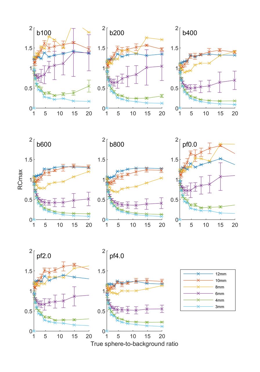
Methods: A wall-less phantom was prepared with homogenous spheres made of 18F-FDG dissolved in a gel with diameters ranging from 3 to 12 mm. The phantom consisted of two sets of spheres: one was suspended in 11C containing gel, providing a hot local background, whereas the other was suspended in a cold gel. The phantom was scanned for 210 min on a GE Discovery 690 PET/CT and reconstructed in 10 min frames resulting in sphere-to-background ratios (contrast) for the first set of spheres ranging from 0.8 to 35. The frame total count rate ranged more than one magnitude. Images were reconstructed either with Q.Clear (beta range 100 to 800) or OSEM-PSF. Time-of-flight was enabled in all reconstructions. The signal recovery of 4 common SUV metrics was evaluated (SUVmax, SUVmean, SUVpeak and SUV50bg). Results: Signal recoveries were monotonically dependent on sphere diameter regardless of contrast, reconstruction or count rate for SUVmean and SUVpeak. However, SUVmax and SUV50bg were sensitive to PSF artefacts and exhibited a non-monotonical dependence on sphere diameter with maximum recovery for the 8 mm sphere for Q.Clear with beta factors <=400 to 800 depending on contrast. Higher contrast required a higher setting of the beta factor. Similarly, SUVmax and SUV50bg exhibited decreasing signal recovery with decreasing frame count rate for constant contrast, due to the absolute count rate dependence of the penalty term.

Conclusions: For images reconstructed with Q.Clear, signal recovery for small lesions was found to depend on both local contrast and total frame count rate. For 18F-FDG, for low contrast regions, a beta factor of 400 reduced the Gibb’s edge artefact, however, for regions with higher contrast a higher beta factor was need. The results imply that optimization of the Q.Clear penalty strength parameter depends on the organ imaged, the nature of the tracer as well as the injected dose regime. [1] Munk, O. L., Tolbod, L. P., Hansen, S. B., & Bogsrud, T. V. (2017). Point-spread function reconstructed PET images of sub-centimeter lesions are not quantitative. EJNMMI Physics, 4(1), [5].

Figure
  • Download figure
  • Open in new tab
  • Download powerpoint
Previous
Back to top

In this issue

Journal of Nuclear Medicine
Vol. 60, Issue supplement 1
May 1, 2019
  • Table of Contents
  • Index by author
Article Alerts
Sign In to Email Alerts with your Email Address
Email Article

Thank you for your interest in spreading the word on Journal of Nuclear Medicine.

NOTE: We only request your email address so that the person you are recommending the page to knows that you wanted them to see it, and that it is not junk mail. We do not capture any email address.

Enter multiple addresses on separate lines or separate them with commas.
Bayesian Penalized Likelihood Image Reconstruction (Q.Clear) in oncology: Effects of contrast and count-rate dependence.
(Your Name) has sent you a message from Journal of Nuclear Medicine
(Your Name) thought you would like to see the Journal of Nuclear Medicine web site.
Citation Tools
Bayesian Penalized Likelihood Image Reconstruction (Q.Clear) in oncology: Effects of contrast and count-rate dependence.
Lars Tolbod, Nana L. Christensen, Ole Munk
Journal of Nuclear Medicine May 2019, 60 (supplement 1) 1350;

Citation Manager Formats

  • BibTeX
  • Bookends
  • EasyBib
  • EndNote (tagged)
  • EndNote 8 (xml)
  • Medlars
  • Mendeley
  • Papers
  • RefWorks Tagged
  • Ref Manager
  • RIS
  • Zotero
Share
Bayesian Penalized Likelihood Image Reconstruction (Q.Clear) in oncology: Effects of contrast and count-rate dependence.
Lars Tolbod, Nana L. Christensen, Ole Munk
Journal of Nuclear Medicine May 2019, 60 (supplement 1) 1350;
del.icio.us logo Digg logo Reddit logo Twitter logo CiteULike logo Facebook logo Google logo Mendeley logo
  • Tweet Widget
  • Facebook Like
  • Google Plus One
Bookmark this article

Jump to section

  • Article
  • Figures & Data
  • Info & Metrics

Related Articles

  • No related articles found.
  • Google Scholar

Cited By...

  • No citing articles found.
  • Google Scholar

More in this TOC Section

Physics, Instrumentation & Data Sciences

  • 3D Structural Convolutional Sparse Coding for PET Image Reconstruction
  • Exploration of Multi-objective Optimization with Genetic Algorithms for PET Image Reconstruction
  • AI-based methods for nuclear-medicine imaging: Need for objective task-specific evaluation
Show more Physics, Instrumentation & Data Sciences

Image Generation Posters

  • On Enhancing Monte-Carlo Scatter Correction for Y90 Bremsstrahlung SPECT using Guided Filtering
  • Data-driven rigid motion correction of PET brain images using list mode reconstruction
  • Simultaneous Attenuation Correction and Reconstruction of PET Images Using Deep Convolutional Encoder Decoder Networks from Emission Data
Show more Image Generation Posters

Similar Articles

SNMMI

© 2023 Journal of Nuclear Medicine

Powered by HighWire