


Article Figures & Data
Figures
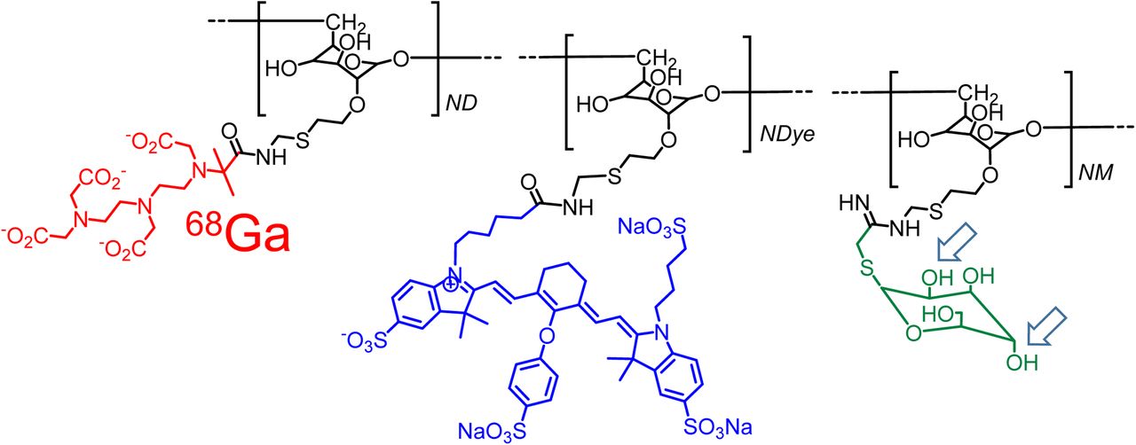
- FIGURE 1.
Bimodal molecular imaging agent, 68Ga-labeled IRDye800-tilmanocept, consists of modified dextran backbone (black), which carries CD206 receptor substrate, mannose (green); metal chelator, DTPA (red); and fluorescent tag, IRDye800CW (blue). Isomeric change of hydroxyls at C2 and C4 carbons of the receptor substrate (arrows) produces glycoconjugate that binds only to asialoglycoprotein receptor, which is specific to hepatocytes.
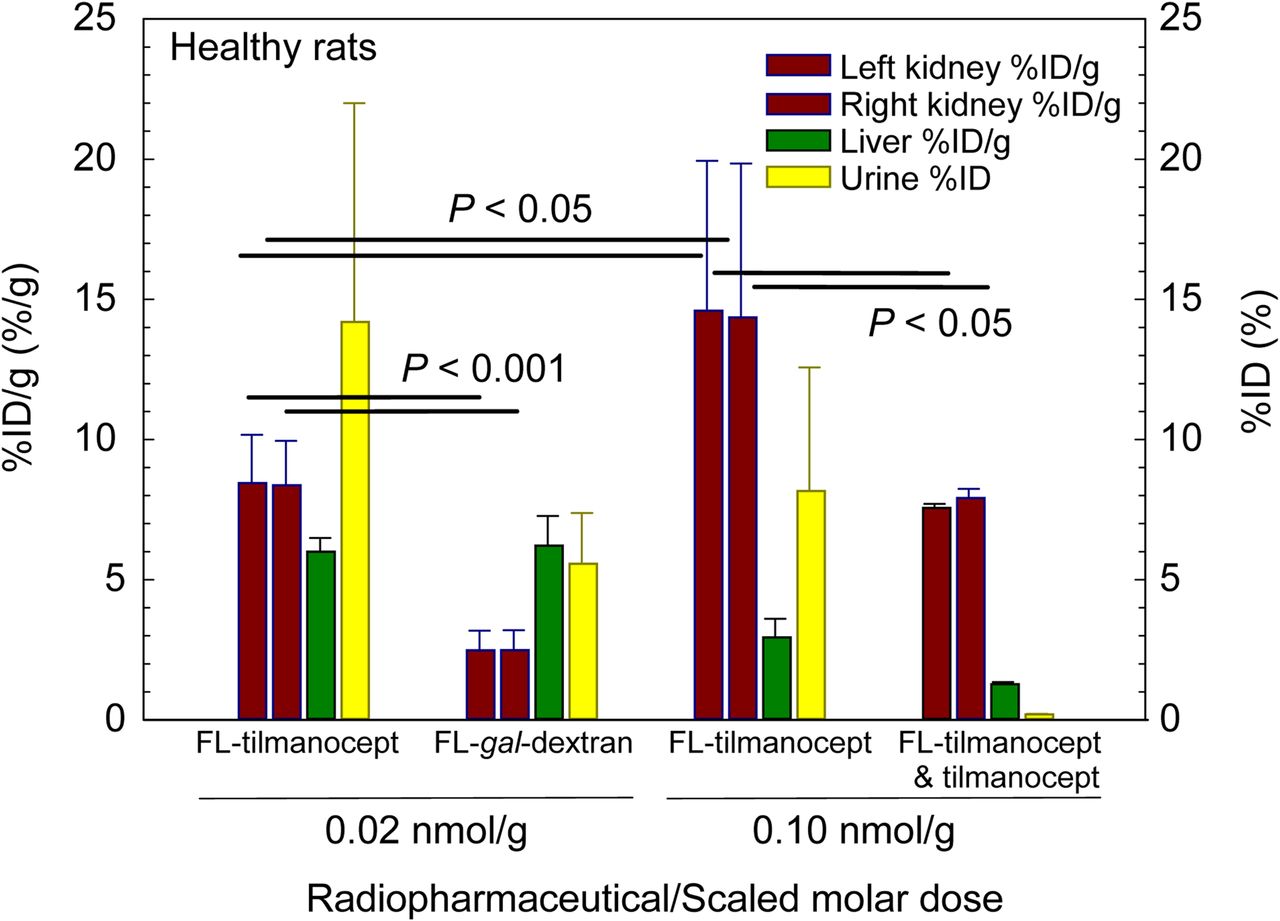
- FIGURE 2.
In vivo demonstration of receptor-mediated binding by tilmanocept. Kidneys, liver, and urine were harvested 40 min after injection of a low scaled molar dose of 68Ga-labeled IRDye800-tilmanocept (FL-tilmanocept) or 68Ga-labeled DTPA-IRDye800-galactosyl-dextran (FL-gal-dextran); injection of a high scaled dose of FL-tilmanocept; or coinjection of FL-tilmanocept and 5.0 nmol/g tilmanocept.
- FIGURE 3.
Renal cortex time–activity curves demonstrated receptor-mediated accumulation of tilmanocept. Left (upper triangles) and right (lower triangles) kidney time–activity curves displayed significantly different shapes after administration of low scaled molar dose of 68Ga-labeled IRDye800-tilmanocept (FL-tilmanocept) or 68Ga-labeled DTPA-IRDye800-galactosyl-dextran (FL-gal-dextran); injection of high scaled dose of FL-tilmanocept; or coinjection of FL-tilmanocept and 5.0 nmol/g tilmanocept.
- FIGURE 4.
PET images demonstrated receptor-mediated accumulation of 68Ga-IRDye800-tilmanocept in renal cortex (red arrows). (A) Scaled high molar dose study demonstrated low liver accumulation (green arrow) compared with kidneys and urinary bladder (yellow arrow). (B) Low molar dose study demonstrated higher liver accumulation and lower kidney accumulation. (C) Coronal cross-sections more clearly delineate activity in renal cortex. (D) Negative control study (maximum-intensity projection) exhibited significantly less radioactivity in kidneys. All images were acquired 20–40 min after injection.
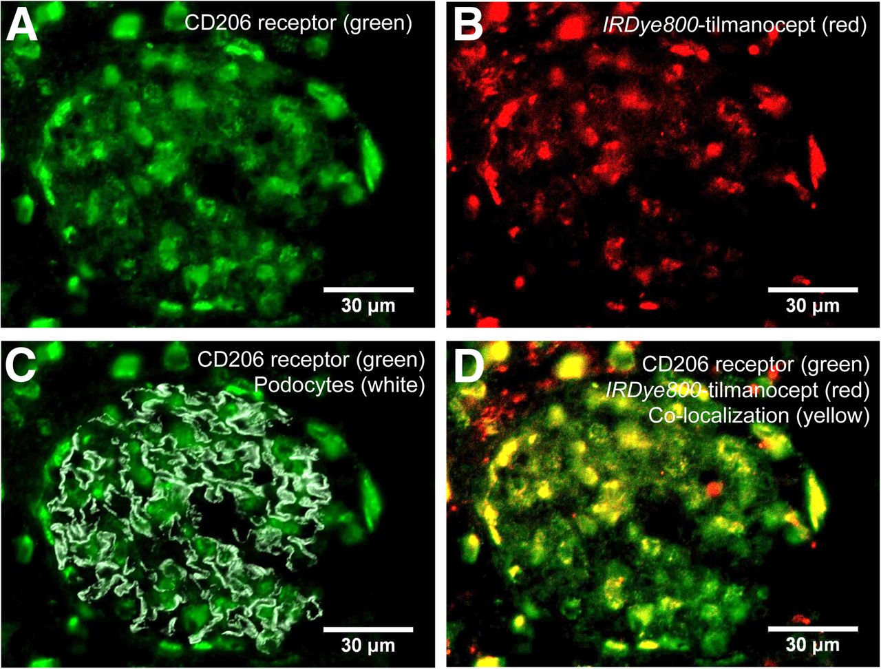
- FIGURE 5.
Histologic demonstration of tilmanocept binding to CD206 receptor within the mesangium. Four histomicrographs are shown from same section of healthy rat kidney excised 40 min after injection of IRDye800CW-tilmanocept. (A) Single channel representing distribution of CD206. (B) Single channel representing distribution of IRDye800-tilmanocept. (C) 2-channel composite, which demonstrates CD206 (green) distribution within glomerular compartment, which is defined by podocytes (gray). (D) 2-channel composite of CD206 (green) and IRDye800-tilmanocept (red) distributions (overlapping stains result in yellow).
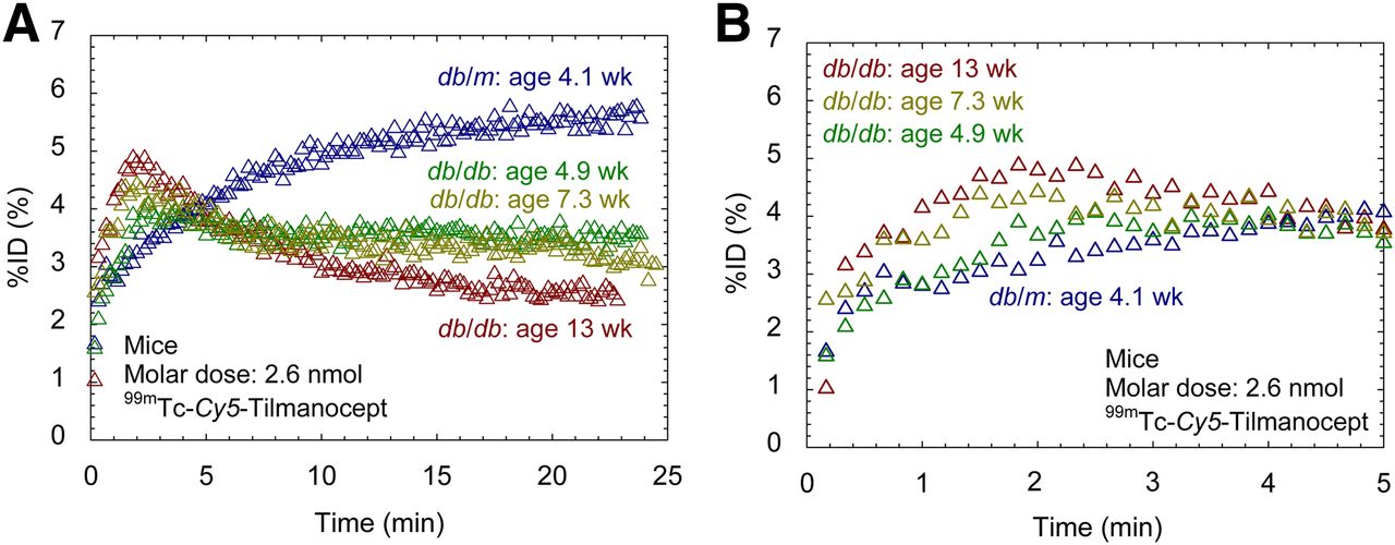
- FIGURE 6.
Studies in nondiabetic and diabetic mice demonstrated sensitivity to renal disease. Nondiabetic mice exhibited higher kidney %ID/g (red bars) and lower urine %ID than diabetic mice at 4.9, 7.3, and 13.3 wk of age. Time–activity curves peaked later in nondiabetic mice (blue bars) than diabetic mice.
- FIGURE 7.
(A) 99mTc-labeled tilmanocept time–activity curves exhibited kinetic sensitivity to chronic kidney disease. Diabetic db/db mice (green, 4.9 wk; yellow, 7.3 wk; red, 13.3 wk) peaked within 2 min after injection. Nondiabetic db/m mouse (blue) peaked 18 min after injection. (B) Expanded view shows progressively faster accumulation with increasing age.
- FIGURE 8.
After delivery to capillary lumen by renal plasma flow (arrow 1), a molecule of radiolabeled IRDye800-tilmanocept can exit glomerulus via renal plasma flow, passively be ultrafiltered into Bowman capsule (inset B, arrow 2), or passively be ultrafiltered into transverse mesangial cell matrix (inset A, arrow 3) and bind to CD206 receptor.


Jump to section
Related Articles
Cited By...
- No citing articles found.