
Main menu

  • Home
  • Content
    • Current
    • Ahead of print
    • Past Issues
    • JNM Supplement
    • SNMMI Annual Meeting Abstracts
    • Continuing Education
    • JNM Podcasts
  • Subscriptions
    • Subscribers
    • Institutional and Non-member
    • Rates
    • Journal Claims
    • Corporate & Special Sales
  • Authors
    • Submit to JNM
    • Information for Authors
    • Assignment of Copyright
    • AQARA requirements
  • Info
    • Reviewers
    • Permissions
    • Advertisers
  • About
    • About Us
    • Editorial Board
    • Contact Information
  • More
    • Alerts
    • Feedback
    • Help
    • SNMMI Journals
  • SNMMI
    • JNM
    • JNMT
    • SNMMI Journals
    • SNMMI

User menu

  • Subscribe
  • My alerts
  • Log in
  • My Cart

Search

  • Advanced search
Journal of Nuclear Medicine
  • SNMMI
    • JNM
    • JNMT
    • SNMMI Journals
    • SNMMI
  • Subscribe
  • My alerts
  • Log in
  • My Cart
Journal of Nuclear Medicine

Advanced Search

  • Home
  • Content
    • Current
    • Ahead of print
    • Past Issues
    • JNM Supplement
    • SNMMI Annual Meeting Abstracts
    • Continuing Education
    • JNM Podcasts
  • Subscriptions
    • Subscribers
    • Institutional and Non-member
    • Rates
    • Journal Claims
    • Corporate & Special Sales
  • Authors
    • Submit to JNM
    • Information for Authors
    • Assignment of Copyright
    • AQARA requirements
  • Info
    • Reviewers
    • Permissions
    • Advertisers
  • About
    • About Us
    • Editorial Board
    • Contact Information
  • More
    • Alerts
    • Feedback
    • Help
    • SNMMI Journals
  • View or Listen to JNM Podcast
  • Visit JNM on Facebook
  • Join JNM on LinkedIn
  • Follow JNM on Twitter
  • Subscribe to our RSS feeds
Research ArticlePulmonary

Should Perfusion Scintigraphy Be Performed to Follow Patients with Acute Pulmonary Embolism? If So, When?

Letizia Marconi, Antonio Palla, Lucia Cestelli, Marco Lazzeretti, Laura Carrozzi, Massimo Pistolesi and Henry Dirk Sostman
Journal of Nuclear Medicine August 2019, 60 (8) 1134-1139; DOI: https://doi.org/10.2967/jnumed.118.222737
Letizia Marconi
1Respiratory Unit, Department of Surgical, Medical and Molecular Pathology, and Critical Care, University of Pisa, Pisa, Italy
  • Find this author on Google Scholar
  • Find this author on PubMed
  • Search for this author on this site
Antonio Palla
1Respiratory Unit, Department of Surgical, Medical and Molecular Pathology, and Critical Care, University of Pisa, Pisa, Italy
  • Find this author on Google Scholar
  • Find this author on PubMed
  • Search for this author on this site
Lucia Cestelli
1Respiratory Unit, Department of Surgical, Medical and Molecular Pathology, and Critical Care, University of Pisa, Pisa, Italy
  • Find this author on Google Scholar
  • Find this author on PubMed
  • Search for this author on this site
Marco Lazzeretti
1Respiratory Unit, Department of Surgical, Medical and Molecular Pathology, and Critical Care, University of Pisa, Pisa, Italy
  • Find this author on Google Scholar
  • Find this author on PubMed
  • Search for this author on this site
Laura Carrozzi
1Respiratory Unit, Department of Surgical, Medical and Molecular Pathology, and Critical Care, University of Pisa, Pisa, Italy
  • Find this author on Google Scholar
  • Find this author on PubMed
  • Search for this author on this site
Massimo Pistolesi
2Section of Respiratory Medicine, Department of Experimental and Clinical Medicine, University of Florence, Florence, Italy
  • Find this author on Google Scholar
  • Find this author on PubMed
  • Search for this author on this site
Henry Dirk Sostman
3Houston Methodist Hospital, Houston, Texas
4Weill Cornell Medicine, New York, New York
5Methodist Research Institute, Houston, Texas; and
6Houston Methodist Hospital System, Houston, Texas
  • Find this author on Google Scholar
  • Find this author on PubMed
  • Search for this author on this site
  • Article
  • Figures & Data
  • Info & Metrics
  • PDF
Loading

Article Figures & Data

Figures

  • Tables
  • FIGURE 1.
    • Download figure
    • Open in new tab
    • Download powerpoint
    FIGURE 1.

    Reading scheme reporting anatomic distribution of lung segments. Right lung (1–10): upper lobe (apical [1]; posterior [2]; anterior [3]); middle lobe (lateral [4]; medial [5]); lower lobe (superior [6]; medial-basal [7; 7 is not shown in the figure because the anterior medial segment could only be visualized on the oblique anterior projection, which is not used in this article]; posterior-basal [8]; lateral-basal [9]; anterior-basal [10]). Left lung (11–18): upper lobe (apical-posterior [11]; anterior [12]); lingula (superior [13]; inferior [14]); lower lobe (superior [15]; medial-basal [16]; lateral-basal [17]; posterior-basal [18]). ANT = anterior; POST = posterior; R LAT = right lateral; L LAT = left lateral; RPO = right posterior oblique; LPO = left posterior oblique.
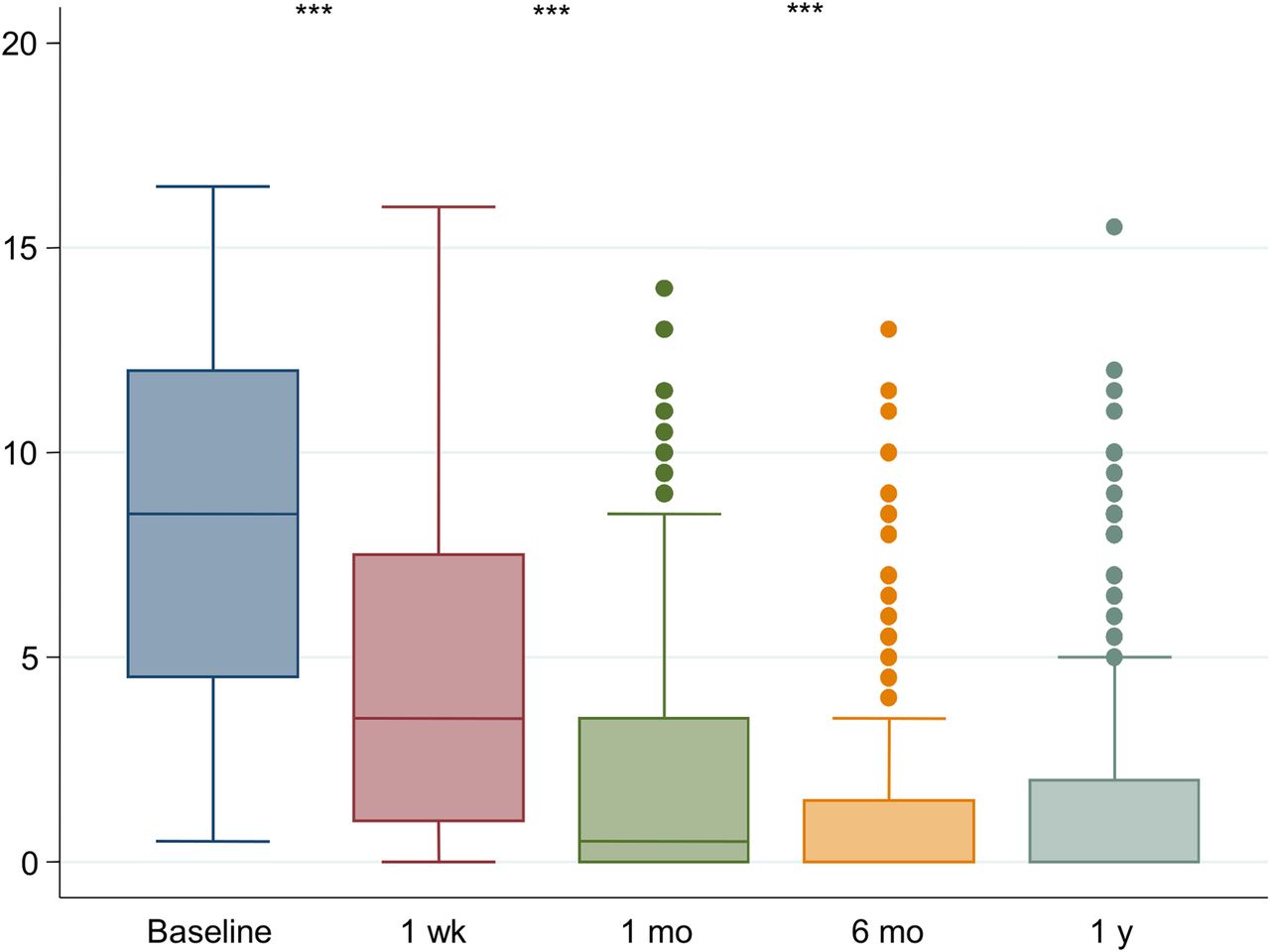
  • FIGURE 2.
    • Download figure
    • Open in new tab
    • Download powerpoint
    FIGURE 2.

    Values of PD score at different follow-up times. ***P < 0.001.
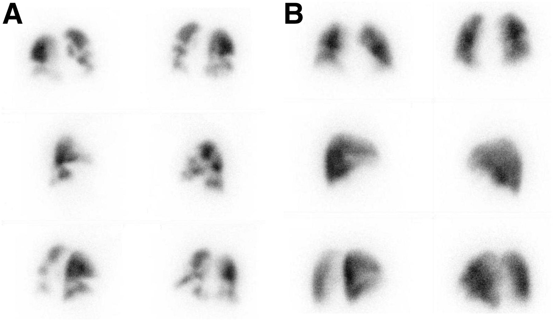
  • FIGURE 3.
    • Download figure
    • Open in new tab
    • Download powerpoint
    FIGURE 3.

    Changes in PD and hyperperfusion in 6 different views: anterior and posterior (top), right and left lateral (middle), and right and left posterior oblique (bottom). (A) Baseline PLS at acute PE shows several segmental PDs and shift of perfusion from posterior–inferior to anterior–superior lung regions (hyperperfusion) (PD score = 5.5, hyperperfusion in upper and middle lobes). (B) PLS 6 mo later shows marked improvement (PD score = 1.0, hyperperfusion in upper and middle lobes).
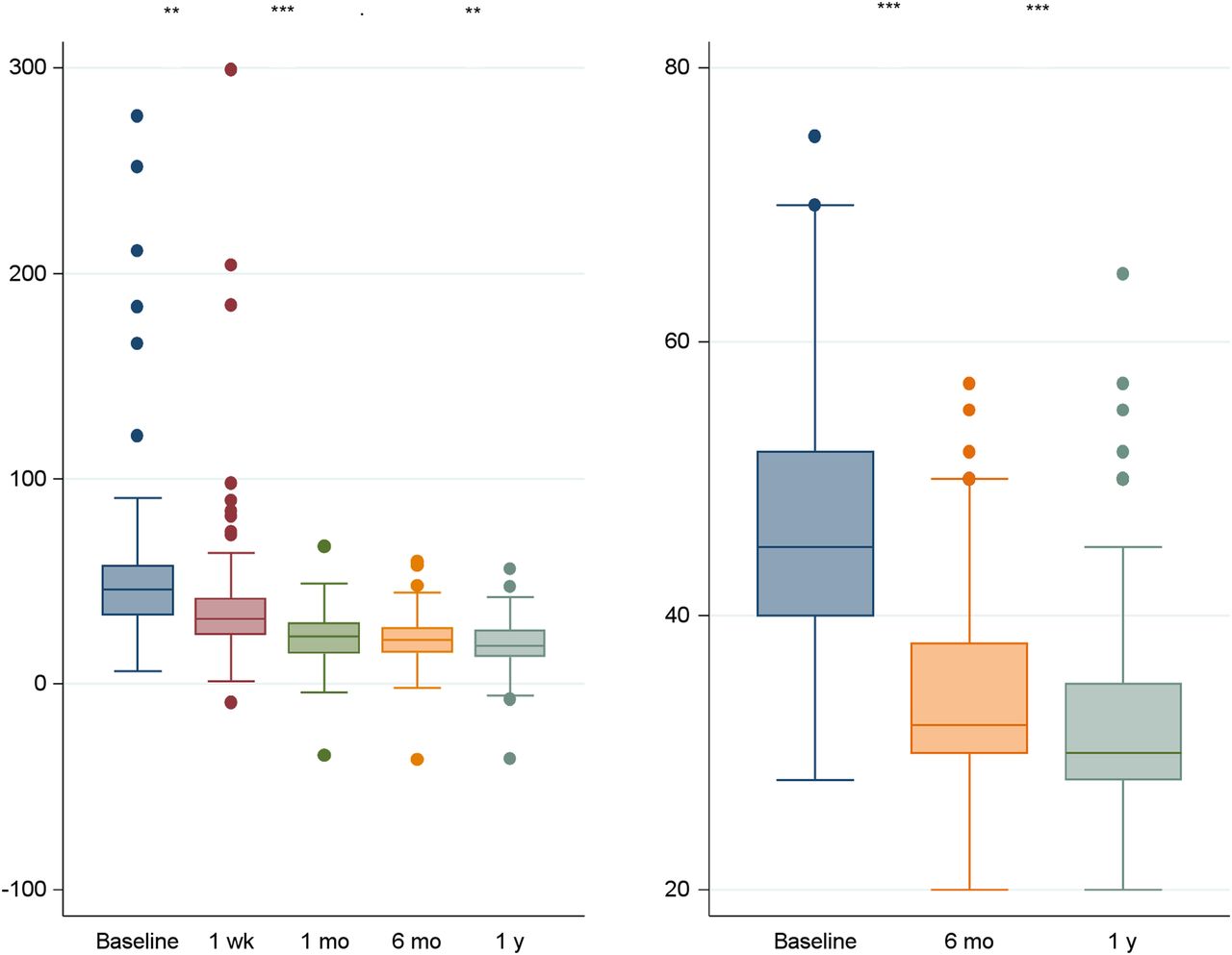
  • FIGURE 4.
    • Download figure
    • Open in new tab
    • Download powerpoint
    FIGURE 4.

    Values of PA-aO2 (A) and PAsP (B) at different follow-up times. **P < 0.01. ***P < 0.001.
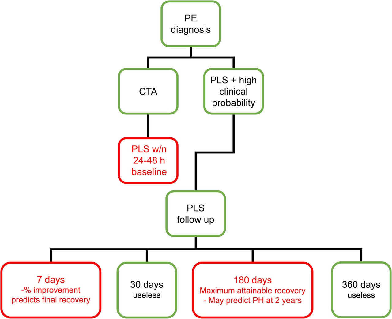
  • FIGURE 5.
    • Download figure
    • Open in new tab
    • Download powerpoint
    FIGURE 5.

    Suggested practical use of PLS in follow-up of acute PE. PH = pulmonary hypertension. Red boxes indicate suggested follow-up controls.

Tables

  • Figures
    • View popup
    TABLE 1

    Baseline Characteristics of Patients

    CharacteristicData
    Mean age ± SD (y)68.56 ± 13.82
    Male79 (43.2)
    Deep-vein thrombosis69 (37.7)
    Previous venous thromboembolism15 (8.2)
    Cardiovascular disease104 (56.8)
    Chronic obstructive pulmonary disease29 (15.8)
    Cancer-associated PE33 (18.0)
    Transient provoked PE48 (26.2)
    Unprovoked PE102 (55.7)
    • Data are n followed by percentage in parentheses, except for age.

    • View popup
    TABLE 2

    Correlation of PD Versus PA-aO2 and PAsP at Different Follow-up Times

    CorrelationBaseline1 wk1 mo6 mo1 y
    PD vs. PA-aO20.29 (P < 0.001)0.19 (P < 0.05)0.24 (P < 0.05)0.20 (P < 0.05)0.34 (P < 0.0001)
    PD vs. PAsP0.04 (P = 0.68)——0.36 (P < 0.001)0.25 (P < 0.05)
    • View popup
    TABLE 3

    Variables Associated with Recovery at 1 and 6 Months of Follow-up

    Recovery at 1 moRecovery at 6 mo
    VariableIncompleteCompletePIncompleteCompleteP
    Male45 (46.9)30 (37.0)0.18732 (50.8)38 (40.9)0.221
    Age (y)70.7 ± 13.665.6 ± 13.9<0.0573.2 ± 11.265.5 ± 14.2<0.001
    Cardiovascular disease54 (56.3)46 (56.8)0.94241 (65.1)54 (58.1)0.378
    Chronic obstructive pulmonary disease19 (19.8)9 (11.1)0.11516 (25.4)11 (11.8)0.028
    Deep-vein thrombosis41 (42.7)28 (34.6)0.26925 (39.7)36 (38.7)0.903
    Active cancer13 (13.5)17 (20.9)0.1888 (12.7%)19 (20.4)0.210
    Unprovoked PE65 (67.7)35 (43.2)<0.0544 (69.8)46 (49.5)<0.05
    Transient provoked PE18 (18.8)29 (35.8)<0.0511 (17.5)28 (30.1)0.073
    PD at baseline9.6 ± 4.26.9 ± 4.2<0.00019.9 ± 4.16.9 ± 4.3<0.0001
    1-wk percent recovery (%)30.0 ± 30.770.9 ± 27.9<0.00128.9 ± 27.464.9 ± 29.8<0.001
    PA-aO2 at baseline (mm Hg)52.7 ± 27.649.4 ± 37.90.71750.1 ± 28.649.9 ± 37.50.978
    PAsP at baseline (mm Hg)47.7 ± 10.143.9 ± 9.6<0.0548.8 ± 10.444.9 ± 10.30.05
    • Univariate analysis: P < 0.05 was considered significant. Qualitative data are expressed as numbers followed by percentages in parentheses; continuous data are expressed as mean ± SD.

    • View popup
    TABLE 4

    Variables Associated with Complete Recovery at 1 and 6 Months of Follow-up

    1 mo6 mo
    VariableOdds ratioPOdds ratioP
    PD baseline0.84 (0.76–0.92)<0.00010.80 (0.72–0.89)<0.0001
    1-wk percent recovery1.05 (1.03–1.06)<0.00011.04 (1.02–1.05)<0.0001
    Unprovoked PE2.90 (1.26–6.69)<0.05——
    Age——0.97 (0.93–0.99)0.046
    • Multivariate analysis: P < 0.05 was considered significant. Data in parentheses are 95% confidence intervals.

PreviousNext
Back to top

In this issue

Journal of Nuclear Medicine: 60 (8)
Journal of Nuclear Medicine
Vol. 60, Issue 8
August 1, 2019
  • Table of Contents
  • Table of Contents (PDF)
  • About the Cover
  • Index by author
Print
Download PDF
Article Alerts
Sign In to Email Alerts with your Email Address
Email Article

Thank you for your interest in spreading the word on Journal of Nuclear Medicine.

NOTE: We only request your email address so that the person you are recommending the page to knows that you wanted them to see it, and that it is not junk mail. We do not capture any email address.

Enter multiple addresses on separate lines or separate them with commas.
Should Perfusion Scintigraphy Be Performed to Follow Patients with Acute Pulmonary Embolism? If So, When?
(Your Name) has sent you a message from Journal of Nuclear Medicine
(Your Name) thought you would like to see the Journal of Nuclear Medicine web site.
Citation Tools
Should Perfusion Scintigraphy Be Performed to Follow Patients with Acute Pulmonary Embolism? If So, When?
Letizia Marconi, Antonio Palla, Lucia Cestelli, Marco Lazzeretti, Laura Carrozzi, Massimo Pistolesi, Henry Dirk Sostman
Journal of Nuclear Medicine Aug 2019, 60 (8) 1134-1139; DOI: 10.2967/jnumed.118.222737

Citation Manager Formats

  • BibTeX
  • Bookends
  • EasyBib
  • EndNote (tagged)
  • EndNote 8 (xml)
  • Medlars
  • Mendeley
  • Papers
  • RefWorks Tagged
  • Ref Manager
  • RIS
  • Zotero
Share
Should Perfusion Scintigraphy Be Performed to Follow Patients with Acute Pulmonary Embolism? If So, When?
Letizia Marconi, Antonio Palla, Lucia Cestelli, Marco Lazzeretti, Laura Carrozzi, Massimo Pistolesi, Henry Dirk Sostman
Journal of Nuclear Medicine Aug 2019, 60 (8) 1134-1139; DOI: 10.2967/jnumed.118.222737
Twitter logo Facebook logo LinkedIn logo Mendeley logo
  • Tweet Widget
  • Facebook Like
  • Google Plus One
Bookmark this article

Jump to section

  • Article
    • Abstract
    • MATERIALS AND METHODS
    • RESULTS
    • DISCUSSION
    • CONCLUSION
    • DISCLOSURE
    • Footnotes
    • REFERENCES
  • Figures & Data
  • Info & Metrics
  • PDF

Related Articles

  • This Month in JNM
  • PubMed
  • Google Scholar

Cited By...

  • No citing articles found.
  • Google Scholar

More in this TOC Section

Pulmonary

  • Effectiveness of D-dimer level for screening pulmonary embolism in COVID era: a single institution experience
  • VQ SPECT/CT in the follow up of recovered COVID-19 patients, with matched perfusion abnormalities and corresponding mosaic hypoattenuation on CT
  • Lung perfusion quantification using a SPECT/CT-based semi-automated segmentation workflow rather than planar perfusion scintigraphy
Show more Pulmonary

Clinical

  • Dual PET Imaging in Bronchial Neuroendocrine Neoplasms: The NETPET Score as a Prognostic Biomarker
  • Addition of 131I-MIBG to PRRT (90Y-DOTATOC) for Personalized Treatment of Selected Patients with Neuroendocrine Tumors
  • SUVs Are Adequate Measures of Lesional 18F-DCFPyL Uptake in Patients with Low Prostate Cancer Disease Burden
Show more Clinical

Similar Articles

Keywords

  • pulmonary embolism
  • pulmonary lung scintigraphy
  • follow-up
SNMMI

© 2025 SNMMI

Powered by HighWire