
Main menu

  • Home
  • Content
    • Current
    • Ahead of print
    • Past Issues
    • JNM Supplement
    • SNMMI Annual Meeting Abstracts
    • Continuing Education
    • JNM Podcasts
  • Subscriptions
    • Subscribers
    • Institutional and Non-member
    • Rates
    • Journal Claims
    • Corporate & Special Sales
  • Authors
    • Submit to JNM
    • Information for Authors
    • Assignment of Copyright
    • AQARA requirements
  • Info
    • Reviewers
    • Permissions
    • Advertisers
  • About
    • About Us
    • Editorial Board
    • Contact Information
  • More
    • Alerts
    • Feedback
    • Help
    • SNMMI Journals
  • SNMMI
    • JNM
    • JNMT
    • SNMMI Journals
    • SNMMI

User menu

  • Subscribe
  • My alerts
  • Log in
  • My Cart

Search

  • Advanced search
Journal of Nuclear Medicine
  • SNMMI
    • JNM
    • JNMT
    • SNMMI Journals
    • SNMMI
  • Subscribe
  • My alerts
  • Log in
  • My Cart
Journal of Nuclear Medicine

Advanced Search

  • Home
  • Content
    • Current
    • Ahead of print
    • Past Issues
    • JNM Supplement
    • SNMMI Annual Meeting Abstracts
    • Continuing Education
    • JNM Podcasts
  • Subscriptions
    • Subscribers
    • Institutional and Non-member
    • Rates
    • Journal Claims
    • Corporate & Special Sales
  • Authors
    • Submit to JNM
    • Information for Authors
    • Assignment of Copyright
    • AQARA requirements
  • Info
    • Reviewers
    • Permissions
    • Advertisers
  • About
    • About Us
    • Editorial Board
    • Contact Information
  • More
    • Alerts
    • Feedback
    • Help
    • SNMMI Journals
  • View or Listen to JNM Podcast
  • Visit JNM on Facebook
  • Join JNM on LinkedIn
  • Follow JNM on Twitter
  • Subscribe to our RSS feeds
Research ArticleTheranostics

Discovery of 18F-JK-PSMA-7, a PET Probe for the Detection of Small PSMA-Positive Lesions

Boris D. Zlatopolskiy, Heike Endepols, Philipp Krapf, Mehrab Guliyev, Elizaveta A. Urusova, Raphael Richarz, Melanie Hohberg, Markus Dietlein, Alexander Drzezga and Bernd Neumaier
Journal of Nuclear Medicine June 2019, 60 (6) 817-823; DOI: https://doi.org/10.2967/jnumed.118.218495
Boris D. Zlatopolskiy
1Institute of Radiochemistry and Experimental Molecular Imaging, University Hospital of Cologne, Cologne, Germany
2Institute of Neuroscience and Medicine, INM-5: Nuclear Chemistry, Forschungszentrum Jülich GmbH, Jülich, Germany
3Max Planck Institute for Metabolism Research, Cologne, Germany; and
  • Find this author on Google Scholar
  • Find this author on PubMed
  • Search for this author on this site
Heike Endepols
1Institute of Radiochemistry and Experimental Molecular Imaging, University Hospital of Cologne, Cologne, Germany
2Institute of Neuroscience and Medicine, INM-5: Nuclear Chemistry, Forschungszentrum Jülich GmbH, Jülich, Germany
4Department of Nuclear Medicine, University Hospital of Cologne, Cologne, Germany
  • Find this author on Google Scholar
  • Find this author on PubMed
  • Search for this author on this site
Philipp Krapf
1Institute of Radiochemistry and Experimental Molecular Imaging, University Hospital of Cologne, Cologne, Germany
2Institute of Neuroscience and Medicine, INM-5: Nuclear Chemistry, Forschungszentrum Jülich GmbH, Jülich, Germany
  • Find this author on Google Scholar
  • Find this author on PubMed
  • Search for this author on this site
Mehrab Guliyev
1Institute of Radiochemistry and Experimental Molecular Imaging, University Hospital of Cologne, Cologne, Germany
  • Find this author on Google Scholar
  • Find this author on PubMed
  • Search for this author on this site
Elizaveta A. Urusova
1Institute of Radiochemistry and Experimental Molecular Imaging, University Hospital of Cologne, Cologne, Germany
2Institute of Neuroscience and Medicine, INM-5: Nuclear Chemistry, Forschungszentrum Jülich GmbH, Jülich, Germany
  • Find this author on Google Scholar
  • Find this author on PubMed
  • Search for this author on this site
Raphael Richarz
1Institute of Radiochemistry and Experimental Molecular Imaging, University Hospital of Cologne, Cologne, Germany
2Institute of Neuroscience and Medicine, INM-5: Nuclear Chemistry, Forschungszentrum Jülich GmbH, Jülich, Germany
  • Find this author on Google Scholar
  • Find this author on PubMed
  • Search for this author on this site
Melanie Hohberg
4Department of Nuclear Medicine, University Hospital of Cologne, Cologne, Germany
  • Find this author on Google Scholar
  • Find this author on PubMed
  • Search for this author on this site
Markus Dietlein
4Department of Nuclear Medicine, University Hospital of Cologne, Cologne, Germany
  • Find this author on Google Scholar
  • Find this author on PubMed
  • Search for this author on this site
Alexander Drzezga
4Department of Nuclear Medicine, University Hospital of Cologne, Cologne, Germany
  • Find this author on Google Scholar
  • Find this author on PubMed
  • Search for this author on this site
Bernd Neumaier
1Institute of Radiochemistry and Experimental Molecular Imaging, University Hospital of Cologne, Cologne, Germany
2Institute of Neuroscience and Medicine, INM-5: Nuclear Chemistry, Forschungszentrum Jülich GmbH, Jülich, Germany
3Max Planck Institute for Metabolism Research, Cologne, Germany; and
  • Find this author on Google Scholar
  • Find this author on PubMed
  • Search for this author on this site
  • Article
  • Figures & Data
  • Supplemental
  • Info & Metrics
  • PDF
Loading

Article Figures & Data

Figures

  • Tables
  • Additional Files
  • FIGURE 1.
    • Download figure
    • Open in new tab
    • Download powerpoint
    FIGURE 1.

    Previously known PSMA-specific PET tracers (68Ga-1, 18F-2, and 18F-3) and emerging probes (18F-JK-4-11) investigated in this study.
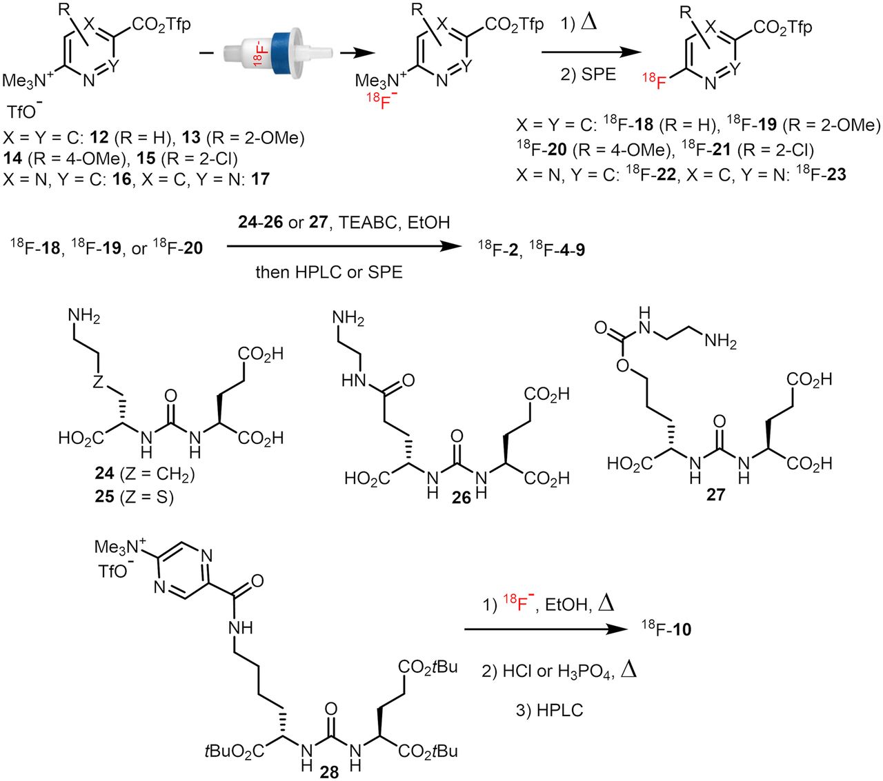
  • FIGURE 2.
    • Download figure
    • Open in new tab
    • Download powerpoint
    FIGURE 2.

    Preparation of 18F-2 and 18F-4-10. TEABC = tetraethylammonium bicarbonate.
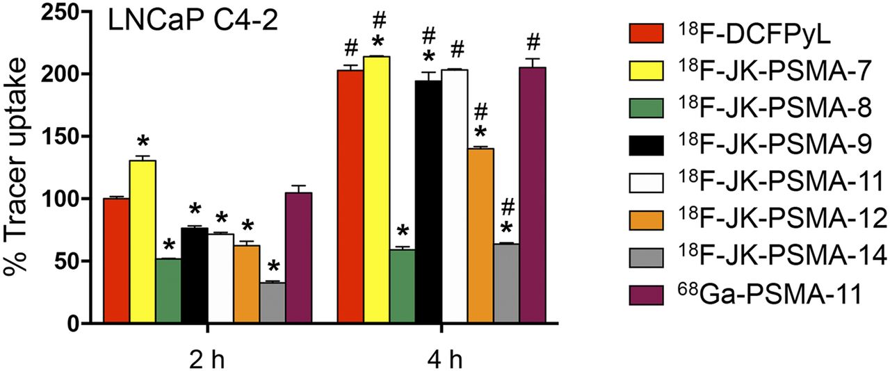
  • FIGURE 3.
    • Download figure
    • Open in new tab
    • Download powerpoint
    FIGURE 3.

    Tracer uptake in LNCaP C4-2 cells in relation to 18F-DCFPyL. 18F-DCFPyL uptake after 2 h was normalized to 100%. Only 18F-JK-PSMA-7 shows higher uptake than 18F-DCFPyL. *Significantly different from 18F-DCFPyL at same time point. F7,38 = 1,136; P < 0.0001 for factor “tracer,” post hoc P < 0.05. #Significantly higher than 2-h uptake of same tracer. F1,38 = 6,981; P < 0.0001 for factor “time,” post hoc P < 0.05.

  • FIGURE 4.
    • Download figure
    • Open in new tab
    • Download powerpoint
    FIGURE 4.

    TBR of PSMA tracers. (A) Sagittal section of PSMA image with SCG and background (Bg) volumes of interest. (B) Graph showing that TBR (SCG/Bg) 60–120 min after injection did not significantly differ among groups (F4,10 = 2.95, P = 0.0756). (C) TBR analyzed for four 30-min frames. *TBE significantly higher for 18F-JK-PSMA-7 than for 68Ga-PSMA-11 (F4,40 = 5.97, P = 0.0102, for factor “tracer,” post hoc P < 0.05). #TBR significantly higher for 18F-JK-PSMA-7 than for 18F-JK-PSMA-8 or 68Ga-PSMA-11 (P < 0.05). TBR of 18F-JK-PSMA-7 and 18F-PSMA-1007 increased significantly over time (F3,30 = 9.12, P = 0.0002, for factor “frame”). †For 18F-JK-PSMA-7, frame 4 was significantly different from frames 1 and 2 (P < 0.05). ‡For 18F-PSMA-1007, frame 4 was significantly different from frame 1 (P < 0.05).
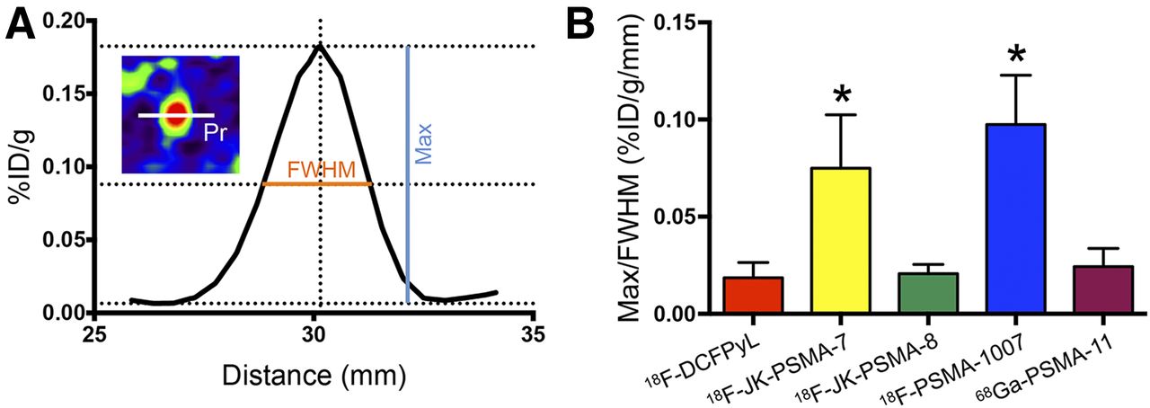
  • FIGURE 5.
    • Download figure
    • Open in new tab
    • Download powerpoint
    FIGURE 5.

    Acutance (measured for SCG) using different PSMA tracers. (A) Profile plot of 1-pixel profile (Pr in inset) through SCG center. Peak against adjacent background (max) and FWHM were measured. (B) Ratio max/FWHM roughly represents slope of profile plot and reflects acutance. *18F-JK-PSMA-7 and 18F-PSMA-1007 show significantly higher acutance than 18F-DCFPyL, 18F-JK-PSMA-8, or 68Ga-PSMA-11 (F4,10 = 12.77, P = 0.0006; post hoc P < 0.05). %ID = percentage injected dose.
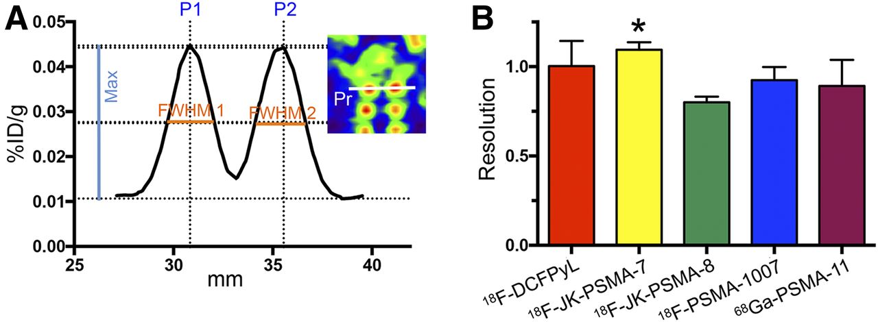
  • FIGURE 6.
    • Download figure
    • Open in new tab
    • Download powerpoint
    FIGURE 6.

    Resolution of images acquired with different tracers. (A) Profile plot of 1-pixel profile (Pr in inset) through apical pair of DRG. Peak against adjacent background (max) and FWHM were measured for each ganglion. P2–P1 was distance between peaks. (B) Formula R = 2(P2 − P1)/1.7(FWHM1 + FWHM2) yields image resolution. *Image resolution was significantly higher for 18F-JK-PSMA-7 than for 18F-JK-PSMA-8 (F4,10 = 3.80, P = 0.0396; post hoc P < 0.05).
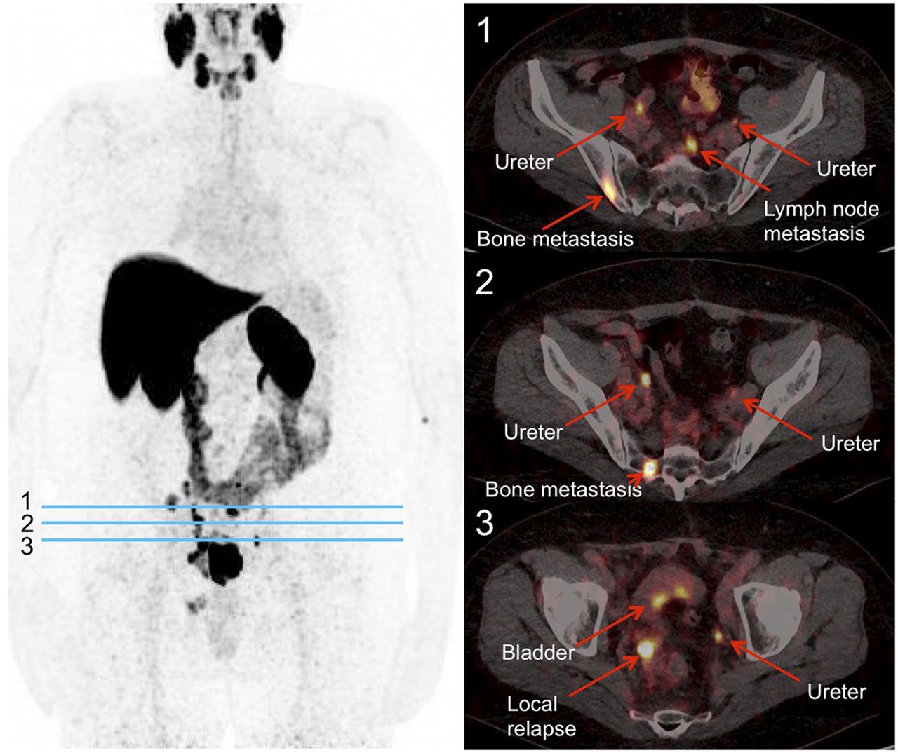
  • FIGURE 7.
    • Download figure
    • Open in new tab
    • Download powerpoint
    FIGURE 7.

    18F-JK-PSMA-7 scan in patient with relapsed PCa. This 64-y-old patient had serum prostate-specific antigen level of 130 ng/mL. Scan was started 233 min after injection of 384 MBq of 18F-JK-PSMA-7. (Left) Maximum-intensity projection of PET image. (Right) PET/CT images at transverse levels indicated by blue lines.

Tables

  • Figures
  • Additional Files
    • View popup
    TABLE 1

    Comparison of Quality of PET Images Obtained with Different Tracers (60–120 Minutes After Injection, n = 3)

    TracerAcutanceResolutionTBR
    18F-DCFPyL0.019 ± 0.0081.002 ± 0.1416.38 ± 1.87
    18F-JK-PSMA-70.075 ± 0.027*1.095 ± 0.042†8.15 ± 1.71
    18F-JK-PSMA-80.021 ± 0.0050.800 ± 0.0324.60 ± 1.30
    18F-PSMA-10070.097 ± 0.025*0.924 ± 0.0746.23 ± 1.88
    68Ga-PSMA-110.024 ± 0.0090.892 ± 0.1464.46 ± 0.08
    • ↵* Significantly higher than 18F-DCFPyL, 18F-JK-PSMA-8, and 68Ga-PSMA-11 (F4,10 = 12.77, P = 0.0006; post hoc P < 0.05).

    • ↵† Significantly higher than 18F-JK-PSMA-8 (F4,10 = 3.80, P = 0.0396; post hoc P < 0.05).

    • View popup
    TABLE 2

    Tracer Uptake (SUVBW) in Different Tissues 60–120 Minutes After Injection (n = 3 Each)

    TracerSCGSalivary glandLiverBloodBoneBackground
    18F-DCFPyL20.9 ± 8.213.2 ± 5.378.4 ± 29.412.9 ± 3.010.5 ± 3.53.2 ± 0.7
    18F-JK-PSMA-731.3 ± 10.514.4 ± 2.4119.3 ± 8.3¶16.3 ± 4.29.9 ± 1.94.0 ± 1.0
    18F-JK-PSMA-814.4 ± 2.69.0 ± 2.529.0 ± 7.28.6 ± 0.319.2 ± 2.13.2 ± 0.4
    18F-PSMA-100794.8 ± 19.6*62.1 ± 14.2†50.7 ± 4.362.6 ± 10.7‖33.2 ± 9.5#15.9 ± 1.7**
    68Ga-PSMA-1141.0 ± 3.438.4 ± 10.2‡43.7 ± 14.439.6 ± 4.6§18.5 ± 0.69.2 ± 0.6**
    • ↵* Significantly higher than any other (F4,10 = 26.52, P < 0.0001; post hoc P < 0.05).

    • ↵† Significantly higher than any other (F4,10 = 22.06, P < 0.0001; post hoc P < 0.05).

    • ↵‡ Significantly higher than 18F-DCFPyL (F4,10 = 22.06, P < 0.0001; post hoc P < 0.05).

    • ↵¶ Significantly higher than 18F-JK-PSMA-8 (18F-PSMA-1007 and 68Ga-PSMA-11. F4,10 = 7.83, P = 0.0040; post hoc P < 0.05).

    • ↵‖ Significantly higher than any other (F4,10 = 47.59, P < 0.0001; post hoc P < 0.05).

    • ↵§ Significantly higher than 18F-DCFPyL, 18F-JK-PSMA-7, and 18F-JK-PSMA-8 (F4,10 = 47.59, P < 0.0001; post hoc P < 0.05).

    • ↵# Significantly higher than any other (F4,10 = 12.19, P = 0.0007; post hoc P < 0.05).

    • ↵** Significantly different from all others (F4,10 = 93.81, P < 0.0001; post hoc P < 0.05).

Additional Files

  • Figures
  • Tables
  • Supplemental Data

    Files in this Data Supplement:

    • Supplemental Data
PreviousNext
Back to top

In this issue

Journal of Nuclear Medicine: 60 (6)
Journal of Nuclear Medicine
Vol. 60, Issue 6
June 1, 2019
  • Table of Contents
  • Table of Contents (PDF)
  • About the Cover
  • Index by author
Print
Download PDF
Article Alerts
Sign In to Email Alerts with your Email Address
Email Article

Thank you for your interest in spreading the word on Journal of Nuclear Medicine.

NOTE: We only request your email address so that the person you are recommending the page to knows that you wanted them to see it, and that it is not junk mail. We do not capture any email address.

Enter multiple addresses on separate lines or separate them with commas.
Discovery of 18F-JK-PSMA-7, a PET Probe for the Detection of Small PSMA-Positive Lesions
(Your Name) has sent you a message from Journal of Nuclear Medicine
(Your Name) thought you would like to see the Journal of Nuclear Medicine web site.
Citation Tools
Discovery of 18F-JK-PSMA-7, a PET Probe for the Detection of Small PSMA-Positive Lesions
Boris D. Zlatopolskiy, Heike Endepols, Philipp Krapf, Mehrab Guliyev, Elizaveta A. Urusova, Raphael Richarz, Melanie Hohberg, Markus Dietlein, Alexander Drzezga, Bernd Neumaier
Journal of Nuclear Medicine Jun 2019, 60 (6) 817-823; DOI: 10.2967/jnumed.118.218495

Citation Manager Formats

  • BibTeX
  • Bookends
  • EasyBib
  • EndNote (tagged)
  • EndNote 8 (xml)
  • Medlars
  • Mendeley
  • Papers
  • RefWorks Tagged
  • Ref Manager
  • RIS
  • Zotero
Share
Discovery of 18F-JK-PSMA-7, a PET Probe for the Detection of Small PSMA-Positive Lesions
Boris D. Zlatopolskiy, Heike Endepols, Philipp Krapf, Mehrab Guliyev, Elizaveta A. Urusova, Raphael Richarz, Melanie Hohberg, Markus Dietlein, Alexander Drzezga, Bernd Neumaier
Journal of Nuclear Medicine Jun 2019, 60 (6) 817-823; DOI: 10.2967/jnumed.118.218495
Twitter logo Facebook logo LinkedIn logo Mendeley logo
  • Tweet Widget
  • Facebook Like
  • Google Plus One
Bookmark this article

Jump to section

  • Article
    • Abstract
    • MATERIALS AND METHODS
    • RESULTS
    • DISCUSSION
    • CONCLUSION
    • DISCLOSURE
    • Acknowledgments
    • Footnotes
    • REFERENCES
  • Figures & Data
  • Supplemental
  • Info & Metrics
  • PDF

Related Articles

  • This Month in JNM
  • PubMed
  • Google Scholar

Cited By...

  • Intraindividual Comparison of 18F-PSMA-1007 with Renally Excreted PSMA Ligands for PSMA PET Imaging in Patients with Relapsed Prostate Cancer
  • An 18F-Labeled PSMA Ligand for PET/CT of Prostate Cancer: First-in-Humans Observational Study and Clinical Experience with 18F-JK-PSMA-7 During the First Year of Application
  • Development of Novel PSMA Ligands for Imaging and Therapy with Copper Isotopes
  • Google Scholar

More in this TOC Section

Theranostics

  • Determination of the Intralesional Distribution of Theranostic 124I-Omburtamab Convection-Enhanced Delivery in Treatment of Diffuse Intrinsic Pontine Glioma
  • Evidence-Based Clinical Protocols to Monitor Efficacy of [177Lu]Lu-PSMA Radiopharmaceutical Therapy in Metastatic Castration-Resistant Prostate Cancer Using Real-World Data
  • 177Lu-Labeled Anticlaudin 6 Monoclonal Antibody for Targeted Therapy in Esophageal Cancer
Show more Theranostics

Basic

  • Dopamine D1 Receptor Agonist PET Tracer Development: Assessment in Nonhuman Primates
  • Optical Navigation of the Drop-In γ-Probe as a Means to Strengthen the Connection Between Robot-Assisted and Radioguided Surgery
  • Synthesis and Preclinical Evaluation of a 68Ga-Labeled Adnectin, 68Ga-BMS-986192, as a PET Agent for Imaging PD-L1 Expression
Show more Basic

Similar Articles

Keywords

  • prostate carcinoma
  • PSMA
  • radiofluorination
  • Positron Emission Tomography
  • imaging
  • preclinical model
SNMMI

© 2025 SNMMI

Powered by HighWire