


Article Figures & Data
Figures
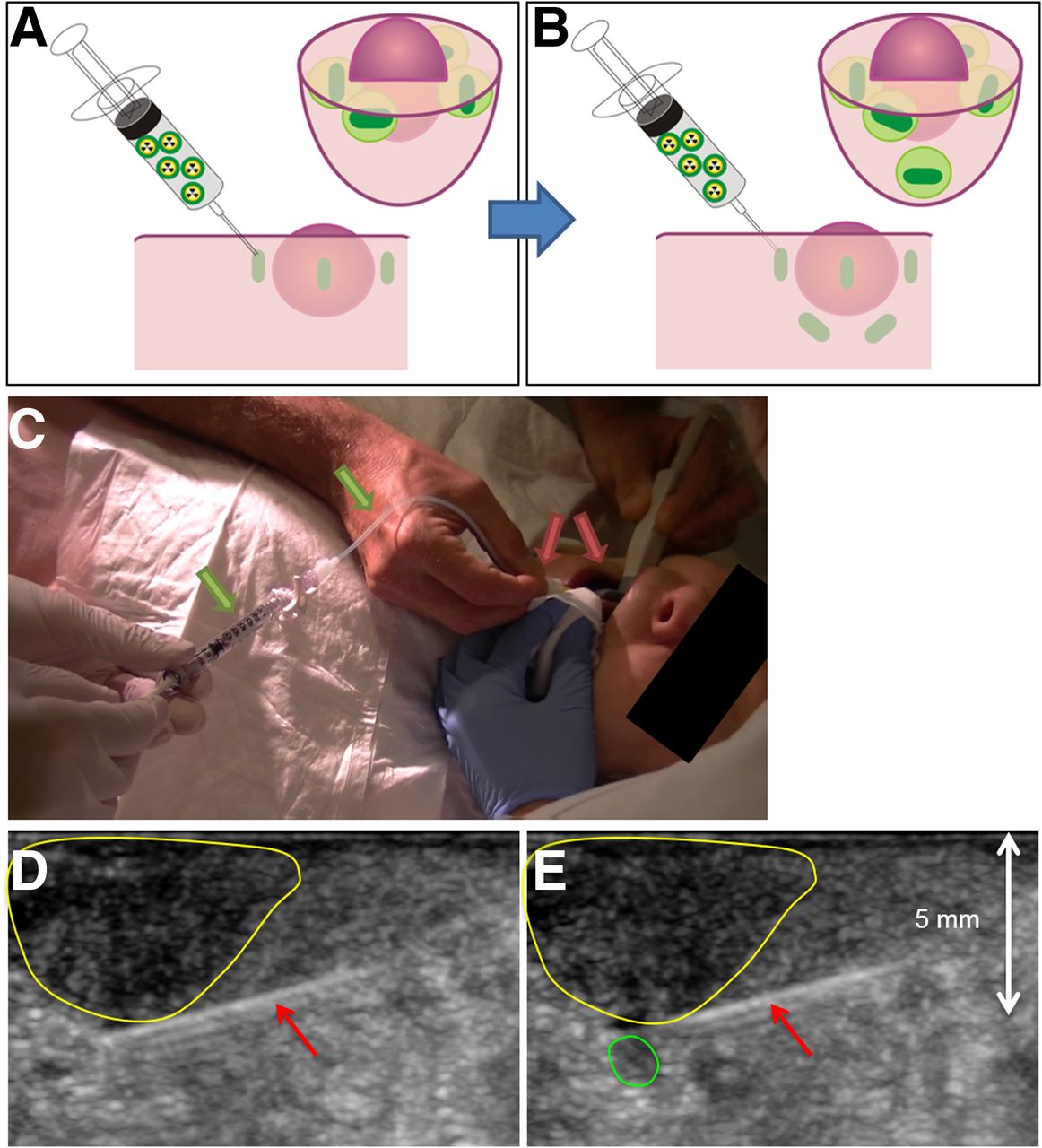
- FIGURE 1.
Injection method. (A) Standard SN procedure consists of 3–4 superficial injections surrounding tumor. (B) In this study, 12 patients received additional injections, placed deep behind tumor using ultrasound guidance. (C) Radiologist inserts injection needle under ultrasound guidance (red arrows). Nuclear physician then injects ICG-99mTc-nanocolloid via syringe-tubing system (green arrows). (D) Radiologist keeps needle (red arrows) 1–2 mm next to tumor border (yellow area) while nuclear physician injects tracer. (E) Deposit of ICG-99mTc-nanocolloid can be visualized on ultrasound (green area).
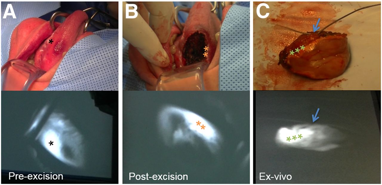
- FIGURE 2.
White light (top) and fluorescence (bottom) imaging during procedure. (A) Preexcision image of tumor. (B) Postexcision image of same tumor. (C) Tumor tissue ex vivo. Stitch in excised tissue marks anterior side (arrow). *Tracer deposit. **Remaining fluorescence signal. ***Tracer deposit surrounding tumor.
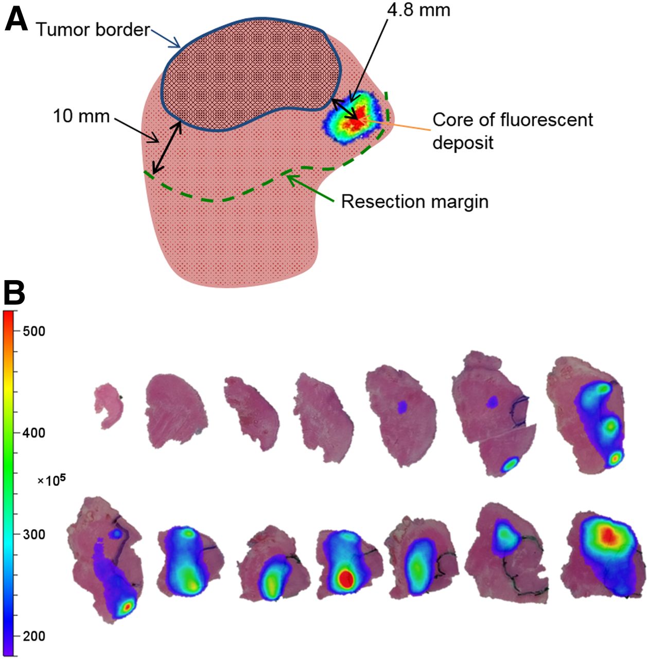
- FIGURE 3.
Schematic overview of pathologic analysis. (A) Tumor has been marked on H&E-stained slides with indelible ink. Overlay of fluorescence signal is projected onto original image. Distance has been measured between core of fluorescent spots and tumor border as drawn on slides. (B) H&E-stained slides from patient in tumor demarcation group with overlay of fluorescence signal. Fluorescent deposits in excised tumor tissue can be visualized, and distance between fluorescent cores and tumor border was measured.


Tables
Parameter Tumor tattooing Standard SN procedure P Mean administered dose (MBq) ± SD 100.0 ± 34.8 (median, 95.9) 78.7 ± 7.3 (median, 79.0) 0.059 Preoperative SN-related hot spots (n) Lymphoscintigraphy 34 (median per patient, 2) 50 (median per patient, 2) 0.674 SPECT/CT 34 (median per patient, 2) 52 (median per patient, 3) 0.836 Mean intraoperative SN results Fluorescent, in vivo 4.0 (median, 3) 2.7 (median, 2.5) 0.192 Fluorescent, ex vivo 3.8 (median, 3.5) 3.2 (median, 3) 0.365 Radioactive, in vivo 2.9 (median, 3) 2.7 (median, 2.5) 0.745 Radioactive, ex vivo 4.0 (median, 4) 3.0 (median, 3) 0.159 Total excised SNs (n) 54 (mean, 4.5; median, 4) 60 (mean, 3.2; median, 3) 0.090 Total excised non-SNs (n) 11 (mean, 0.9; median, 0.5) 9 (mean, 0.5; median, 0) 0.215 Total excised nodes (n) 65 69 Pathology (n) SNs 84 83 0.072 Non-SNs 32 7 0.306 Total nodes 116 90 0.060 Tumor-positive SNs 5 (in 3 patients) 5 (in 4 patients) Tumor-positive non-SNs 1* (in 1 patient) 0 Tumor-positive nodes 6 (in 3 patients) 5 (in 4 patients) Resection status (n) R0 11 (91.7%) 19 (100.0%) R1 1 (8.3%) 0 ↵* In this patient, 1 SN was also found tumor-positive.
Parameter Data Fluorescent spots on tissue section with tumor tissue (n) 62 Fluorescence signal outside tumor tissue (n) 58/62 (93.5%) Fluorescence signal within 10-mm margin 56/58 (96.6%) Fluorescence signal outside 10-mm margin 2/58 (3.4%) Fluorescence signal inside tumor tissue (n) 4/62 (6.5%) Patients with all fluorescent spots 0–10 mm outside tumor border (n) 8/12 (66.7%) Patients with fluorescent spot inside tumor (n) 4/12 (33.3%) Distance from tumor border to fluorescence signal outside tumor (mm) Median 2.8 Mean 3.2 Range 0.2–10.2 (IQR, 1.3–4.3) Distance from tumor border to fluorescence signal inside tumor (mm) Median 1.2 Mean 1.7 Range 0.2–4.2 IQR = interquartile range.
Supplemental Data
Files in this Data Supplement:


Jump to section
Related Articles
Cited By...
- No citing articles found.