


Article Figures & Data
Figures
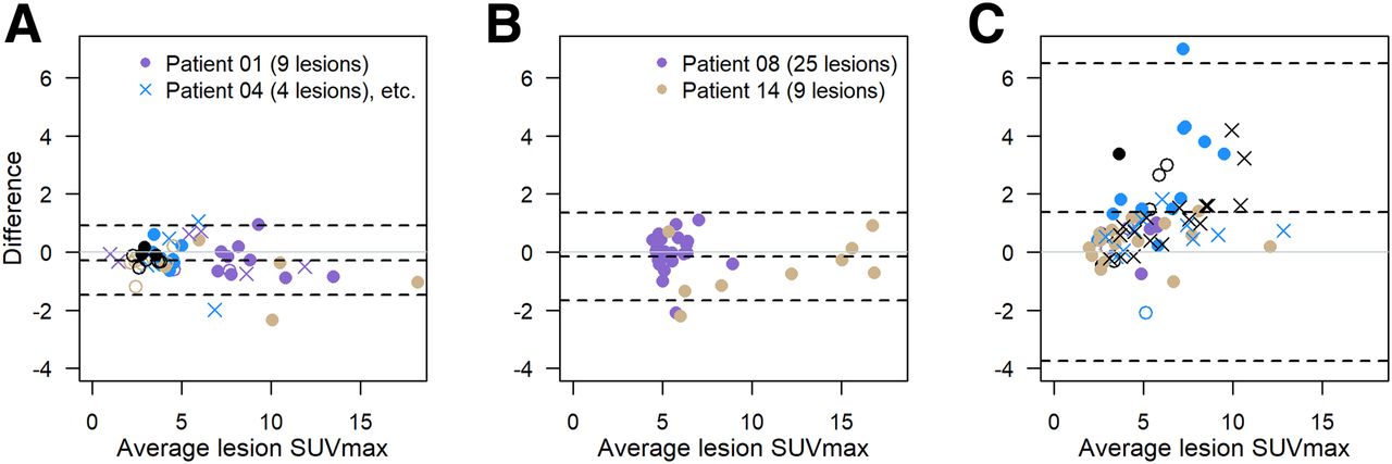
- FIGURE 1.
Bland–Altman plots of difference in SUVmax vs. average SUVmax: 10 patients (51 lesions) with repeat scans using same scanner (A); 2 patients (34 lesions) using different scanners from same academic institution (B); and 11 patients (77 lesions) using different scanners from different sites (C). Within each panel, plotting character/color is same for multiple lesions in single patient. Dashed lines = average difference and 95% limits of agreement. The 2 lesions from melanoma patient (SUVmax, 38.3 and 25.0 on first scan and 19.2 and 16.4 on second scan) are not shown in C but contribute to limits-of-agreement calculations.
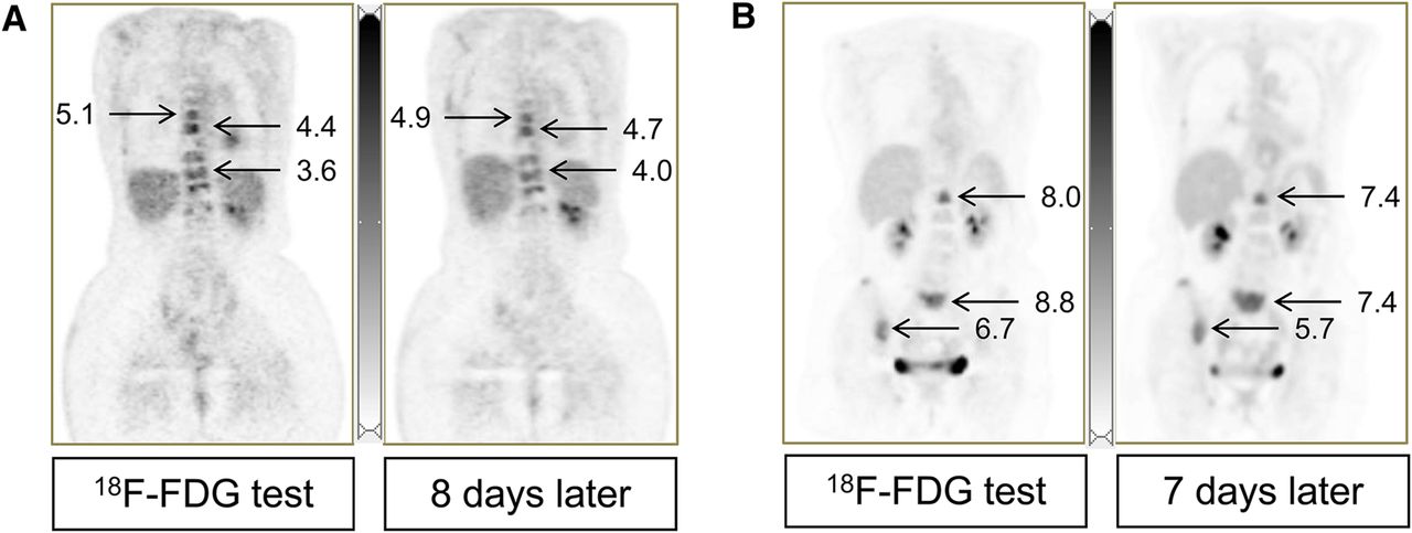
- FIGURE 2.
(A) Coronal images from 60-y-old woman with stage IV ductal breast carcinoma (blue circles in Fig. 1A, same scanner). SUVmax for 9 evaluable lesions ranged from 3.4 to 5.1 (average, 4.0) for first scan and from 3.1 to 4.9 (average, 4.2) for second scan. Percentage difference was −16% to +16% (average, 3.9%); SUV unit difference was −0.62 to +0.64 (average, 0.15). (B) A 73-y-old woman with stage IV mixed ductal/lobular breast carcinoma (yellow circles in Fig. 1C, different institutions). SUVmax for 17 evaluable lesions was 2.0–12.2 (average, 4.8) for first scan and 1.9–12.0 (average, 4.4) for second scan. Percentage difference was −24% to +25% (average, −7.1%); SUV unit difference was −1.4 to +1.0 (average, −0.39). Normal liver SUVmean was 2.5 (A) and 2.6 (B) in both scans.
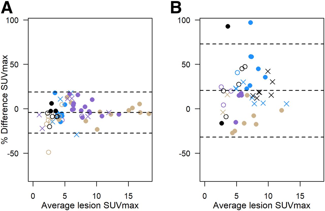
- FIGURE 3.
Percentage difference in SUVmax vs. average SUVmax: 12 patients (85 lesions) with repeat scans using same scanner or different scanners from same unit (combined data from Figs. 1A and 1B) (A); 11 patients (77 lesions) using different scanners from different sites (B). Plotting character/color identifies multiple lesions in single patient, as for Figure 1. Dashed lines = average percentage difference and 95% limits of agreement.
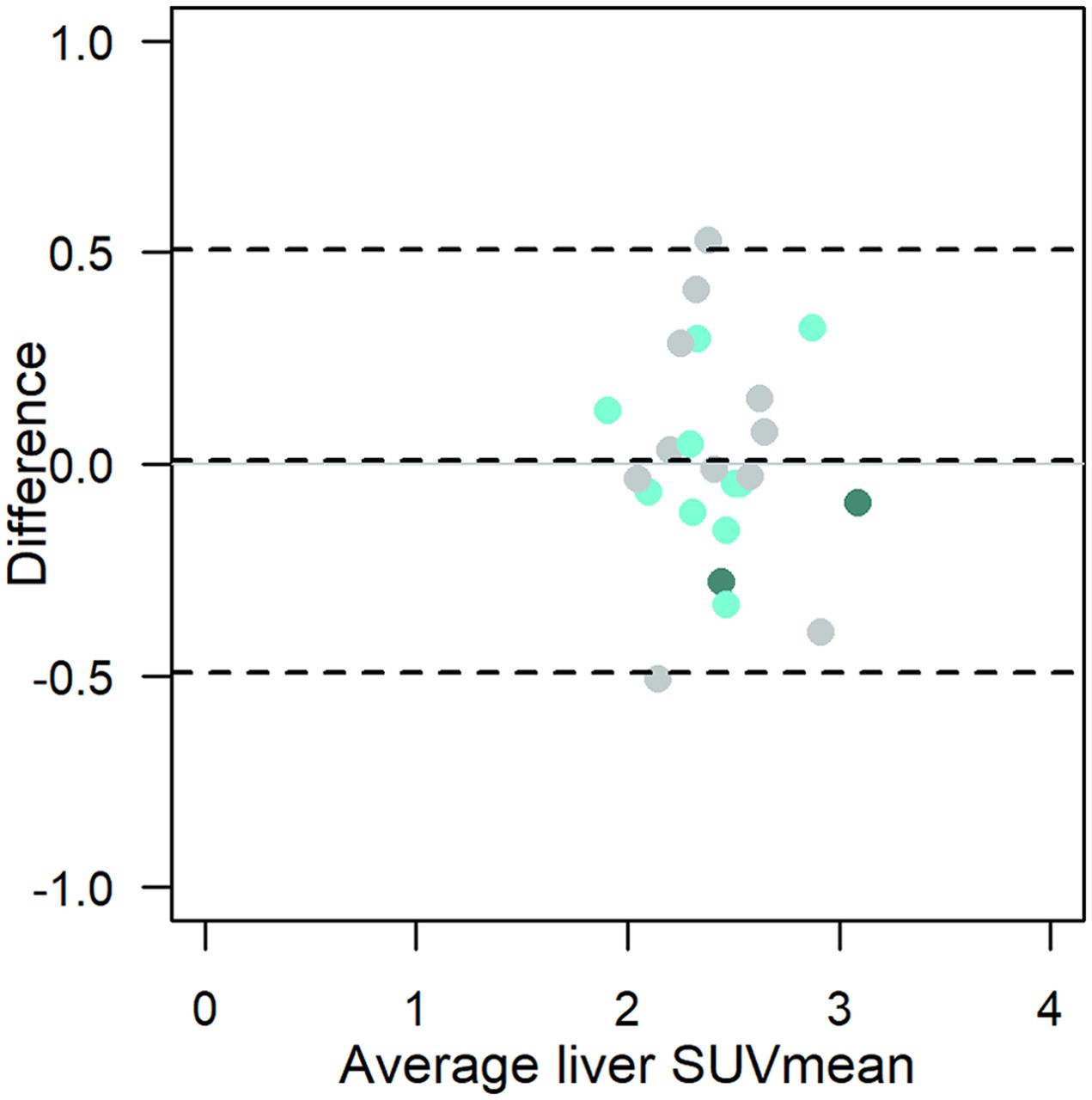
- FIGURE 4.
Bland–Altman plot of liver SUVmean (n = 23). Light-green circles = same scanner; dark-green circles = different scanners from same site; gray circles = different scanner models from different sites; dashed lines = average difference and 95% limits of agreement.


Tables
Characteristic (PET emission images) Discovery STE (both) Gemini TF 64 Biograph 20 mCT Biograph 6 N 17 3D, 18 2D 6 2 3 Slice thickness (mm) 3.27 4 5 5 Pixel size (mm) 5.47 4 4.07 4.06 Pixel volume (cm3) 0.098 0.064 0.083 0.083 Reconstruction diameter (mm) 700 576 815 683 Array size (pixels) 128 × 128 144 × 144 200 × 200 168 × 168 Bed-position duration (min) 5, 7 4 2, 3.5 3 Average coverage (total cm/total min) 2.1, 2.4 2.3 2.8, 4.9 4.0, 4.1 Reconstruction method OSEM 3D/2D BLOB-OS-TOF PSF+TOF 2i21s PSF, 3i24s Scatter correction method Model-based SS-SIMUL Model-based Model-based 3D = 3-dimensional; 2D = 2-dimensional; OSEM = ordered-subset expectation maximization; TOF = time-of-flight; PSF = point-spread function; 2i21s = 2 iterations, 21 subsets; 3i24s = 3 iterations, 24 subsets; SS-SIMUL = single-scatter simulation.
All scans were in inferior-to-superior direction.
Characteristic Same site/scanner (n = 10) Same institution, different scanner (n = 2) Different site/scanner (n = 11) All patients (n = 23) Age (y) 53.5 (32–67) 58 (45–71) 66 (43–76) 60 (32–76) Body mass index at scan 1 (kg/m2) 28.3 (18.3–37.6) 38.6 (31.4–45.7) 28.4 (22.5–43.2) 28.4 (18.3–45.7) Time between scans (d) 8 (2–15) 7 (1–13) 10 (7–14) 9 (1–15) Lesions* (n) 5 (1–9) 17 (9–25) 5 (1–17) 5 (1–25) Sex (n) Male – – 3 3 (13%) Female 10 2 8 20 (87%) Diagnosis (n) Breast cancer 10 2 6 18 (78%) Other† – – 5 5 (22%) Ongoing treatment between scans (n) None 2 – 5 7 (30%) Bisphosphonates or biologic only 1 1 1 3 (13%) Endocrine therapy‡ 4 – 3 7 (30%) Chemotherapy§ 3 1 2 6 (26%) PET/CT scanner (n) Discovery STE (both) 10 2 – 12 (52%) Ingenuity TF – – 6 6 (26%) Biograph 6 – – 3 3 (13%) Biograph 20 mCT – – 2 2 (9%) ↵* All identified, but ≤25 lesions/patient used in analysis.
↵† 1 each: colorectal, head/neck, stage IV lung, stage III melanoma, neuroendocrine/Merkel cell cancer.
↵‡ 2 also bisphosphonates; 3 also biologic.
↵§ 4 also biologic; 1 also endocrine. Biologics: erlotinib, trastuzumab, everolimus, pertuzumab, denosumab, ado-trastuzumab emtansine. Cytotoxic agents: capecitabine, cyclophosphamide, doxorubicin.
Continuous data are expressed as median and range.
Model Fitted % difference in repeat scans 95% confidence interval Model 1 (SUVmax)* A. Same scanner (n = 10) 8% 6%–11% B. Same institution, different scanner (n = 2) 6% 3%–11% C. Different institution and scanner (n = 11) 18% 13%–24% Model 2 (SUVmax)† Same scanner or institution 8% 6%–10% Different institution and scanner 18% 13%–24% Model 3 (liver SUVmean)‡ Same scanner or institution 5% 3%–10% Different scanner and institution 6% 3%–11% ↵* C > A (P = 0.0015), C > B (P = 0.003), A and B not different on average (P = 0.66) (Tukey–Kramer adjustment for pairwise comparisons).
↵† P < 0.001, Wald test.
↵‡ P = 0.85, Wald test (n = 23).
Group differences are back-transformed from log(absolute percentage difference + 1); n = 162 tumors in 23 patients.
Supplemental Data
Files in this Data Supplement:


