


Article Figures & Data
Figures
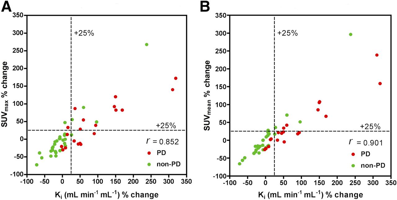
- FIGURE 1.
Lesion analysis: Scatterplot for percentage changes in Ki against percentage changes in SUVmax and SUVmean for PD and non-PD 8 wk after start of treatment.
- FIGURE 2.
Patient basis: Scatterplot for percentage changes in Ki against percentage changes in SUVmax and SUVmean for PD and non-PD 8 wk after start of treatment.


Tables
Baseline 8 wk Mean % change Mean tumor volume Ki SUVmax SUVmean Mean tumor volume Ki SUVmax SUVmean Ki SUVmax SUVmean 6.8 0.067 35.1 18.8 6.6 0.08 38.3 20.8 35.1 16.0 17.2 Data are for 52 tumors. Units are cm3 for tumor volume, mL min−1 mL−1 for Ki, and g/mL for SUV.
Correlation coefficient % change Comparison Baseline 8 wk All patients PD patients Non-PD patients Ki vs. SUVmax 0.632 (P < 0.001) 0.830 (P < 0.001) 0.852 (P < 0.001) 0.811 (P < 0.001) 0.863 (P < 0.001) Ki vs. SUVmean 0.784 (P < 0.001) 0.901 (P < 0.001) 0.901 (P < 0.001) 0.904 (P < 0.001) 0.933 (P < 0.001) Data are for 52 tumors in 12 patients. Units are mL min−1 mL−1 for Ki and g/mL for SUV.
- TABLE 3
Comparison of Tumor Parameters at Baseline and at 8 Weeks in Individual PD and non-PD Patients
Baseline 8 wk % change Patient type Patient no. Ki SUVmax SUVmean Ki SUVmax SUVmean Ki SUVmax SUVmean PD 1 0.033 25.7 12.0 0.075 37.1 16.9 145.0 58.8 51.8 2 0.071 32.6 20.2 0.091 32.6 19.6 38.6 2.1 3.3 3 0.047 49.6 21.9 0.121 85.9 46.8 140.2 63.2 98.4 4 0.083 30.7 19.4 0.110 44.0 23.1 34.9 43.0 20.4 Non-PD 1 0.067 25.8 11.7 0.063 25.74 12.72 −4.7 −4.1 9.4 2 0.145 60.9 34.4 0.121 46.96 26.64 −14.5 −20.0 −20.7 3 0.078 37.6 21.7 0.076 26.70 16.72 −1.3 −28.0 −23.1 4 0.045 38.8 19.8 0.036 30.38 15.47 −19.8 −21.7 −21.8 5 0.051 26.1 14.4 0.051 27.35 15.40 5.4 12.3 11.4 6 0.040 12.4 25.3 0.079 37.59 18.77 97.5 48.2 50.8 7 0.055 25.0 14.8 0.063 34.73 18.31 15.6 32.5 23.0 8 0.059 27.6 13.4 0.051 23.68 14.02 10.1 30.1 30.5 P* P = <0.01 P = 0.067 P = 0.153 ↵* PD vs. non-PD.
PD data are for 20 tumors; non-PD data are for 32 tumors. Units are mL min−1 mL−1 for Ki and g/mL for SUV.
Supplemental Data
Files in this Data Supplement:


