


Article Figures & Data
Figures
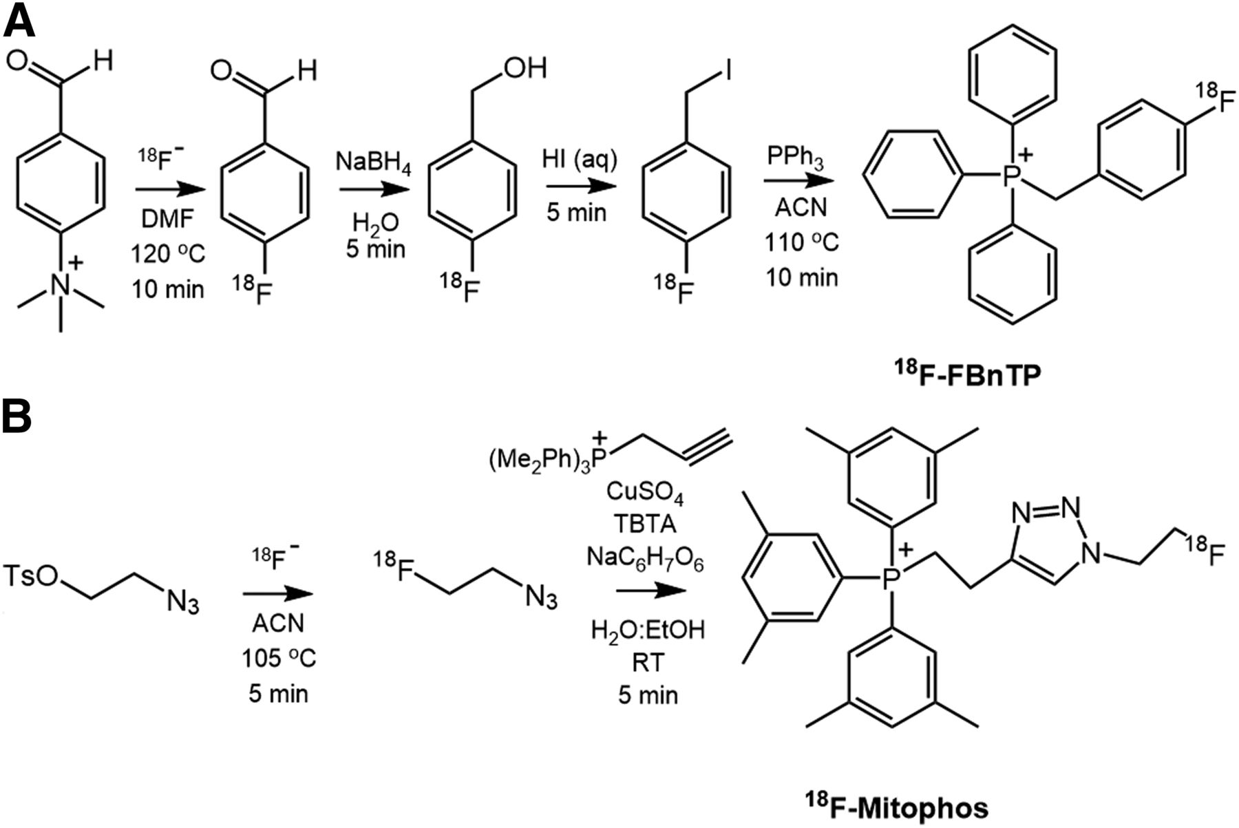
- FIGURE 1.
Radiosynthetic pathway for PET tracers: 18F-FBnTP (A) and 18F-MitoPhos (B). ACN = acetonitrile; DMF = dimethylformamide; RT = room temperature; TBTA = tris(benzyltriazolylmethyl)amine.
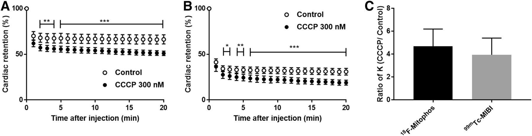
- FIGURE 2.
(A and B) Cardiac retention of percentage injected dose of 18F-MitoPhos (A) and 99mTc-sestamibi (B) in Langendorff-perfused hearts in control and CCCP (300 nM)-treated animals. (C) Relative increase in heart washout rate (K) for 18F-MitoPhos and 99mTc-sestamibi. Statistical analysis was 2-way ANOVA and Dunnett post hoc analysis (A and B) and 2-tailed t test (C). *P < 0.05. **P < 0.01. ***P < 0.001.
- FIGURE 3.
Uptake of 18F-MitoPhos in control and doxorubicin-treated animals (mean ± SD). (A) Ex vivo biodistribution of collected cardiac tissue (60 min after injection). (B) Average SUV from 30 to 60 min from left ventricle, lung, and liver time–activity curve, derived from dynamic PET/CT scan. Statistical analysis was 1-way ANOVA and Dunnett post hoc analysis. *P < 0.05. ***P < 0.001.
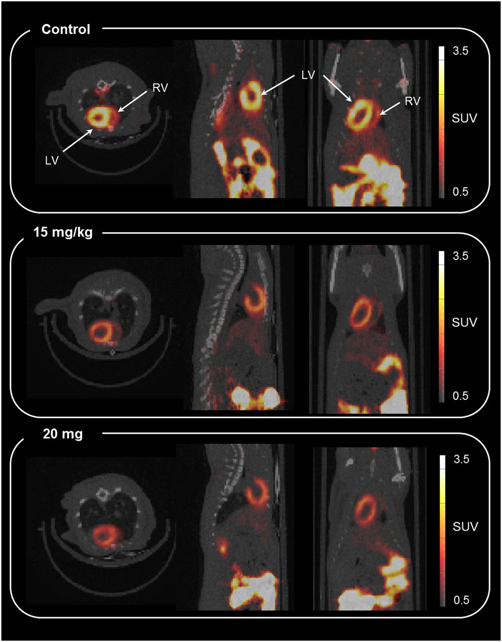
- FIGURE 4.
Representative axial, sagittal, and coronal coregistered PET/CT image (summed from 30 to 60 min) for 18F-MitoPhos in Sprague–Dawley rats 48 h after doxorubicin dose. Shown are control (top), 15 mg/kg (middle), and 20 mg/kg (bottom). LV = left ventricle; RV = right ventricle.
- FIGURE 5.
Time–activity curves (average SUV ± SD) for 18F-MitoPhos in Sprague–Dawley rats for left ventricle. SUVs are displayed over duration of scan (A) and for first minute (B). Significant decrease in cardiac uptake for both 15 mg/kg and 20 mg/kg dose in myocardium is observed. Statistical analysis was 2-way ANOVA and Dunnett post hoc analysis. ***P < 0.001 for control compared with doxorubicin (DOX), 20 mg/kg. ††P < 0.01 for control compared with doxorubicin, 15 mg/kg.


Tables
- TABLE 1
Selected Ex Vivo Biodistribution Data for 18F-FBnTP and 18F-MitoPhos and Heart-to-Tissue Ratios
Tissue 5 min 15 min 30 min 60 min 18F-FBnTP Blood 0.12 (0.22) 0.16 (0.06) 0.12 (0.06) 0.04 (0.01) Plasma 0.56 (0.51) 0.08 (0.06) 0.05 (0.04) 0.02 (0.00) Muscle 0.30 (0.12) 0.22 (0.12) 0.21 (0.04) 0.34 (0.14) Spleen 5.63 (0.28) 4.58 (1.22) 3.17 (1.65) 2.49 (0.76) Liver 3.71 (0.93) 3.57 (0.65) 3.58 (1.06) 3.59 (0.73) Adrenals 6.87 (9.16) 9.84 (1.74) 11.33 (5.33) 7.00 (2.49) Kidney 17.57 (5.27) 30.08 (3.81) 32.24 (3.44) 25.81 (5.92) Lung 2.09 (0.38) 2.03 (0.12) 2.09 (0.27) 1.63 (0.47) Heart 7.42 (0.43) 7.32 (0.69) 7.93 (2.45) 6.75 (1.54) Brain 0.07 (0.03) 0.05 (0.01) 0.06 (0.02) 0.03 (0.01) Heart-to-plasma 13.2 (12.0) 95.6 (70.9) 153.1 (135.2) 422.7 (142.5) Heart-to-lung 3.6 (0.7) 3.6 (0.4) 3.8 (1.3) 4.1 (1.5) Heart-to-liver 2.0 (0.5) 2.1 (0.4) 2.2 (0.9) 1.9 (0.6) 18F-MitoPhos Blood 0.35 (0.17) 0.16 (0.02) 0.14 (0.02) 0.06 (0.02) Plasma 0.19 (0.11) 0.06 (0.01) 0.07 (0.02) 0.02 (0.01) Muscle 0.30 (0.04) 0.22 (0.02) 0.24 (0.06) 0.22 (0.06) Spleen 6.67 (1.87) 6.04 (0.36) 7.28 (0.55) 4.231 (0.83) Liver 3.91 (0.69) 2.25 (0.30) 1.53 (0.30) 0.463 (0.12) Adrenals 12.50 (4.52) 11.32 (1.31) 21.16 (6.47) 14.30 (4.25) Kidney 20.77 (1.63) 17.76 (0.28) 21.69 (1.37) 18.64 (4.57) Lung 2.36 (0.15) 2.97 (1.42) 1.43 (0.25) 1.37 (0.36) Heart 5.40 (0.61) 4.60 (0.32) 5.17 (0.56) 5.17 (0.86) Brain 0.08 (0.01) 0.07 (0.00) 0.06 (0.01) 0.05 (0.01) Heart-to-plasma 28.3 (16.6) 82.2 (15.7) 95.7 (36.9) 303.9 (185.8) Heart-to-lung 2.3 (0.3) 1.6 (0.7) 3.6 (0.7) 3.8 (0.6) Heart-to-liver 1.4 (0.5) 2.0 (0.2) 3.4 (0.8) 11.2 (1.2) Data are average SUV (followed by SD in parentheses) determined by γ-counter measurement for each time point/min.
- TABLE 2
Histologic Assessment of Hematoxylin- and Eosin-Stained Tissue Sections of Control and Doxorubicin-Treated Myocardium
Assessor 1 Assessor 2 Dose Average median Range Average median Range Control 0 0–1 0.5 0–2 10 mg/kg 2 0–3 2.5 0–3 15 mg/kg 2.5 1–3 2.75 2–3 20 mg/kg 2.25 1–3 2.5 1–3 Average median denotes median score of each heart (n = 3 sections) averaged over treatment group. Range denotes total range of sections within treatment group.
Supplemental Data
Files in this Data Supplement:


