


Article Figures & Data
Figures
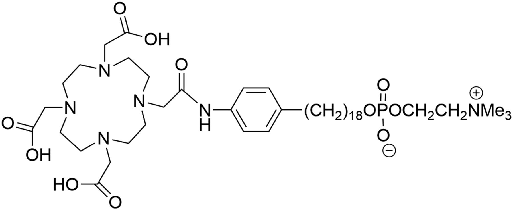
- FIGURE 1.
Schematic representation of NM600.
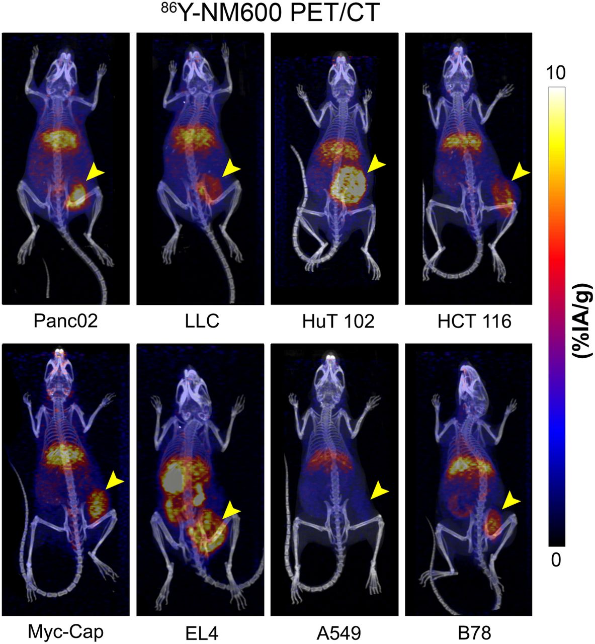
- FIGURE 2.
Volume rendering of PET overlaid on CT at final time point for each tumor model. Data are decay-corrected, and tumors are on right flank.
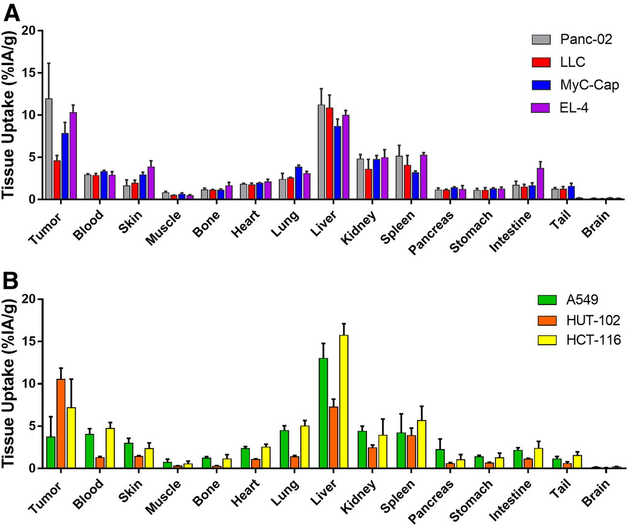
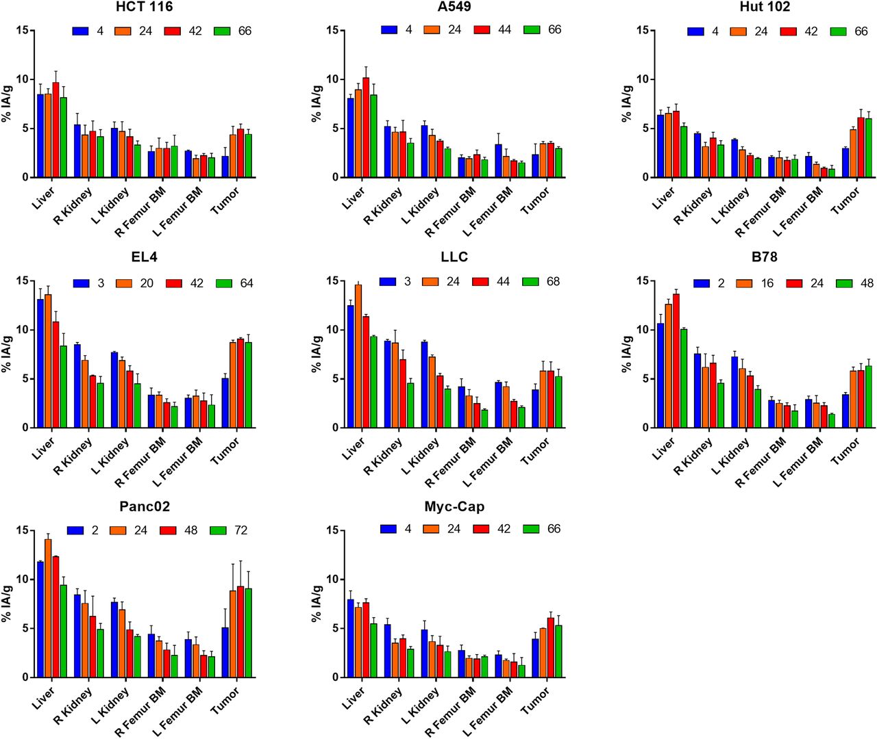
- FIGURE 3.
In vivo biodistribution. Mean (±SD) %IA/g of each VOI is charted for each tumor model (n = 3). In vivo biodistribution measured via PET imaging showed consistent tumor accumulation and retention across all tumor models investigated, with little off-target retention of NM600 except in liver, as is characteristic of hepatobiliary clearance. At early time points in distribution phase, there was elevated uptake of NM600 in highly perfused tissues such as kidneys. In later time points in clearance phase, NM600 washed out of highly perfused tissues.
- FIGURE 4.
Ex vivo biodistribution was performed after final imaging time point at 66–68 h after injection. (A) Results of syngeneic models. (B) Results of human xenograft models. Trends in biodistribution are like that of in vivo analysis. Numeric data are presented as %IA/g (mean ± SD).
- FIGURE 5.
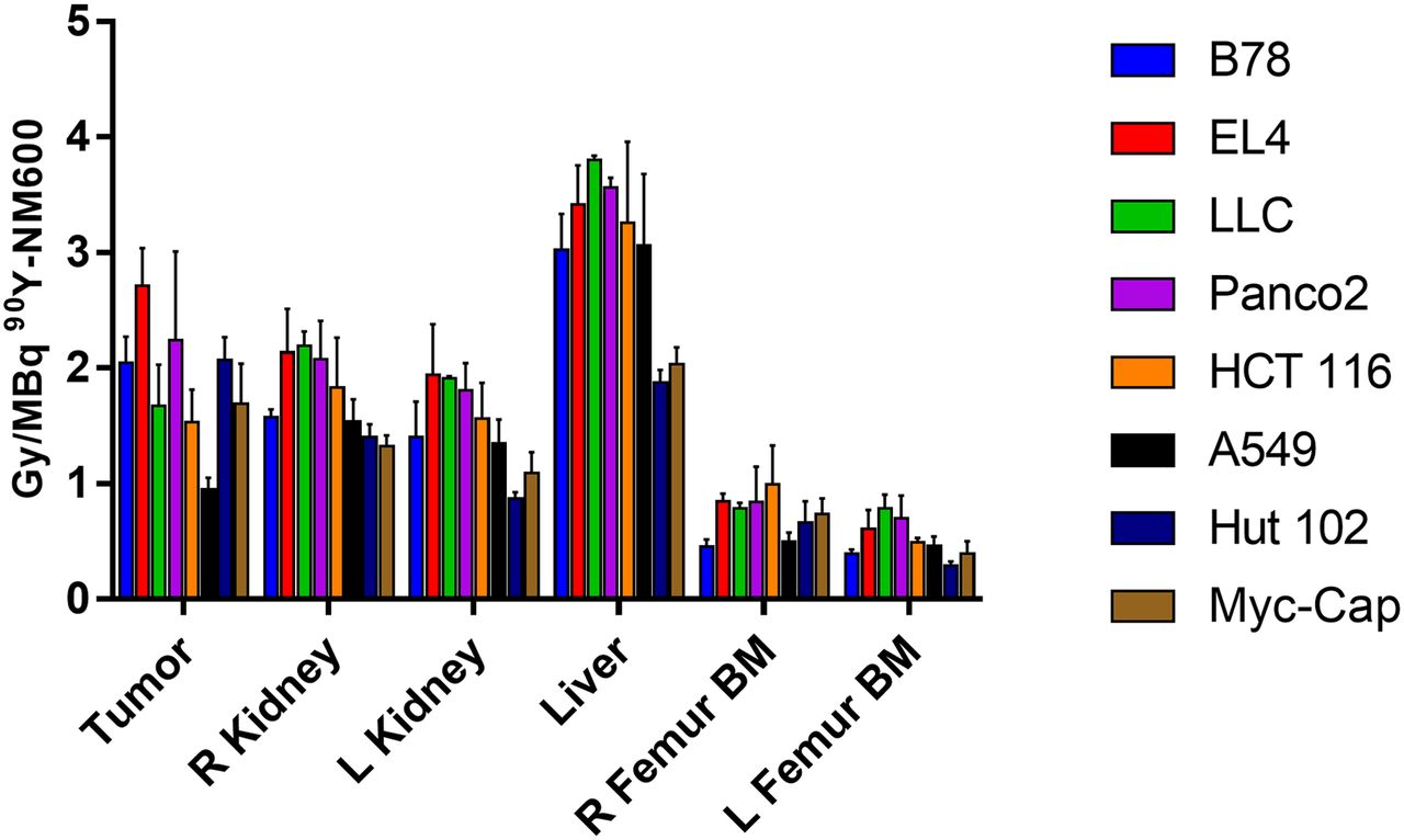
Dosimetry summary for tumor and organs at risk. Mean (± SD) absorbed dose per injected activity of 90Y-NM600 is shown for each mouse tumor model.
- FIGURE 6.
Toxicity assessment in naïve C57BL/6 mice administered 90Y-NM600. (A) Hematoxylin and eosin staining of potential organs at risk was performed for mice given 9.25 MBq of 90Y-NM600 IA at days 5, 10, and 28 after injection. Day 5 shows reduction in cellularity in white pulp of spleen and bone marrow, which were largely recovered at day 10. Images are displayed at ×20 magnification. (B) Complete blood count panel at days 5, 10, and 28 normalized by control group, which was not treated. White blood cells (WBC) and lymphocytes (LYM) decrease out to day 10 below control group but drastically increase at day 28—a finding that does not appear to be induced by radiation toxicity. However, platelets appear to be reduced at day 10 but recover by day 28. Red blood cells (RBC) and hemoglobin (HGB) do not appear to be affected by 90Y-NM600.
- FIGURE 7.
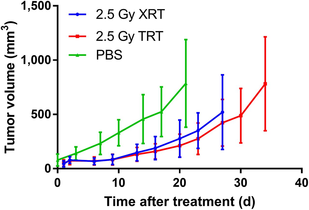
Tumor response of 2.5 Gy of 90Y-NM600 or XRT compared with no treatment, phosphate-buffered saline (PBS). There is not much evidence of significant difference between XRT and TRT. However, volume slope of XRT was significantly lower than PBS slope (β = −15.63, SE = 3.97, P = 0.0001). Numeric data are presented as mm3 (mean ± SD).


Tables
Cell type Half-life (h) Peak uptake (%IA/g) HCT 116 40.5 5.0 A549 39.8 3.5 EL4 26.7 9.1 B78 28.6 6.3 LLC 33.1 5.8 Hut 102 29.7 6.1 Panc02 38.8 9.3 Myc-Cap 27.6 6.1 Average 33.1 (SD, 5.8) 6.4 (SD, 1.9)
Supplemental Data
Files in this Data Supplement:


