
Main menu

  • Home
  • Content
    • Current
    • Ahead of print
    • Past Issues
    • JNM Supplement
    • SNMMI Annual Meeting Abstracts
    • Continuing Education
    • JNM Podcasts
  • Subscriptions
    • Subscribers
    • Institutional and Non-member
    • Rates
    • Journal Claims
    • Corporate & Special Sales
  • Authors
    • Submit to JNM
    • Information for Authors
    • Assignment of Copyright
    • AQARA requirements
  • Info
    • Reviewers
    • Permissions
    • Advertisers
  • About
    • About Us
    • Editorial Board
    • Contact Information
  • More
    • Alerts
    • Feedback
    • Help
    • SNMMI Journals
  • SNMMI
    • JNM
    • JNMT
    • SNMMI Journals
    • SNMMI

User menu

  • Subscribe
  • My alerts
  • Log in
  • My Cart

Search

  • Advanced search
Journal of Nuclear Medicine
  • SNMMI
    • JNM
    • JNMT
    • SNMMI Journals
    • SNMMI
  • Subscribe
  • My alerts
  • Log in
  • My Cart
Journal of Nuclear Medicine

Advanced Search

  • Home
  • Content
    • Current
    • Ahead of print
    • Past Issues
    • JNM Supplement
    • SNMMI Annual Meeting Abstracts
    • Continuing Education
    • JNM Podcasts
  • Subscriptions
    • Subscribers
    • Institutional and Non-member
    • Rates
    • Journal Claims
    • Corporate & Special Sales
  • Authors
    • Submit to JNM
    • Information for Authors
    • Assignment of Copyright
    • AQARA requirements
  • Info
    • Reviewers
    • Permissions
    • Advertisers
  • About
    • About Us
    • Editorial Board
    • Contact Information
  • More
    • Alerts
    • Feedback
    • Help
    • SNMMI Journals
  • View or Listen to JNM Podcast
  • Visit JNM on Facebook
  • Join JNM on LinkedIn
  • Follow JNM on Twitter
  • Subscribe to our RSS feeds
Research ArticleOncology

18F-FDG PET/MRI for Therapy Response Assessment of Isolated Limb Perfusion in Patients with Soft-Tissue Sarcomas

Johannes Grueneisen, Benedikt Schaarschmidt, Aydin Demircioglu, Michal Chodyla, Ole Martin, Stefanie Bertram, Axel Wetter, Sebastian Bauer, Wolfgang Peter Fendler, Lars Podleska, Michael Forsting, Ken Herrmann and Lale Umutlu
Journal of Nuclear Medicine November 2019, 60 (11) 1537-1542; DOI: https://doi.org/10.2967/jnumed.119.226761
Johannes Grueneisen
1Department of Diagnostic and Interventional Radiology, University Hospital Essen, University of Duisburg–Essen, Essen, Germany
  • Find this author on Google Scholar
  • Find this author on PubMed
  • Search for this author on this site
Benedikt Schaarschmidt
1Department of Diagnostic and Interventional Radiology, University Hospital Essen, University of Duisburg–Essen, Essen, Germany
  • Find this author on Google Scholar
  • Find this author on PubMed
  • Search for this author on this site
Aydin Demircioglu
1Department of Diagnostic and Interventional Radiology, University Hospital Essen, University of Duisburg–Essen, Essen, Germany
  • Find this author on Google Scholar
  • Find this author on PubMed
  • Search for this author on this site
Michal Chodyla
1Department of Diagnostic and Interventional Radiology, University Hospital Essen, University of Duisburg–Essen, Essen, Germany
  • Find this author on Google Scholar
  • Find this author on PubMed
  • Search for this author on this site
Ole Martin
2Department of Diagnostic and Interventional Radiology, University Hospital Dusseldorf, University of Dusseldorf, Dusseldorf, Germany
  • Find this author on Google Scholar
  • Find this author on PubMed
  • Search for this author on this site
Stefanie Bertram
3Institute of Pathology, University Hospital Essen, University of Duisburg–Essen, Essen, Germany
  • Find this author on Google Scholar
  • Find this author on PubMed
  • Search for this author on this site
Axel Wetter
1Department of Diagnostic and Interventional Radiology, University Hospital Essen, University of Duisburg–Essen, Essen, Germany
  • Find this author on Google Scholar
  • Find this author on PubMed
  • Search for this author on this site
Sebastian Bauer
4Sarcoma Center, Western German Cancer Center, University of Duisburg–Essen, Essen, Germany
  • Find this author on Google Scholar
  • Find this author on PubMed
  • Search for this author on this site
Wolfgang Peter Fendler
5Department of Nuclear Medicine, University Hospital Essen, University of Duisburg–Essen, Essen, Germany; and
  • Find this author on Google Scholar
  • Find this author on PubMed
  • Search for this author on this site
Lars Podleska
6Sarcoma Surgery Division, Department of General, Visceral, and Transplantation Surgery, University Hospital Essen, University of Duisburg–Essen, Essen, Germany
  • Find this author on Google Scholar
  • Find this author on PubMed
  • Search for this author on this site
Michael Forsting
1Department of Diagnostic and Interventional Radiology, University Hospital Essen, University of Duisburg–Essen, Essen, Germany
  • Find this author on Google Scholar
  • Find this author on PubMed
  • Search for this author on this site
Ken Herrmann
5Department of Nuclear Medicine, University Hospital Essen, University of Duisburg–Essen, Essen, Germany; and
  • Find this author on Google Scholar
  • Find this author on PubMed
  • Search for this author on this site
Lale Umutlu
1Department of Diagnostic and Interventional Radiology, University Hospital Essen, University of Duisburg–Essen, Essen, Germany
  • Find this author on Google Scholar
  • Find this author on PubMed
  • Search for this author on this site
  • Article
  • Figures & Data
  • Supplemental
  • Info & Metrics
  • PDF
Loading

Article Figures & Data

Figures

  • Tables
  • Additional Files
  • FIGURE 1.
    • Download figure
    • Open in new tab
    • Download powerpoint
    FIGURE 1.

    A 56-y-old patient with a leiomyosarcoma of the left limb (arrows). The tumor manifestation does not show significant changes in tumor size (diameter: from 243 to 256 mm), contrast-enhancing tumor parts and metabolic activity (SUVpeak: from 3.9 to 3.6). Histopathologic analysis revealed a regression grade 5 (histopathologic nonresponder).

  • FIGURE 2.
    • Download figure
    • Open in new tab
    • Download powerpoint
    FIGURE 2.

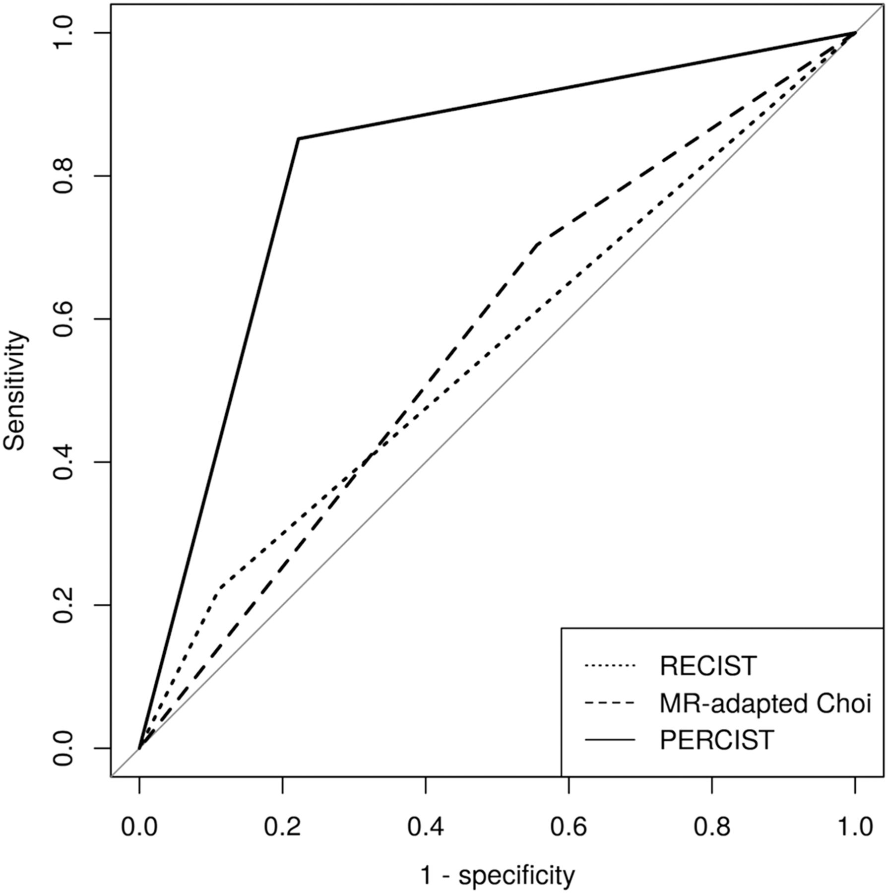
    Receiver-operating-characteristic curves of ratings according to RECIST, Choi criteria, and PERCIST.

  • FIGURE 3.
    • Download figure
    • Open in new tab
    • Download powerpoint
    FIGURE 3.

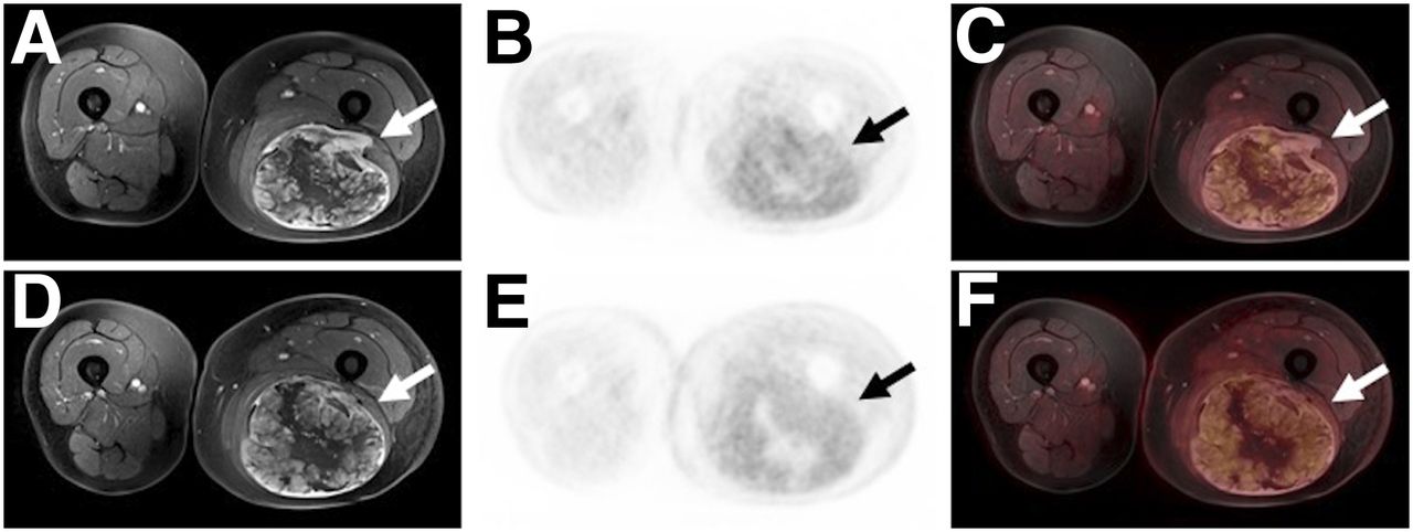
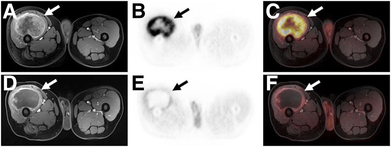
    A 60-y-old patient with an undifferentiated pleomorphic sarcoma of the right limb (arrows). The tumor reveals an extensive tumor necrosis and a significant reduction of 18F-FDG uptake (SUVpeak: from 15.3 to 4.1) between pre- (A–C) and posttherapeutic (D–F) examinations, whereas tumor size remains substantially stable (diameter: from 99 to 97 mm). Histopathologic analysis revealed a regression grade 3 (histopathologic responder).

  • FIGURE 4.
    • Download figure
    • Open in new tab
    • Download powerpoint
    FIGURE 4.

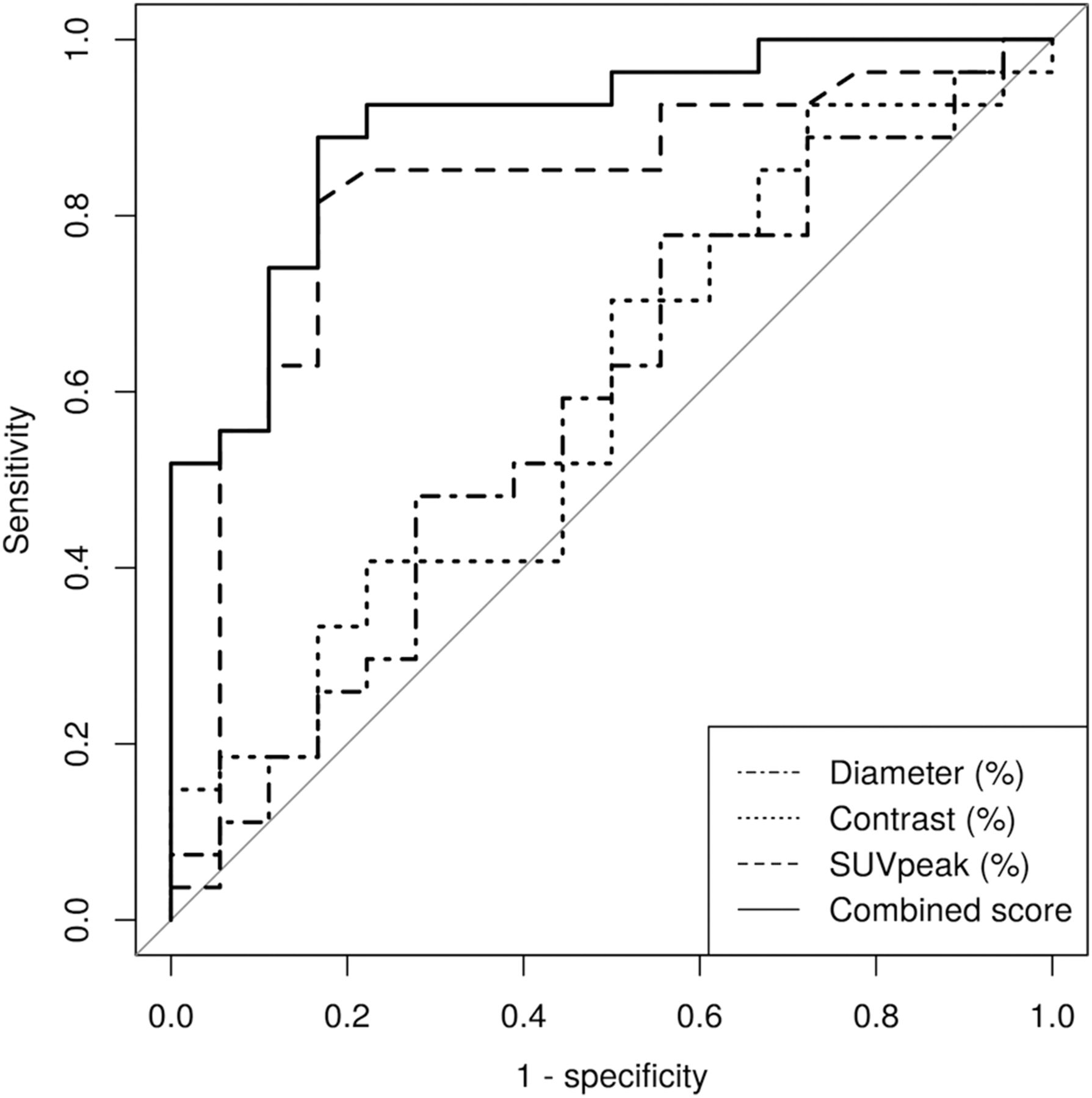
    Receiver-operating-characteristic curves of quantitative variables for assessment of therapeutic response to ILP.

Tables

  • Figures
  • Additional Files
    • View popup
    TABLE 1

    Distribution of Histologic Subtypes of STS

    Histologic subtypePatients (n)
    Undifferentiated pleomorphic sarcoma9
    Synovial sarcoma9
    Undifferentiated spindle cell sarcoma8
    Myxofibrosarcoma7
    Liposarcoma4
    Epitheloid sarcoma2
    Leiomyosarcoma2
    Clear cell sarcoma1
    Angiosarcoma1
    Fibrosarcoma1
    Malignant peripheral nerve sheath tumor1
    Total45
    • View popup
    TABLE 2

    Tumor Grades of All Patients, According to 6-Stage Salzer-Kuntschik Regression Scale

    Tumor gradePatients (n)
    I6
    II8
    III13
    IV10
    V7
    VI1
    • View popup
    TABLE 3

    Sensitivity, Specificity, Positive Predictive Value, Negative Predictive Value, Diagnostic Accuracy, and AUC of the 3 Response Criteria for Differentiation Between Responders and Nonresponders

    Response criteriaSensitivitySpecificityPositive predictive valueNegative predictive valueAccuracyAUC
    RECIST22.2%88.9%75.0%43.2%48.9%0.556
    Choi criteria70.4%44.4%65.5%50.0%60.0%0.574
    PERCIST85.2%77.8%85.2%77.8%82.2%0.815
    • View popup
    TABLE 4

    P Values for Comparison of RECIST, Choi criteria, and PERCIST

    ParameterRECIST vs. ChoiRECIST vs. PERCISTChoi vs. PERCIST
    Sensitivity0.0016*0.0003*0.2482
    Specificity0.0140*0.31730.0286*
    Positive predictive value0.82050.82050.0202*
    Negative predictive value0.48210.0014*0.0995
    Accuracy0.39720.0057*0.0727
    AUC0.81970.0016*0.0069*
    • ↵* Statistically significant difference.

Additional Files

  • Figures
  • Tables
  • Supplemental Data

    Files in this Data Supplement:

    • Supplemental Data
PreviousNext
Back to top

In this issue

Journal of Nuclear Medicine: 60 (11)
Journal of Nuclear Medicine
Vol. 60, Issue 11
November 1, 2019
  • Table of Contents
  • Table of Contents (PDF)
  • About the Cover
  • Index by author
Print
Download PDF
Article Alerts
Sign In to Email Alerts with your Email Address
Email Article

Thank you for your interest in spreading the word on Journal of Nuclear Medicine.

NOTE: We only request your email address so that the person you are recommending the page to knows that you wanted them to see it, and that it is not junk mail. We do not capture any email address.

Enter multiple addresses on separate lines or separate them with commas.
18F-FDG PET/MRI for Therapy Response Assessment of Isolated Limb Perfusion in Patients with Soft-Tissue Sarcomas
(Your Name) has sent you a message from Journal of Nuclear Medicine
(Your Name) thought you would like to see the Journal of Nuclear Medicine web site.
Citation Tools
18F-FDG PET/MRI for Therapy Response Assessment of Isolated Limb Perfusion in Patients with Soft-Tissue Sarcomas
Johannes Grueneisen, Benedikt Schaarschmidt, Aydin Demircioglu, Michal Chodyla, Ole Martin, Stefanie Bertram, Axel Wetter, Sebastian Bauer, Wolfgang Peter Fendler, Lars Podleska, Michael Forsting, Ken Herrmann, Lale Umutlu
Journal of Nuclear Medicine Nov 2019, 60 (11) 1537-1542; DOI: 10.2967/jnumed.119.226761

Citation Manager Formats

  • BibTeX
  • Bookends
  • EasyBib
  • EndNote (tagged)
  • EndNote 8 (xml)
  • Medlars
  • Mendeley
  • Papers
  • RefWorks Tagged
  • Ref Manager
  • RIS
  • Zotero
Share
18F-FDG PET/MRI for Therapy Response Assessment of Isolated Limb Perfusion in Patients with Soft-Tissue Sarcomas
Johannes Grueneisen, Benedikt Schaarschmidt, Aydin Demircioglu, Michal Chodyla, Ole Martin, Stefanie Bertram, Axel Wetter, Sebastian Bauer, Wolfgang Peter Fendler, Lars Podleska, Michael Forsting, Ken Herrmann, Lale Umutlu
Journal of Nuclear Medicine Nov 2019, 60 (11) 1537-1542; DOI: 10.2967/jnumed.119.226761
Twitter logo Facebook logo LinkedIn logo Mendeley logo
  • Tweet Widget
  • Facebook Like
  • Google Plus One
Bookmark this article

Jump to section

  • Article
    • Abstract
    • MATERIALS AND METHODS
    • RESULTS
    • DISCUSSION
    • CONCLUSION
    • DISCLOSURE
    • KEY POINTS
    • Footnotes
    • REFERENCES
  • Figures & Data
  • Supplemental
  • Info & Metrics
  • PDF

Related Articles

  • This Month in JNM
  • PubMed
  • Google Scholar

Cited By...

  • Evaluation of 18F-FDG PET and DWI Datasets for Predicting Therapy Response of Soft-Tissue Sarcomas Under Neoadjuvant Isolated Limb Perfusion
  • Google Scholar

More in this TOC Section

Oncology

  • Comparative PSMA expression at early (1hour) vs late (2hour) in primary and secondary sites of involvement in prostate cancer.
  • Imaging spectrum of peritoneal carcinomatosis associated with various etiologies on 18F-FDG PET/CT
  • Hybrid Imaging Features of Musculoskeletal Tumors
Show more Oncology

Clinical

  • TauIQ: A Canonical Image Based Algorithm to Quantify Tau PET Scans
  • Dual PET Imaging in Bronchial Neuroendocrine Neoplasms: The NETPET Score as a Prognostic Biomarker
  • Addition of 131I-MIBG to PRRT (90Y-DOTATOC) for Personalized Treatment of Selected Patients with Neuroendocrine Tumors
Show more Clinical

Similar Articles

Keywords

  • PET/MRI
  • Soft-tissue sarcoma
  • isolated limb perfusion
  • therapy response assessment
SNMMI

© 2025 SNMMI

Powered by HighWire