


Article Figures & Data
Figures
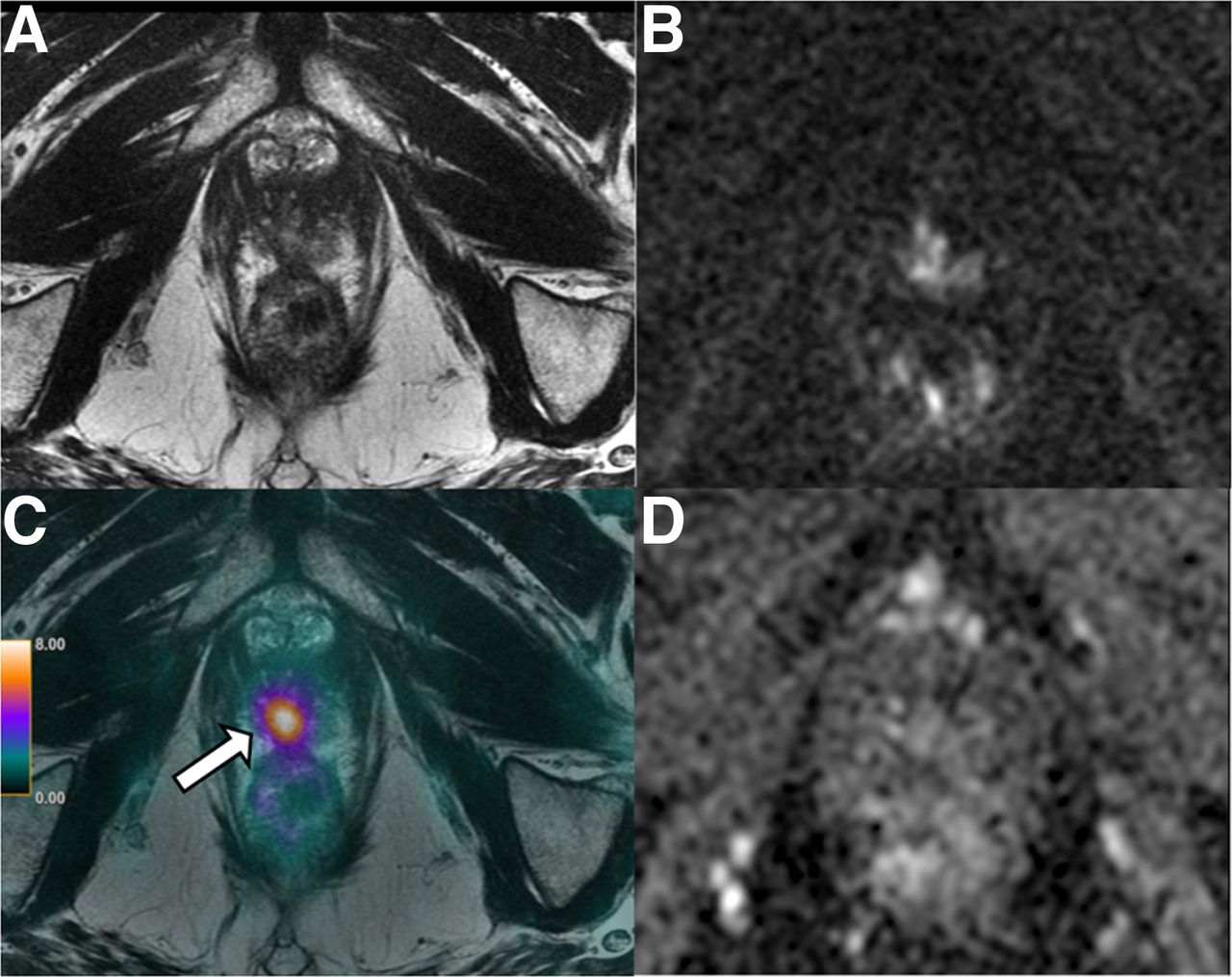
- FIGURE 1.
A 75-y-old patient with local recurrence of Gleason 3 + 4 PCa (arrows) treated with radical prostatectomy. (A–C) mpMRI shows masslike T2-hypointense thickening at right lateral vesicourethral anastomosis (A) with diffusion restriction on apparent diffusion coefficient image (B) and early arterial enhancement on dynamic contrast-enhanced image (C). (D) 18F-fluciclovine PET/CT shows asymmetric increased radiotracer uptake at that site (SUVmax, 5.5).
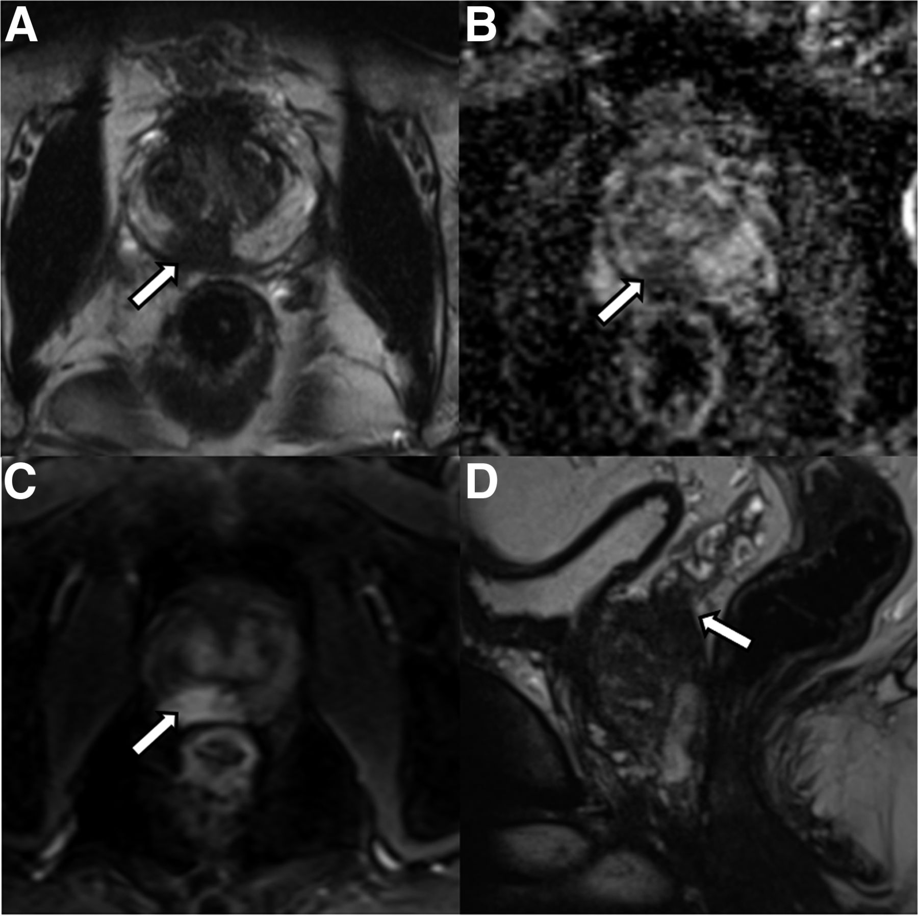
- FIGURE 2.
A 53-y-old patient with abnormal digital rectal findings, elevated PSA level, and lesion (arrows) found on imaging. (A–C) mpMRI shows circumscribed T2-hypointense lesion in right posterior base to mid gland peripheral zone (A) with marked diffusion restriction (B) and early arterial enhancement (C). Lesion measured 1.9 cm and involved the central zone and base of seminal vesicles. Final surgical pathology revealed 3 + 4 Gleason PCa with extracapsular extension and seminal vesicle invasion.
- FIGURE 3.
A 69-y-old patient with history of Gleason 7 PCa treated with high-intensity focus ultrasound therapy 1 year previously. (A, B, and D) Surveillance mpMRI does not reveal any findings concerning for recurrence. (C) On 68Ga-PSMA PET/MRI, area of focal intense uptake localized to right apex (arrow) is seen, corresponding to subsequently biopsy-proven recurrent Gleason 4 + 4 tumor.
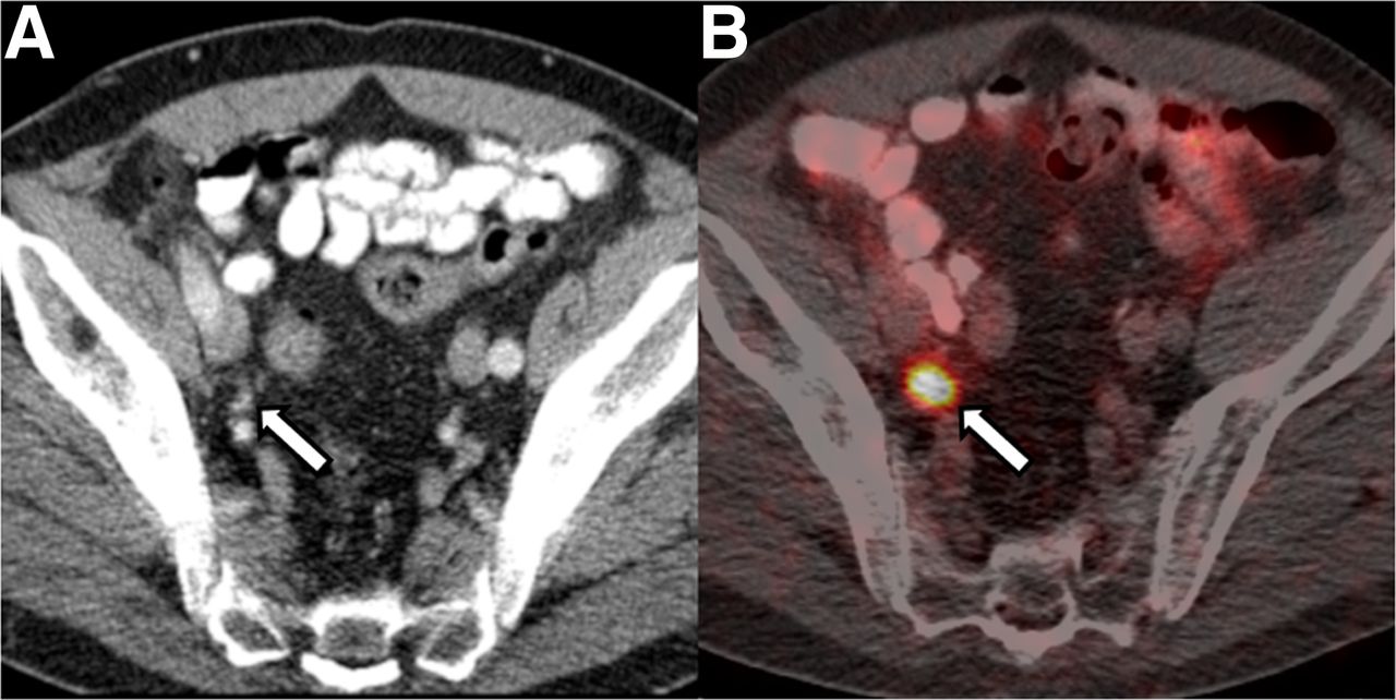
- FIGURE 4.
A 68-y-old patient with BCR of Gleason 3 + 4 PCa after radical prostatectomy and pelvic LN dissection 11 years previously. PSA was 1.4 ng/mL at time of imaging. (A) CT portion of study does not reveal any abnormal LNs. (B) Fused 68Ga-PSMA PET/CT shows focal avid uptake in right external iliac region (arrow) corresponding to subcentimeter external iliac LN (arrow in A), suggestive of LN metastasis, which was confirmed on subsequent excisional biopsy.


Additional Files
Supplemental Data
Files in this Data Supplement:


Jump to section
Related Articles
Cited By...
- No citing articles found.