


Article Figures & Data
Figures
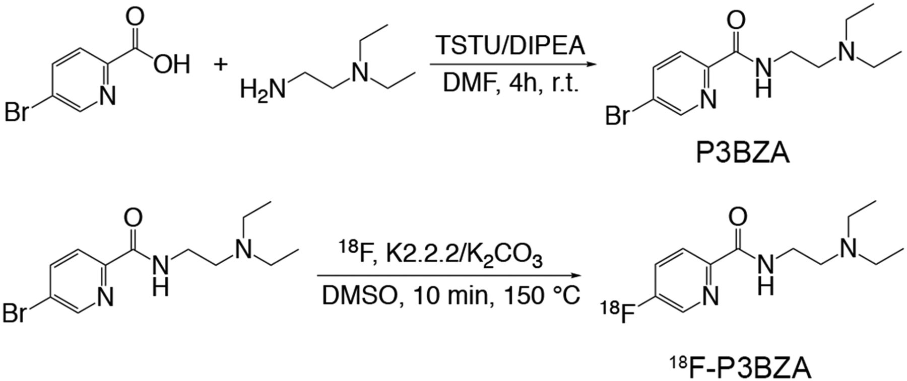
- FIGURE 1.
18F-P3BZA synthesis scheme.
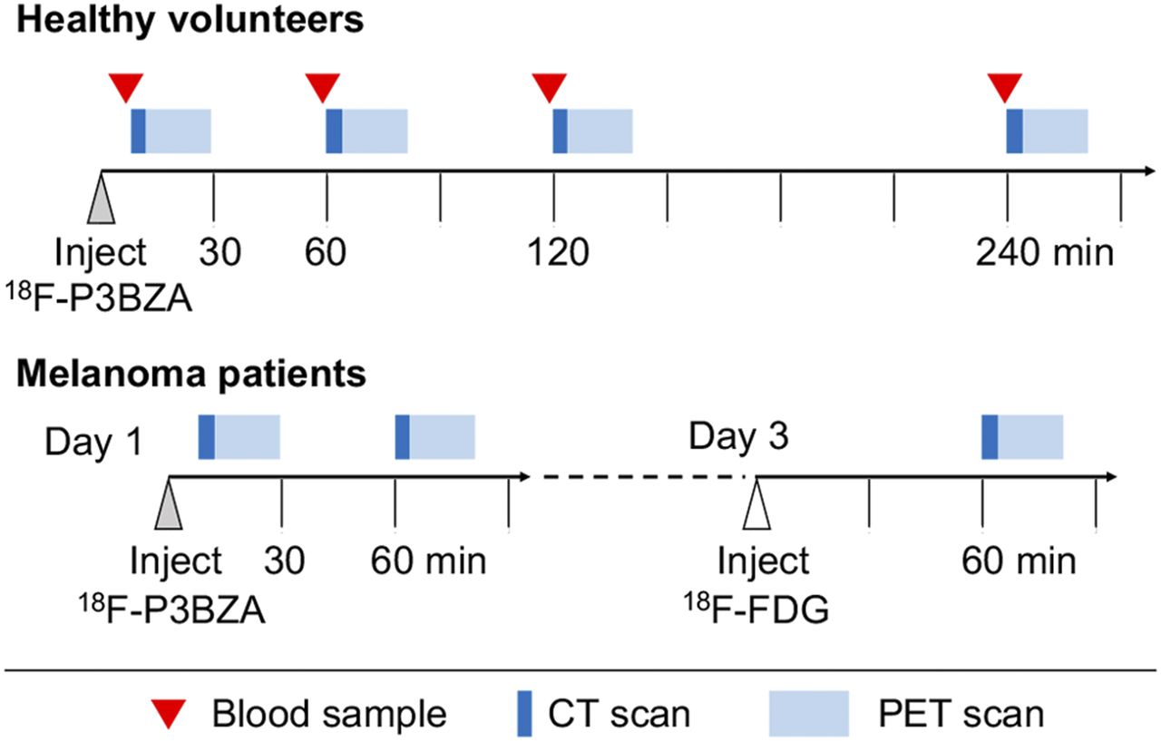
- FIGURE 2.
Timeline of the dosimetry study (upper part) and melanoma (lower part) PET/CT imaging.
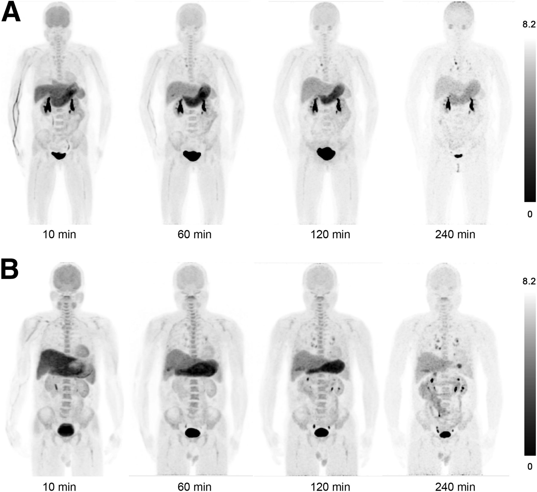
- FIGURE 3.
Maximum-intensity-projection PET images at different time points after 18F-P3BZA injection in female subject (A) and male subject (B).
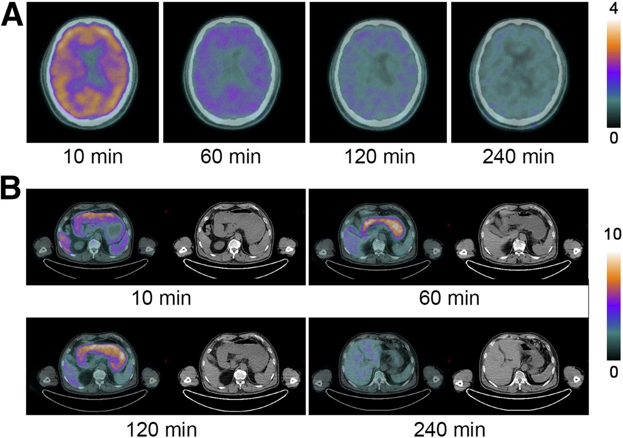
- FIGURE 4.
Transverse images of brain (A) and stomach (B) at different time points after 18F-P3BZA injection.
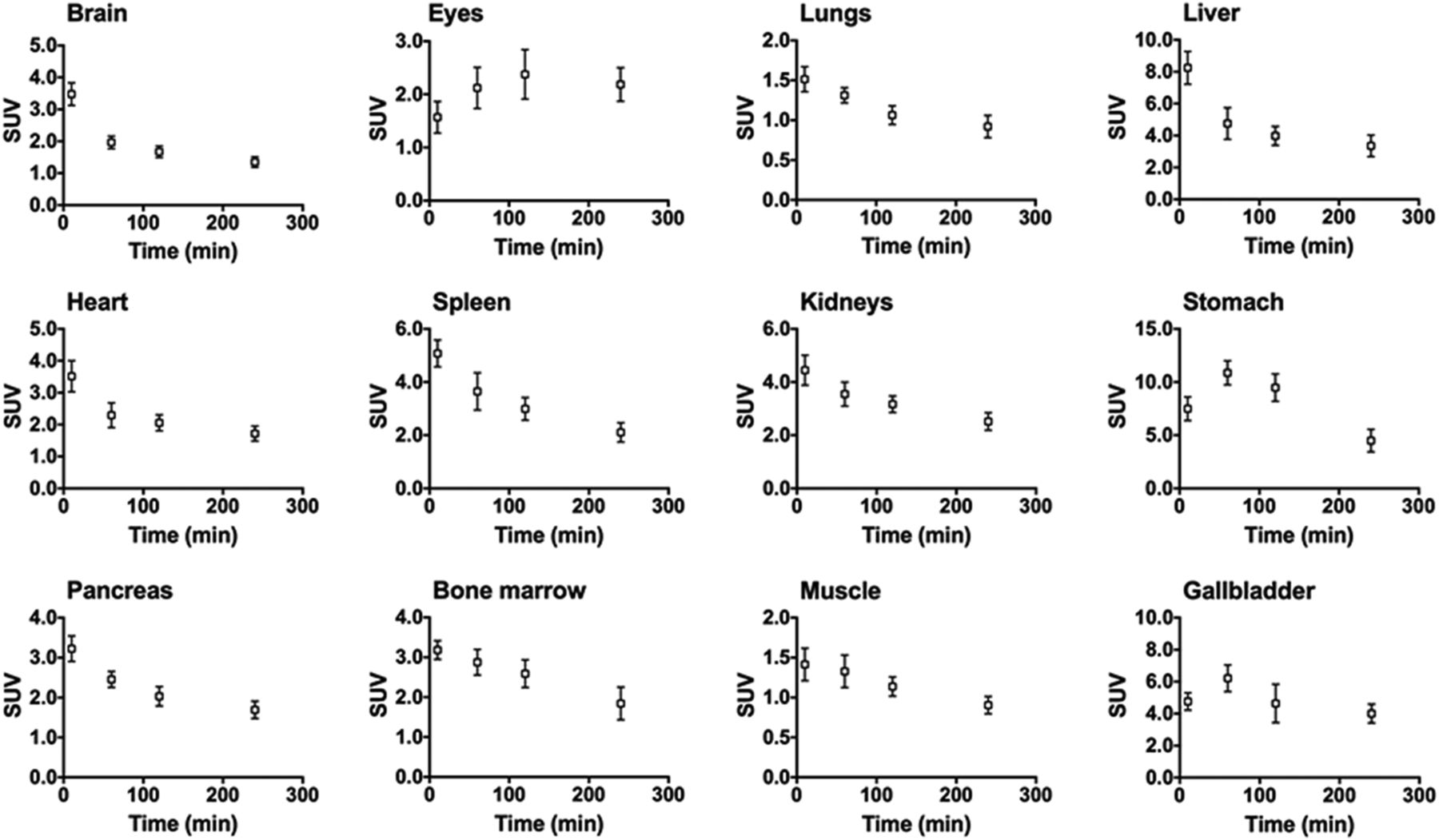
- FIGURE 5.
Average time–SUVmean curves (n = 6) for 12 major organs (error bars indicate ±SD).
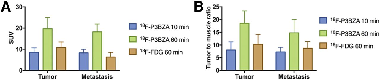
- FIGURE 6.
Average SUVmean (A) and tumor-to-muscle ratio (B) of melanoma tumors and metastases in patients (n = 5).
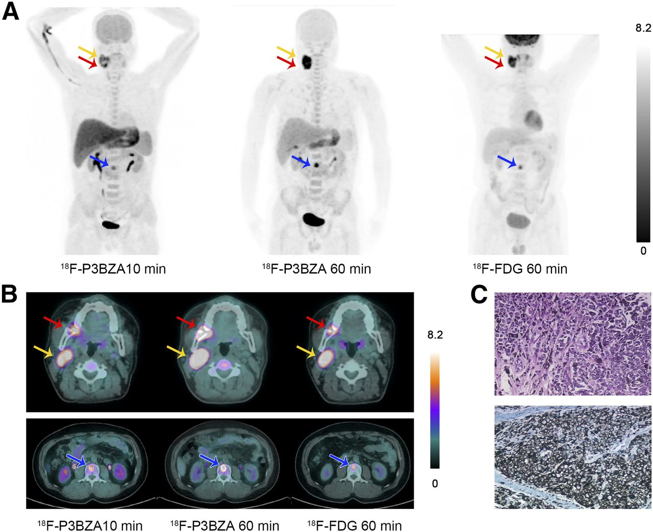
- FIGURE 7.
Sample images of a melanoma patient. (A) Maximum-intensity-projection PET images at 10 and 60 min after 18F-P3BZA injection and at 60 min after 18F-FDG injection. (B) Transverse images of primary melanoma (red arrow), lymph node metastasis (yellow arrow), and bone metastasis (blue arrow). (C) H&E (upper part) and immunohistochemistry (lower part) images of primary melanoma.

Tables
Patient no. Age (y) Sex Weight (kg) Injected dose (MBq) 1 55 F 55 187 2 61 F 59 210 3 42 F 51 204 4 49 M 75 229 5 63 M 66 214 6 57 M 70 226 Mean 54.5 ± 7.8 — 62.6 ± 9.2 211.7 ± 15.4 Organ Dose (mSv/MBq) Adrenals 0.0150 Brain 0.0147 Breasts 0.0075 Gallbladder wall 0.0158 Lower large intestine wall 0.0134 Small intestine 0.0207 Stomach wall 0.0219 Upper large intestine wall 0.0228 Heart wall 0.0166 Kidneys 0.0218 Liver 0.0407 Lungs 0.0121 Muscle 0.0120 Ovaries (n = 3) 0.0109 Pancreas 0.0224 Red marrow 0.0122 Osteogenic cells 0.0085 Skin 0.0034 Spleen 0.0247 Testes (n = 3) 0.0086 Thymus 0.0051 Thyroid 0.0110 Urinary bladder wall 0.1200 Uterus 0.0141 Eyes 0.0128 Total body 0.0099 Effective dose 0.0193
Supplemental Data
Files in this Data Supplement:


Jump to section
Related Articles
Cited By...
- Melanin-Targeting Radiotracers and Their Preclinical, Translational, and Clinical Status: From Past to Future
- 18F-PFPN PET: A New and Attractive Imaging Modality for Patients with Malignant Melanoma
- Advances in Receptor-Targeted Radiolabeled Peptides for Melanoma Imaging and Therapy
- Ultrasensitive detection of malignant melanoma using PET molecular imaging probes
- N-(2-(Dimethylamino)Ethyl)-4-18F-Fluorobenzamide: A Novel Molecular Probe for High-Contrast PET Imaging of Malignant Melanoma