
Main menu

  • Home
  • Content
    • Current
    • Ahead of print
    • Past Issues
    • JNM Supplement
    • SNMMI Annual Meeting Abstracts
    • Continuing Education
    • JNM Podcasts
  • Subscriptions
    • Subscribers
    • Institutional and Non-member
    • Rates
    • Journal Claims
    • Corporate & Special Sales
  • Authors
    • Submit to JNM
    • Information for Authors
    • Assignment of Copyright
    • AQARA requirements
  • Info
    • Reviewers
    • Permissions
    • Advertisers
  • About
    • About Us
    • Editorial Board
    • Contact Information
  • More
    • Alerts
    • Feedback
    • Help
    • SNMMI Journals
  • SNMMI
    • JNM
    • JNMT
    • SNMMI Journals
    • SNMMI

User menu

  • Subscribe
  • My alerts
  • Log in
  • My Cart

Search

  • Advanced search
Journal of Nuclear Medicine
  • SNMMI
    • JNM
    • JNMT
    • SNMMI Journals
    • SNMMI
  • Subscribe
  • My alerts
  • Log in
  • My Cart
Journal of Nuclear Medicine

Advanced Search

  • Home
  • Content
    • Current
    • Ahead of print
    • Past Issues
    • JNM Supplement
    • SNMMI Annual Meeting Abstracts
    • Continuing Education
    • JNM Podcasts
  • Subscriptions
    • Subscribers
    • Institutional and Non-member
    • Rates
    • Journal Claims
    • Corporate & Special Sales
  • Authors
    • Submit to JNM
    • Information for Authors
    • Assignment of Copyright
    • AQARA requirements
  • Info
    • Reviewers
    • Permissions
    • Advertisers
  • About
    • About Us
    • Editorial Board
    • Contact Information
  • More
    • Alerts
    • Feedback
    • Help
    • SNMMI Journals
  • View or Listen to JNM Podcast
  • Visit JNM on Facebook
  • Join JNM on LinkedIn
  • Follow JNM on Twitter
  • Subscribe to our RSS feeds
Meeting ReportInstrumentation & Data Analysis Track

A Novel Penalized Joint Image Reconstruction Method for Tau-PET Imaging

Alvin Ihsani, Joyita Dutta, J. Alex Becker, Keith Alan Johnson, Jinsong Ouyang and Georges El Fakhri
Journal of Nuclear Medicine May 2018, 59 (supplement 1) 99;
Alvin Ihsani
1Massachusetts General Hospital Boston MA United States
2Massachusetts General Hospital Boston MA United States
  • Find this author on Google Scholar
  • Find this author on PubMed
  • Search for this author on this site
Joyita Dutta
4MGH / Harvard / UMass Lowell Boston MA United States
  • Find this author on Google Scholar
  • Find this author on PubMed
  • Search for this author on this site
J. Alex Becker
1Massachusetts General Hospital Boston MA United States
2Massachusetts General Hospital Boston MA United States
  • Find this author on Google Scholar
  • Find this author on PubMed
  • Search for this author on this site
Keith Alan Johnson
1Massachusetts General Hospital Boston MA United States
2Massachusetts General Hospital Boston MA United States
  • Find this author on Google Scholar
  • Find this author on PubMed
  • Search for this author on this site
Jinsong Ouyang
1Massachusetts General Hospital Boston MA United States
2Massachusetts General Hospital Boston MA United States
  • Find this author on Google Scholar
  • Find this author on PubMed
  • Search for this author on this site
Georges El Fakhri
3Massachusetts General Hospital, Harvard Medical Sc Boston MA United States
  • Find this author on Google Scholar
  • Find this author on PubMed
  • Search for this author on this site
  • Article
  • Figures & Data
  • Info & Metrics
Loading

Abstract

99

Introduction: A strong correlation between the distribution of paired helical filament (PHF) Tau protein deposition in the brain and the appearance of cognitive decline typical of dementia and Alzheimer Disease (AD) has recently been established. The advent of the Positron Emission Tomography (PET) tracer [18F]AV-1451 has enabled in vivo imaging of PHF Tau. To develop preventive treatments for AD and to quickly refine said treatments, it is imperative to detect the disease at the prodromal stage (before symptoms appear). The conventional method to estimate PHF Tau buildup in the brain is to scan a patient at least two times every 1-2 years using [18F]AV-1451 in PET, independently reconstruct images of each scan, and analyze the difference images between scans. The drawback of this approach is the lower limit it imposes on detection of Tau burden due to increased variance in the difference image. We propose a novel penalized longitudinal Tau PET image reconstruction method where the difference image is reconstructed directly from projections and therefore exhibits less variation compared to the conventional approach. In turn, the lowered variation allows for more sensitivity to small changes, and therefore more discernibility between subject groups. Validating the Proposed Longitudinal Reconstruction Method We validated the proposed reconstruction approach in two ways: (1) we used synthetic lesions to determine the "discernibility" (sensitivity index) of a known lesion to surrounding tissues, and (2) we compared a set of regions of interest in a small sample of normal/control (NC) subjects and subjects with mild cognitive impairment (MCI).

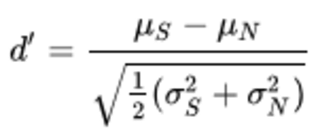
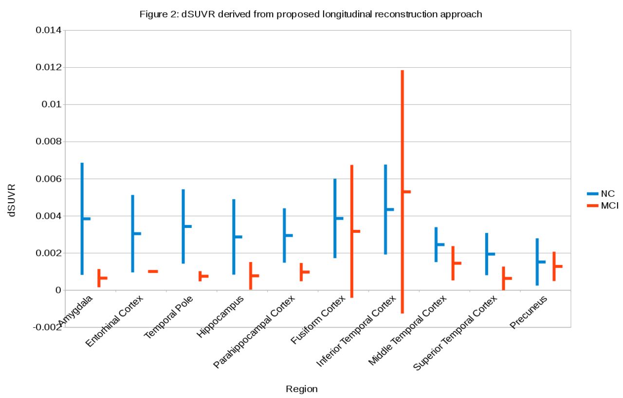
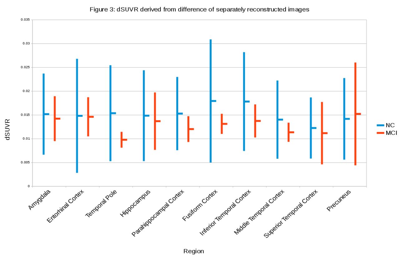
Results: Figure 1 shows the sensitivity index (d’) computed as and plotted as a function of the percent intensity of the inserted lesion (see Figure 1). Here, µS and µN are the mean intensities of the lesion and the surrounding tissue respectively, while σS and σN are the standard deviations. It is apparent that d’ is retained better at smaller lesion intensities using the proposed approach. Figures 2 and 3 show the annualized dSUVR (difference standardized uptake value ratios) on a per-region basis for a small group of subjects (8 NC, 2 MCI) for the proposed and conventional methods. It is apparent that the variance in our proposed method is much lower allowing for more signification separation of dSUVR by group compared to the conventional approach despite the negative bias in the proposed

Figure
  • Download figure
  • Open in new tab
  • Download powerpoint
Figure
  • Download figure
  • Open in new tab
  • Download powerpoint

Methods: Figure 4 shows two example dSUVR images reconstructed with the proposed and the conventional approach from each group. Summary Preliminary assessment of the proposed penalized longitudinal Tau PET image reconstruction shows promising results both in terms of detectability of lesions at low contrast, and separability of groups by regional uptake.

Figure
  • Download figure
  • Open in new tab
  • Download powerpoint
Figure
  • Download figure
  • Open in new tab
  • Download powerpoint
Figure
  • Download figure
  • Open in new tab
  • Download powerpoint
Previous
Back to top

In this issue

Journal of Nuclear Medicine
Vol. 59, Issue supplement 1
May 1, 2018
  • Table of Contents
  • Index by author
Article Alerts
Sign In to Email Alerts with your Email Address
Email Article

Thank you for your interest in spreading the word on Journal of Nuclear Medicine.

NOTE: We only request your email address so that the person you are recommending the page to knows that you wanted them to see it, and that it is not junk mail. We do not capture any email address.

Enter multiple addresses on separate lines or separate them with commas.
A Novel Penalized Joint Image Reconstruction Method for Tau-PET Imaging
(Your Name) has sent you a message from Journal of Nuclear Medicine
(Your Name) thought you would like to see the Journal of Nuclear Medicine web site.
Citation Tools
A Novel Penalized Joint Image Reconstruction Method for Tau-PET Imaging
Alvin Ihsani, Joyita Dutta, J. Alex Becker, Keith Alan Johnson, Jinsong Ouyang, Georges El Fakhri
Journal of Nuclear Medicine May 2018, 59 (supplement 1) 99;

Citation Manager Formats

  • BibTeX
  • Bookends
  • EasyBib
  • EndNote (tagged)
  • EndNote 8 (xml)
  • Medlars
  • Mendeley
  • Papers
  • RefWorks Tagged
  • Ref Manager
  • RIS
  • Zotero
Share
A Novel Penalized Joint Image Reconstruction Method for Tau-PET Imaging
Alvin Ihsani, Joyita Dutta, J. Alex Becker, Keith Alan Johnson, Jinsong Ouyang, Georges El Fakhri
Journal of Nuclear Medicine May 2018, 59 (supplement 1) 99;
Twitter logo Facebook logo LinkedIn logo Mendeley logo
  • Tweet Widget
  • Facebook Like
  • Google Plus One
Bookmark this article

Jump to section

  • Article
  • Figures & Data
  • Info & Metrics

Related Articles

  • No related articles found.
  • Google Scholar

Cited By...

  • No citing articles found.
  • Google Scholar

More in this TOC Section

Instrumentation & Data Analysis Track

  • Deep Learning Based Kidney Segmentation for Glomerular Filtration Rate Measurement Using Quantitative SPECT/CT
  • The Benefit of Time-of-Flight in Digital Photon Counting PET Imaging: Physics and Clinical Evaluation
  • Preclinical validation of a single-scan rest/stress imaging technique for 13NH3 cardiac perfusion studies
Show more Instrumentation & Data Analysis Track

Computer & Instrumentation Council YIA Symposium

  • Brain PET dose reduction using a shallow artificial neural network
  • PET Imaging of Pancreatic β-Cell Mass with 11C-(+)-PHNO: Determination of the appropriate approach for quantitative analysis
  • Characterizing the TOF performance of the PennPET Explorer scanner
Show more Computer & Instrumentation Council YIA Symposium

Similar Articles

SNMMI

© 2025 SNMMI

Powered by HighWire