
Main menu

  • Home
  • Content
    • Current
    • Ahead of print
    • Past Issues
    • JNM Supplement
    • SNMMI Annual Meeting Abstracts
    • Continuing Education
    • JNM Podcasts
  • Subscriptions
    • Subscribers
    • Institutional and Non-member
    • Rates
    • Journal Claims
    • Corporate & Special Sales
  • Authors
    • Submit to JNM
    • Information for Authors
    • Assignment of Copyright
    • AQARA requirements
  • Info
    • Reviewers
    • Permissions
    • Advertisers
  • About
    • About Us
    • Editorial Board
    • Contact Information
  • More
    • Alerts
    • Feedback
    • Help
    • SNMMI Journals
  • SNMMI
    • JNM
    • JNMT
    • SNMMI Journals
    • SNMMI

User menu

  • Subscribe
  • My alerts
  • Log in
  • My Cart

Search

  • Advanced search
Journal of Nuclear Medicine
  • SNMMI
    • JNM
    • JNMT
    • SNMMI Journals
    • SNMMI
  • Subscribe
  • My alerts
  • Log in
  • My Cart
Journal of Nuclear Medicine

Advanced Search

  • Home
  • Content
    • Current
    • Ahead of print
    • Past Issues
    • JNM Supplement
    • SNMMI Annual Meeting Abstracts
    • Continuing Education
    • JNM Podcasts
  • Subscriptions
    • Subscribers
    • Institutional and Non-member
    • Rates
    • Journal Claims
    • Corporate & Special Sales
  • Authors
    • Submit to JNM
    • Information for Authors
    • Assignment of Copyright
    • AQARA requirements
  • Info
    • Reviewers
    • Permissions
    • Advertisers
  • About
    • About Us
    • Editorial Board
    • Contact Information
  • More
    • Alerts
    • Feedback
    • Help
    • SNMMI Journals
  • View or Listen to JNM Podcast
  • Visit JNM on Facebook
  • Join JNM on LinkedIn
  • Follow JNM on Twitter
  • Subscribe to our RSS feeds
Meeting ReportInstrumentation & Data Analysis Track

Radiomic Phenotype of Activated Brown Fat on FDG PET

Richard Wahl, John Crandall and Aria Nazeri
Journal of Nuclear Medicine May 2018, 59 (supplement 1) 648;
Richard Wahl
1Mallinckrodt Institute of Radiology St. Louis MO United States
  • Find this author on Google Scholar
  • Find this author on PubMed
  • Search for this author on this site
John Crandall
3Washington University in St. Louis St. Louis MO United States
  • Find this author on Google Scholar
  • Find this author on PubMed
  • Search for this author on this site
Aria Nazeri
2Diagnostic Radiology Mallinckrodt Institute of Radiology St. Louis MA United States
  • Find this author on Google Scholar
  • Find this author on PubMed
  • Search for this author on this site
  • Article
  • Figures & Data
  • Info & Metrics
Loading

Abstract

648

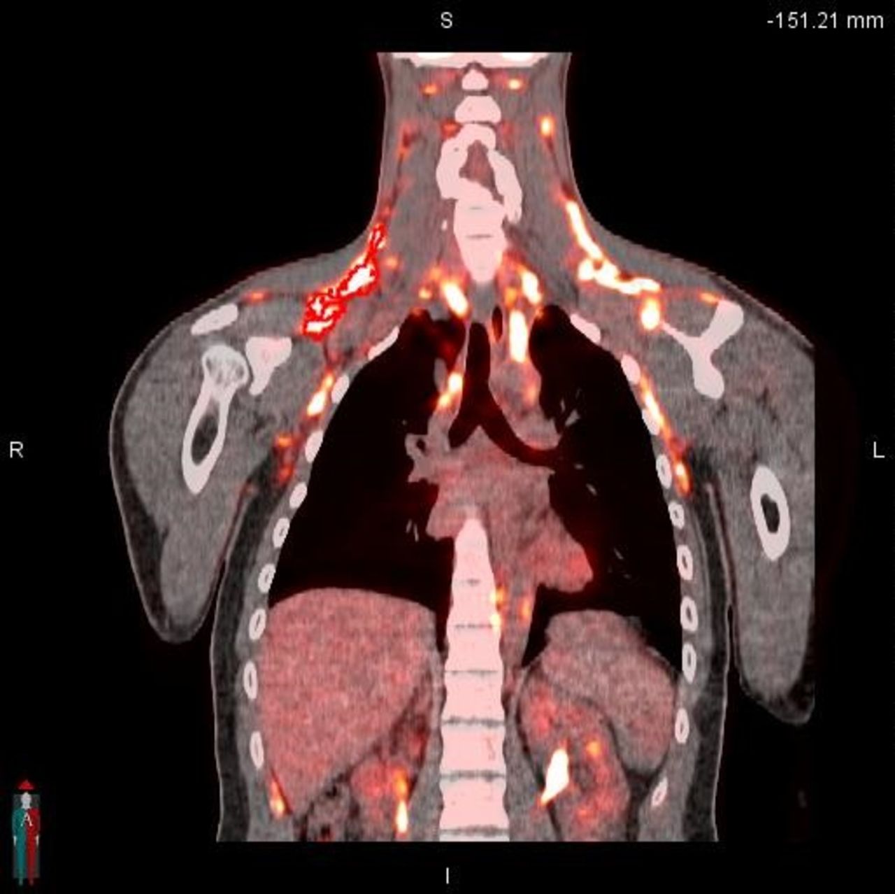
Objectives: Brown Adipose Tissue (BAT) is a metabolically active adipose tissue that plays a major role in shivering-independent thermogenesis via uncoupling protein-1(UCP-1), as well as glucose and lipid homeostasis in adult humans.[1] Emerging radiomic analysis of tomographic imaging can further characterize BAT by providing a diverse set of novel quantitative features. These quantitative radiomic features have been utilized as biomarkers for phenotyping tumors, their response to treatment, and prognosis.[2] We aim to assess BAT radiomics features repeatability using FDG-PET in order to elucidate its radiomics characteristic

Methods: Twenty seven healthy adults (21 women and 6 men; mean age ± SD, 23.94 ± 3.64 y; body mass index, 22.21 ± 1.82 kg/m2) under the age of 35; 26 subjects completed the study protocol. Subjects were cooled via cool water cooling suit for 30 minutes prior to FDG administration and for an additional 60 minutes during FDG uptake phase. Each subject underwent FDG-PET/CT scan after cooling procedure. Participants underwent imaging with the same protocols and identical FDG dose within 2 weeks from the first scan. Brown Adipose Reporting Criteria in Imaging Studies (BARCIST) criteria was employed to identify BAT[3]. Active BAT in right supraclavicular region was selected. 878 radiomic features were extracted using Pyradiomics packages (http://pyradiomics.readthedocs.io) for each region of interest (ROI) on high definition reconstructed PET.[4] Lin's concordance correlation coefficients(CCC) was used to assess repeatability of the extracted radiomic features.[5] To elucidate the association between repeatable QIFs and SUVmax for the sake of characterization of the radiomic signature, a heat map analysis with hierarchical clustering on median centered radiomic features using complete linkage while using Pearson's correlation coefficient as the similarity measure.Results: 61 features(6.94%) had high reproducibility on test-retest (CCC>0.85). Hierarchical clustering of these repeatable features resulted in 4 distinct clusters. SUVmax did not cluster with any repeatable radiomic features.Conclusion:The identified FDG-PET radiomic cluster features provided additional information regarding BAT metabolic activation profile distinct from SUVmax and potentially can be used as quantitative biomarkers of BAT in future studies.

Figure
  • Download figure
  • Open in new tab
  • Download powerpoint
Figure
  • Download figure
  • Open in new tab
  • Download powerpoint
Previous
Back to top

In this issue

Journal of Nuclear Medicine
Vol. 59, Issue supplement 1
May 1, 2018
  • Table of Contents
  • Index by author
Article Alerts
Sign In to Email Alerts with your Email Address
Email Article

Thank you for your interest in spreading the word on Journal of Nuclear Medicine.

NOTE: We only request your email address so that the person you are recommending the page to knows that you wanted them to see it, and that it is not junk mail. We do not capture any email address.

Enter multiple addresses on separate lines or separate them with commas.
Radiomic Phenotype of Activated Brown Fat on FDG PET
(Your Name) has sent you a message from Journal of Nuclear Medicine
(Your Name) thought you would like to see the Journal of Nuclear Medicine web site.
Citation Tools
Radiomic Phenotype of Activated Brown Fat on FDG PET
Richard Wahl, John Crandall, Aria Nazeri
Journal of Nuclear Medicine May 2018, 59 (supplement 1) 648;

Citation Manager Formats

  • BibTeX
  • Bookends
  • EasyBib
  • EndNote (tagged)
  • EndNote 8 (xml)
  • Medlars
  • Mendeley
  • Papers
  • RefWorks Tagged
  • Ref Manager
  • RIS
  • Zotero
Share
Radiomic Phenotype of Activated Brown Fat on FDG PET
Richard Wahl, John Crandall, Aria Nazeri
Journal of Nuclear Medicine May 2018, 59 (supplement 1) 648;
Twitter logo Facebook logo LinkedIn logo Mendeley logo
  • Tweet Widget
  • Facebook Like
  • Google Plus One
Bookmark this article

Jump to section

  • Article
  • Figures & Data
  • Info & Metrics

Related Articles

  • No related articles found.
  • Google Scholar

Cited By...

  • No citing articles found.
  • Google Scholar

More in this TOC Section

Instrumentation & Data Analysis Track

  • Deep Learning Based Kidney Segmentation for Glomerular Filtration Rate Measurement Using Quantitative SPECT/CT
  • The Benefit of Time-of-Flight in Digital Photon Counting PET Imaging: Physics and Clinical Evaluation
  • Preclinical validation of a single-scan rest/stress imaging technique for 13NH3 cardiac perfusion studies
Show more Instrumentation & Data Analysis Track

Radiomics and Quantification in Oncology PET Studies

  • Dependency of a validated radiomics signature on tumor volume and potential corrections
  • FDG-PET radiomic twins often share molecular subtype and hormonal receptor status in breast cancer patients
  • Investigating the radiomic signatures of metastatic and non-metastatic breast cancer patients based on F18-FDG PET/CT images.
Show more Radiomics and Quantification in Oncology PET Studies

Similar Articles

SNMMI

© 2025 SNMMI

Powered by HighWire