
Main menu

  • Home
  • Content
    • Current
    • Ahead of print
    • Past Issues
    • JNM Supplement
    • SNMMI Annual Meeting Abstracts
    • Continuing Education
    • JNM Podcasts
  • Subscriptions
    • Subscribers
    • Institutional and Non-member
    • Rates
    • Journal Claims
    • Corporate & Special Sales
  • Authors
    • Submit to JNM
    • Information for Authors
    • Assignment of Copyright
    • AQARA requirements
  • Info
    • Reviewers
    • Permissions
    • Advertisers
  • About
    • About Us
    • Editorial Board
    • Contact Information
  • More
    • Alerts
    • Feedback
    • Help
    • SNMMI Journals
  • SNMMI
    • JNM
    • JNMT
    • SNMMI Journals
    • SNMMI

User menu

  • Subscribe
  • My alerts
  • Log in
  • My Cart

Search

  • Advanced search
Journal of Nuclear Medicine
  • SNMMI
    • JNM
    • JNMT
    • SNMMI Journals
    • SNMMI
  • Subscribe
  • My alerts
  • Log in
  • My Cart
Journal of Nuclear Medicine

Advanced Search

  • Home
  • Content
    • Current
    • Ahead of print
    • Past Issues
    • JNM Supplement
    • SNMMI Annual Meeting Abstracts
    • Continuing Education
    • JNM Podcasts
  • Subscriptions
    • Subscribers
    • Institutional and Non-member
    • Rates
    • Journal Claims
    • Corporate & Special Sales
  • Authors
    • Submit to JNM
    • Information for Authors
    • Assignment of Copyright
    • AQARA requirements
  • Info
    • Reviewers
    • Permissions
    • Advertisers
  • About
    • About Us
    • Editorial Board
    • Contact Information
  • More
    • Alerts
    • Feedback
    • Help
    • SNMMI Journals
  • View or Listen to JNM Podcast
  • Visit JNM on Facebook
  • Join JNM on LinkedIn
  • Follow JNM on Twitter
  • Subscribe to our RSS feeds
Meeting ReportInstrumentation & Data Analysis Track

Initial Evaluation of Three Novel Quantiative Imaging Utilities from a Newly Developed SPECT Camera in China

Wenrui Zhao, Xinqiang Wang, Yingkui Liang, Feng Guo, Lilei Gao, Yaqiang Liu and Si Chen
Journal of Nuclear Medicine May 2018, 59 (supplement 1) 219;
Wenrui Zhao
3Department of Nuclear Medicine Navy General Hospital of Chinese PLA Beijing China
  • Find this author on Google Scholar
  • Find this author on PubMed
  • Search for this author on this site
Xinqiang Wang
3Department of Nuclear Medicine Navy General Hospital of Chinese PLA Beijing China
  • Find this author on Google Scholar
  • Find this author on PubMed
  • Search for this author on this site
Yingkui Liang
3Department of Nuclear Medicine Navy General Hospital of Chinese PLA Beijing China
  • Find this author on Google Scholar
  • Find this author on PubMed
  • Search for this author on this site
Feng Guo
3Department of Nuclear Medicine Navy General Hospital of Chinese PLA Beijing China
  • Find this author on Google Scholar
  • Find this author on PubMed
  • Search for this author on this site
Lilei Gao
1Beijing Novel Medical Equipment Ltd Beijing China
  • Find this author on Google Scholar
  • Find this author on PubMed
  • Search for this author on this site
Yaqiang Liu
2Key Laboratory of Particle & Radiation Imaging(Tsinghua University), Ministry of Education Beijing China
4Department of Engineering Physics Tsinghua University Beijing China
  • Find this author on Google Scholar
  • Find this author on PubMed
  • Search for this author on this site
Si Chen
1Beijing Novel Medical Equipment Ltd Beijing China
  • Find this author on Google Scholar
  • Find this author on PubMed
  • Search for this author on this site
  • Article
  • Figures & Data
  • Info & Metrics
Loading

Abstract

219

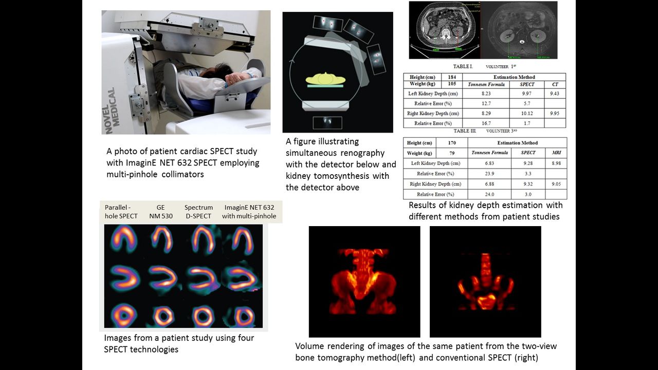
Objectives: A general purpose flexible angle dual head SPECT camera, ImaginE NET 632, was developed by Beijing Novel Medical Equipment Ltd and has been recently approved by China Food and Drug Administration (CFDA) for sale in China. The objective of this study was to evaluate three novel quantitative imaging utilities of this camera for bone,kidney and myocardium respectively.

Methods: The first utility is quantitative bone tomography for a certain axial region of interest (ROI) based on two-view whole body gamma scintigraphy and diagnostic CT images of the same ROI but from a separate scanner. The quantitative bone tomography was achieved by the following steps: first segmented 3D bone structures from CT images automatically, then registered the 3D CT bone segmentations with axial ROIs of whole body gamma planar images by iterative 3D transformation,3D to 2D projection and 2D registration, finally reconstructed gamma bone tomography with CT-based attenuation correction from the two-view gamma projection image ROI by incorporating the CT bone segmentations as prior information. Two patients were studied and the resulted tomography images were compared with the images from conventional SPECT scans. The second utility is quantitative renography featured by patient specific image-based estimation of the kidney depth for attenuation correction to improve the quantitation accuracy. The quantitative renography was achieved through a novel data acquisition procedure and subsequent data processing methods. For data acquisition, we employed the flexible angle option of ImaginE NET 632 SPECT which performed simultaneous dynamic renal imaging with the detector below staying stationary and tomosynthesis of kidneys with the detector above rotating while acquiring projections. For data processing, centroids of kidneys are estimated by segmentation and least square fit of the tomosynthetic data and then applied for kidney depth calculation and attenuation correction in renography. Three patients were studied and the gamma image-based estimations of kidney depths were compared with those from Tonnessen formula(i.e. based on patient’s height and weight) employing CT or MR image-based estimations as golden standard. The third utility is quantitative dynamic myocardium SPECT imaging achieved by V-mode acquisition of dual detector head. Equipped with a pair of multi-pinhole collimators of 24 pinholes in total, two SPECT detector span 180 degrees of sampling angle range on a 18cm-diameter spherical common field of view(CFOV). Phantom studies demonstrated 7mm spatial resolution and 0.06% sensitivity were achieved for the center of CFOV. Four patients were studied and compared with conventional SPECT with parallel-hole collimators and two dedicated cardiac SPECT cameras. Results: In the study of novel quantitative bone tomography, improved image resolution and enhanced detailed features were observed compared with the conventional bone SPECT images. These differences were mainly attributed to the CT prior information,smaller bin size and better statistics of the whole body planar image data compared with the SPECT projection data. In the study of quantitative renography, <6% error was achieved in the kidney depth estimation with the tomosynthesis method compared with >15% error with conventional Tonnessen formula. In the study of quantitative dynamic cardiac SPECT imaging, similar degree of improved defect detection and reduced variation of functional myocardium were observed for ImaginE NET 632 SPECT with dedicated multi-pinhole collimators and two other dedicated cardiac SPECT, compared with the parallel-hole SPECT. Conclusion: The ImaginE NET 632 SPECT camera with three novel imaging utilities has great potential to improve the corresponding bone, kidney and cardiac study applications. Systematic patient studies need to be carried out to further evaluate the utilities and optimize the clinical protocols.

Figure
  • Download figure
  • Open in new tab
  • Download powerpoint
Previous
Back to top

In this issue

Journal of Nuclear Medicine
Vol. 59, Issue supplement 1
May 1, 2018
  • Table of Contents
  • Index by author
Article Alerts
Sign In to Email Alerts with your Email Address
Email Article

Thank you for your interest in spreading the word on Journal of Nuclear Medicine.

NOTE: We only request your email address so that the person you are recommending the page to knows that you wanted them to see it, and that it is not junk mail. We do not capture any email address.

Enter multiple addresses on separate lines or separate them with commas.
Initial Evaluation of Three Novel Quantiative Imaging Utilities from a Newly Developed SPECT Camera in China
(Your Name) has sent you a message from Journal of Nuclear Medicine
(Your Name) thought you would like to see the Journal of Nuclear Medicine web site.
Citation Tools
Initial Evaluation of Three Novel Quantiative Imaging Utilities from a Newly Developed SPECT Camera in China
Wenrui Zhao, Xinqiang Wang, Yingkui Liang, Feng Guo, Lilei Gao, Yaqiang Liu, Si Chen
Journal of Nuclear Medicine May 2018, 59 (supplement 1) 219;

Citation Manager Formats

  • BibTeX
  • Bookends
  • EasyBib
  • EndNote (tagged)
  • EndNote 8 (xml)
  • Medlars
  • Mendeley
  • Papers
  • RefWorks Tagged
  • Ref Manager
  • RIS
  • Zotero
Share
Initial Evaluation of Three Novel Quantiative Imaging Utilities from a Newly Developed SPECT Camera in China
Wenrui Zhao, Xinqiang Wang, Yingkui Liang, Feng Guo, Lilei Gao, Yaqiang Liu, Si Chen
Journal of Nuclear Medicine May 2018, 59 (supplement 1) 219;
Twitter logo Facebook logo LinkedIn logo Mendeley logo
  • Tweet Widget
  • Facebook Like
  • Google Plus One
Bookmark this article

Jump to section

  • Article
  • Figures & Data
  • Info & Metrics

Related Articles

  • No related articles found.
  • Google Scholar

Cited By...

  • No citing articles found.
  • Google Scholar

More in this TOC Section

Instrumentation & Data Analysis Track

  • Deep Learning Based Kidney Segmentation for Glomerular Filtration Rate Measurement Using Quantitative SPECT/CT
  • The Benefit of Time-of-Flight in Digital Photon Counting PET Imaging: Physics and Clinical Evaluation
  • Preclinical validation of a single-scan rest/stress imaging technique for 13NH3 cardiac perfusion studies
Show more Instrumentation & Data Analysis Track

Systems

  • The Benefit of Time-of-Flight in Digital Photon Counting PET Imaging: Physics and Clinical Evaluation
  • NEMA NU2-2012 Performance Measurements of the United-Imaging uPMR790: A HD TOF Simultaneous PET/MR System
  • Evaluation of CZT gamma cameras for human SPECT and small FOV imaging
Show more Systems

Similar Articles

SNMMI

© 2025 SNMMI

Powered by HighWire