


Article Figures & Data
Figures
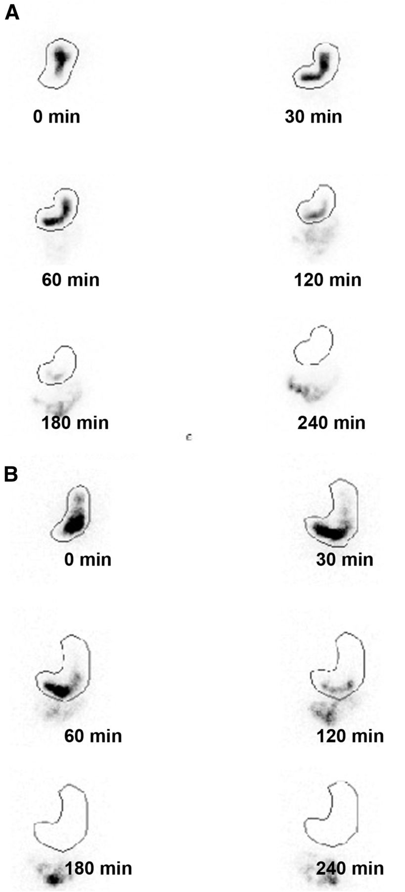
- FIGURE 1.
Examples of normal FA (A) and abnormal FA (B) assessed by GES. In normal FA, most radiolabeled solids appeared in proximal stomach immediately after meal ingestion (time, 0 min). Over time, solids progressed into distal stomach. In abnormal FA, most radiolabeled solids appeared in distal stomach at 0 min.
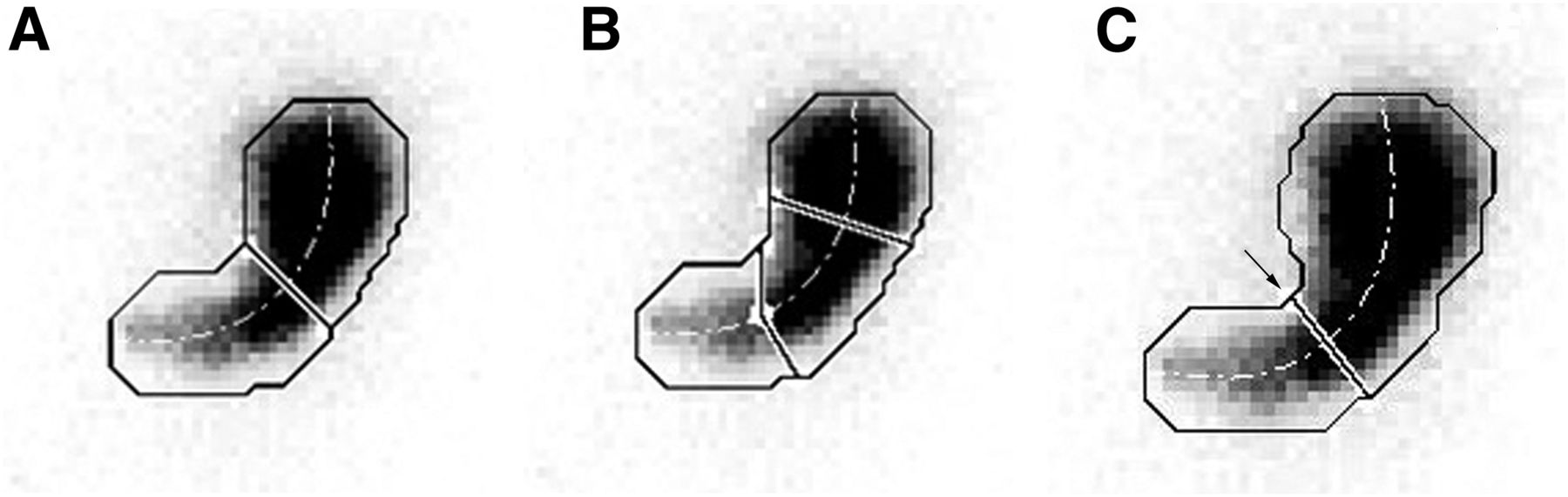
- FIGURE 2.
Three methods for dividing stomach into proximal and distal portions. (A) Illustration of how computer-generated regions of interest (ROIs) for proximal and distal stomach (solid line) were defined by dividing stomach at one-half the distance along long axis of stomach (dotted line). (B) Illustration of how computer-generated ROIs were defined by selecting equal one-third divisions along long axis of stomach. (C) Stomach incisura angularis is site of formation of acute angle on lesser curvature (arrow) to form localized “notch.” Location of incisura varies depending on degree of gastric distension; therefore, consistent localization is difficult.
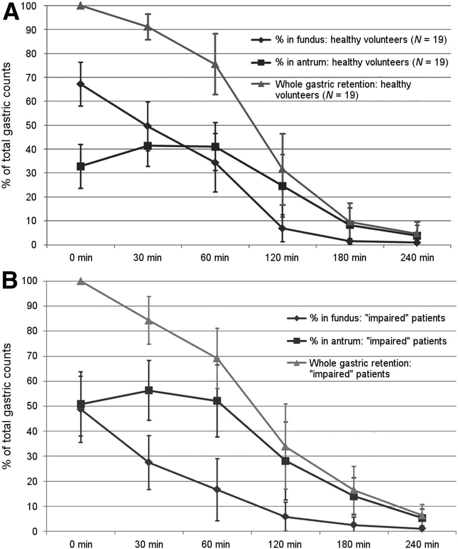
- FIGURE 3.
IMD over time after meal ingestion for normal volunteers (A) and for patients with abnormal FA (B), as indicated by readers’ assessment of FA. Values are means ± 1 SD at each recorded time.
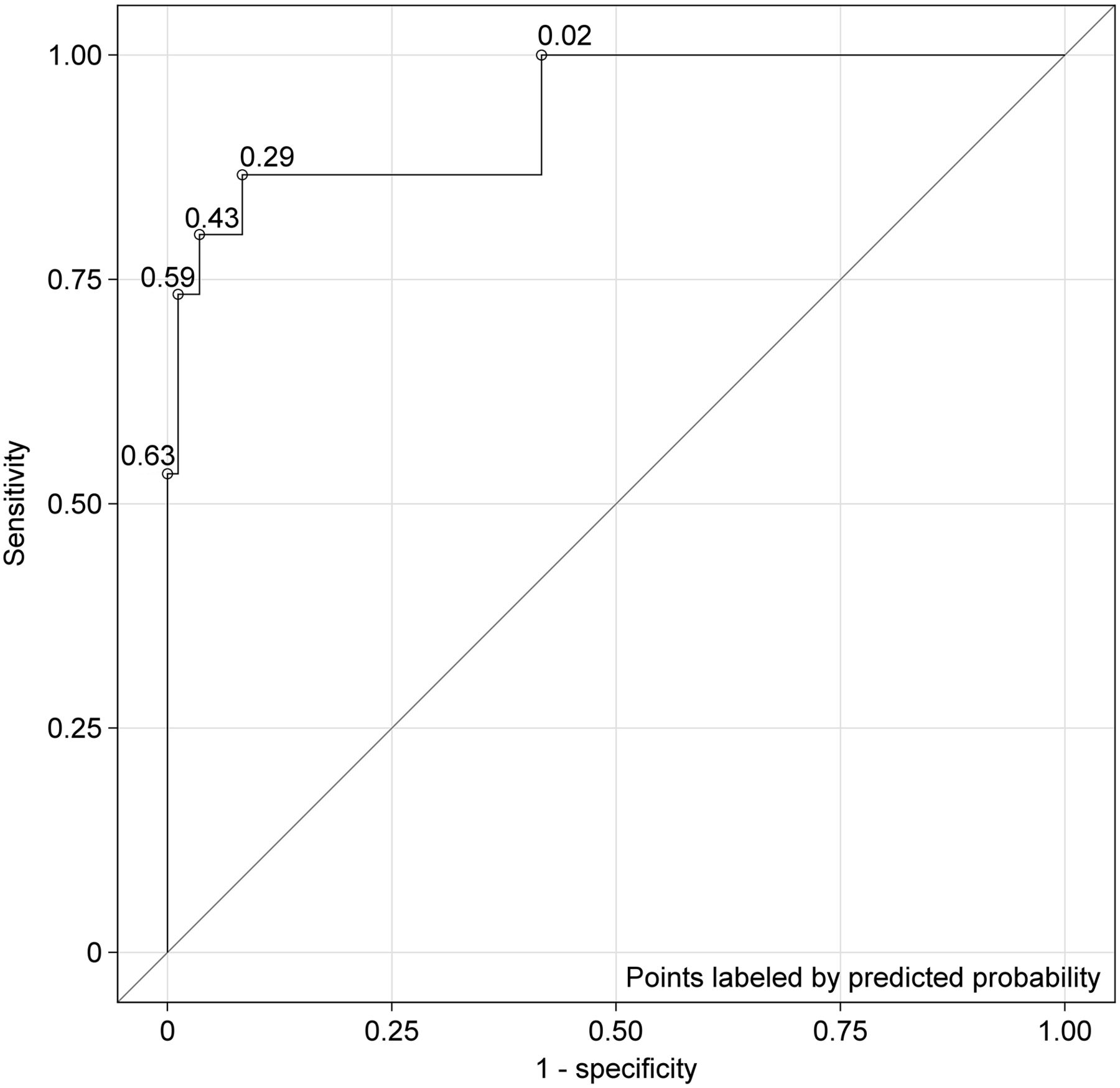
- FIGURE 4.
Logistic regression and receiver-operating-characteristic (ROC) curve for 99 test subjects, with IMD0 or percentage of proximal gastric retention at baseline being used as predictor of abnormal/impaired FA. Area under curve (concordance statistic) for this ROC curve was 0.934; this value implied that corresponding logistic regression model offered excellent fit to data (25).


Tables
- TABLE 1
Agreement of Assessment of FA by 4 Nuclear Medicine and Radiology Physicians Evaluating 99 Subjects
No. of subjects for which positive report was given by: Judgment of entire panel* 4 Readers 3 Readers 2 Readers 1 Reader 0 Reader Positive (impaired/abnormal) 7 8 0 0 0 Negative (normal) 0 0 11 20 53 Overall No. 7 8 11 20 53 Percentage 7.1 8.1 11.1 20.2 53.5 ↵* Images that were classified as positive (impaired/abnormal) by at least 3 of 4 nuclear medicine and radiology physicians were considered true-positive; all others were considered true-negative (normal).
Institutions at which pairs of readers were located Weighted κ (95% CI)* Simple κ (95% CI)† TUH vs. Wake 0.230 (0.122–0.338) 0.230 (0.097–0.364) TUH vs. Stanford 0.410 (0.267–0.554) 0.457 (0.252–0.661) TUH vs. JHH 0.423 (0.246–0.600) 0.476 (0.230–0.722) Wake vs. Stanford 0.438 (0.325–0.551) 0.525 (0.364–0.685) Wake vs. JHH 0.293 (0.190–0.396) 0.381 (0.223–0.539) Stanford vs. JHH 0.483 (0.350–0.616) 0.521 (0.320–0.722) Overall average 0.380 (0.250–0.509) 0.432 (0.248–0.616)
Supplemental Data
Files in this Data Supplement:


Jump to section
Related Articles
Cited By...
- Relationship between intragastric meal distribution, gastric emptying and gastric neuromuscular dysfunction in chronic gastroduodenal disorders
- Distinct subgroups in gastroparesis defined by simultaneous body surface gastric mapping and gastric emptying breath testing
- Gastric emptying study before gastric peroral endoscopic myotomy (G-POEM): can intragastric meal distribution be a predictor of success?
- Actionable biomarkers: the key to resolving disorders of gastrointestinal function
- Proximal and Distal Gastric Retention Patterns in Gastroparesis and the Impact of Gastric Per-Oral Endoscopic Myotomy: A Retrospective Analysis Using Gastric Emptying Scintigraphy
- Enhanced Gastric Emptying Scintigraphy to Assess Fundic Accommodation Using Intragastric Meal Distribution and Antral Contractility