


Article Figures & Data
Figures
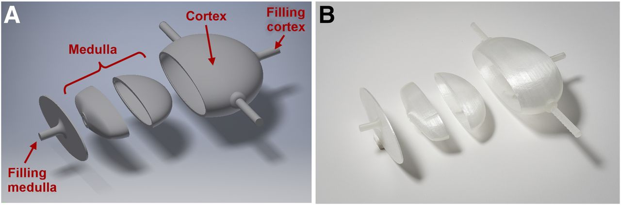
- FIGURE 1.
Division of kidney into 4 separate parts to enable fused deposition modeling 3D printing. (A) CAD model. (B) Printed parts.
- FIGURE 2.
Cross-section through kidney phantom at different stages. (A) CAD model. (B) CT-based segmentation in 3D Slicer. (C) Fused SPECT/CT reconstruction.
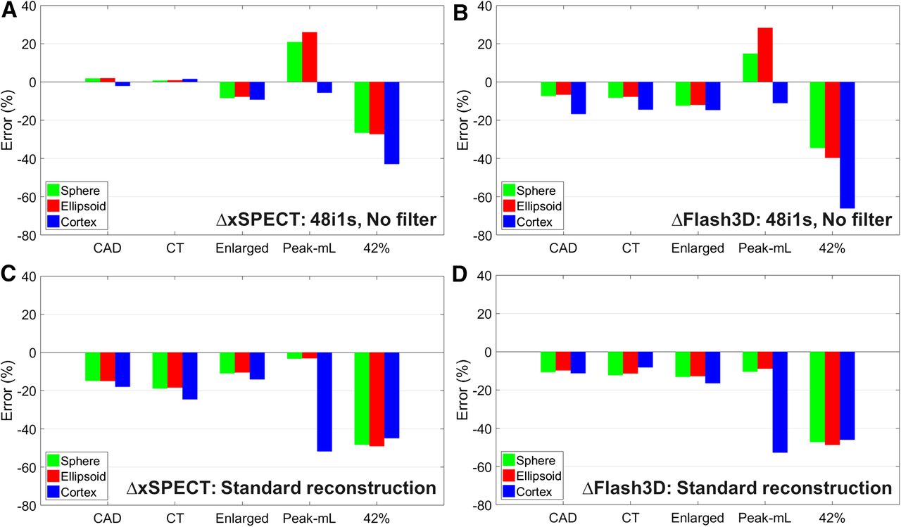
- FIGURE 3.
Accuracy of activity determination with different PVC methods. (A and B) xSPECT/Flash3D: 48 iterations, 1 subset, and no filter. (C and D) xSPECT/Flash3D standard reconstruction: 16/30 iterations, 1/2 subsets, and 20.8-mm gaussian filter.
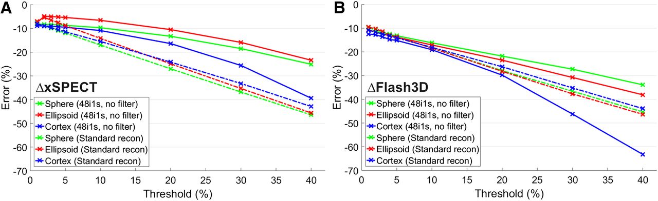
- FIGURE 4.
Threshold-dependent accuracy of activity determined with and without filtering for xSPECT (A) and Flash3D (B). Solid lines are for initial reconstruction (48 iterations, 1 subset, and no filter); dashed–dotted lines are for filtered standard reconstruction (xSPECT/Flash3D: 16/30 iterations, 1/2 subsets, and 20.8-mm gaussian filter).
- FIGURE 5.
Effect of gaussian postfiltering on sphere phantom for xSPECT (A) and Flash3D (B). Shown are fused SPECT/CT images at different stages of reconstruction (left) and horizontal cross-sections (right). Solid lines are for initial reconstruction; dashed–dotted lines are for filtered reconstruction.
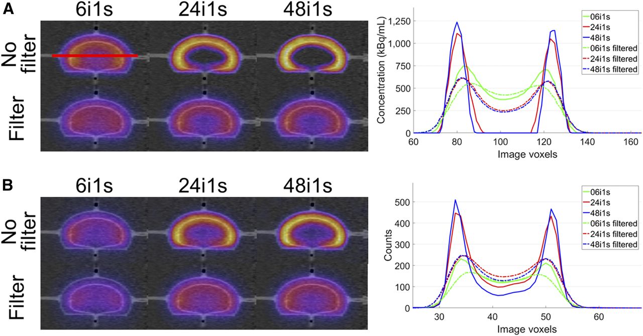
- FIGURE 6.
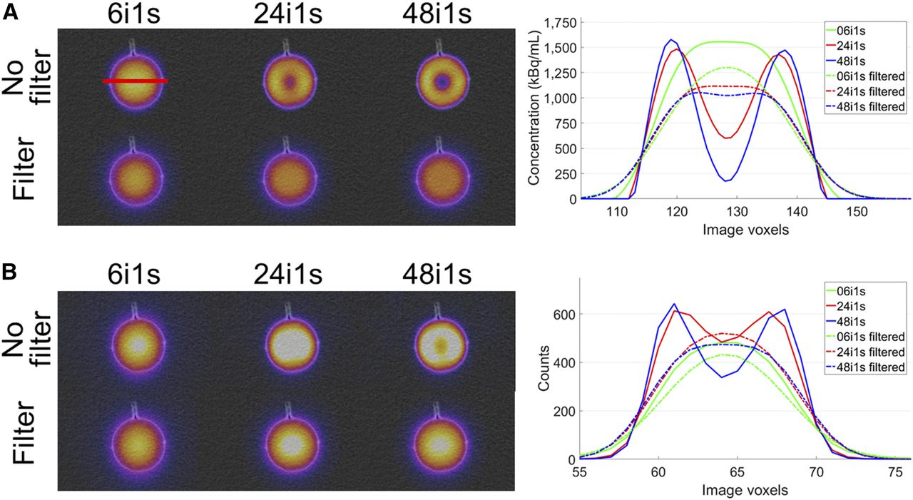
Effect of gaussian postfiltering on kidney phantom for xSPECT (A) and Flash3D (B). Shown are fused SPECT/CT images at different stages of reconstruction (left) and horizontal cross-sections (right). Solid lines are for initial reconstruction; dashed–dotted lines are for filtered reconstruction.


Tables
Volume (cm3) Length (mm) Width (mm) Thickness (mm) Kidney Cortex Medulla 113.5 59.5 44.5 143.57 99.58 43.98 Parameter xSPECT Flash3D 48 iterations, 1 subset, no filtering 9.41 mm 10.35 mm Standard reconstruction 17.25 mm 21.19 mm Parameter Mask Sphere Ellipsoid ΔS-E Cortex ΔS-C xSPECT 48 iterations, 1 subset, no filtering CAD 0.834 0.829 −0.6 0.634 −23.9 CT 0.823 0.820 −0.7 0.602 −38.4 Standard reconstruction CAD 0.716 0.711 −0.4 0.441 −26.9 CT 0.728 0.721 −1.0 0.471 −35.3 Flash3D 48 iterations, 1 subset, no filtering CAD 0.839 0.834 −0.5 0.643 −23.4 CT 0.830 0.828 −1.3 0.617 −38.9 Standard reconstruction CAD 0.665 0.657 −0.2 0.406 −25.7 CT 0.657 0.650 −1.1 0.385 −41.5 ΔS-E = percentage difference between sphere and ellipsoid; ΔS-C = percentage difference between sphere and cortex.
Geometry Parameter −2 mm −1 mm Nominal +1 mm +2 mm xSPECT Resolution (mm) 8.35 9.35 10.35 11.35 12.35 Sphere 
0.865 0.850 0.834 0.819 0.804 Difference (%) 3.8 2.0 — −1.8 −3.6 Ellipsoid 0.861 0.846 0.829 0.813 0.798 Difference (%) 3.9 2.0 — −1.8 −3.7 Cortex 0.697 0.667 0.634 0.606 0.580 Difference (%) 9.9 5.1 — −4.4 −8.6 Flash3D Resolution (mm) 7.41 8.41 9.41 10.41 11.41 Sphere 0.866 0.854 0.839 0.823 0.808 Difference (%) 3.2 1.8 — −1.9 −3.7 Ellipsoid 0.862 0.850 0.834 0.818 0.803 Difference (%) 3.3 1.9 — −2.0 −3.7 Cortex 0.698 0.674 0.643 0.613 0.586 Difference (%) 8.6 4.9 — −4.6 −8.8


Jump to section
Related Articles
Cited By...
- A Deep-Learning-Based Partial-Volume Correction Method for Quantitative 177Lu SPECT/CT Imaging
- A Pipeline for Automated Voxel Dosimetry: Application in Patients with Multi-SPECT/CT Imaging After 177Lu-Peptide Receptor Radionuclide Therapy
- Toward a Patient-Specific Traceable Quantification of SPECT/CT-Based Radiopharmaceutical Distributions
- What You See Is Not What You Get: On the Accuracy of Voxel-Based Dosimetry in Molecular Radiotherapy
- Characterization of Noise and Resolution for Quantitative 177Lu SPECT/CT with xSPECT Quant
- The Relevance of Dosimetry in Precision Medicine