
Main menu

  • Home
  • Content
    • Current
    • Ahead of print
    • Past Issues
    • JNM Supplement
    • SNMMI Annual Meeting Abstracts
    • Continuing Education
    • JNM Podcasts
  • Subscriptions
    • Subscribers
    • Institutional and Non-member
    • Rates
    • Journal Claims
    • Corporate & Special Sales
  • Authors
    • Submit to JNM
    • Information for Authors
    • Assignment of Copyright
    • AQARA requirements
  • Info
    • Reviewers
    • Permissions
    • Advertisers
  • About
    • About Us
    • Editorial Board
    • Contact Information
  • More
    • Alerts
    • Feedback
    • Help
    • SNMMI Journals
  • SNMMI
    • JNM
    • JNMT
    • SNMMI Journals
    • SNMMI

User menu

  • Subscribe
  • My alerts
  • Log in
  • My Cart

Search

  • Advanced search
Journal of Nuclear Medicine
  • SNMMI
    • JNM
    • JNMT
    • SNMMI Journals
    • SNMMI
  • Subscribe
  • My alerts
  • Log in
  • My Cart
Journal of Nuclear Medicine

Advanced Search

  • Home
  • Content
    • Current
    • Ahead of print
    • Past Issues
    • JNM Supplement
    • SNMMI Annual Meeting Abstracts
    • Continuing Education
    • JNM Podcasts
  • Subscriptions
    • Subscribers
    • Institutional and Non-member
    • Rates
    • Journal Claims
    • Corporate & Special Sales
  • Authors
    • Submit to JNM
    • Information for Authors
    • Assignment of Copyright
    • AQARA requirements
  • Info
    • Reviewers
    • Permissions
    • Advertisers
  • About
    • About Us
    • Editorial Board
    • Contact Information
  • More
    • Alerts
    • Feedback
    • Help
    • SNMMI Journals
  • View or Listen to JNM Podcast
  • Visit JNM on Facebook
  • Join JNM on LinkedIn
  • Follow JNM on Twitter
  • Subscribe to our RSS feeds
Research ArticleFocus on Molecular Imaging

In Vivo Imaging of Pro- and Antitumoral Cellular Components of the Tumor Microenvironment

Anne Helfen, Johannes Roth, Tony Ng and Michel Eisenblaetter
Journal of Nuclear Medicine February 2018, 59 (2) 183-188; DOI: https://doi.org/10.2967/jnumed.117.198952
Anne Helfen
1Department of Clinical Radiology, University Hospital Muenster, Muenster, Germany
  • Find this author on Google Scholar
  • Find this author on PubMed
  • Search for this author on this site
Johannes Roth
2Institute of Immunology, University Hospital Muenster, Muenster, Germany
  • Find this author on Google Scholar
  • Find this author on PubMed
  • Search for this author on this site
Tony Ng
3Richard Dimbleby Department of Cancer Research, School of Cancer & Pharmaceutical Sciences, King’s College London, London, United Kingdom
4Breast Cancer Now Research Unit, Department of Research Oncology, Guy’s Hospital, King’s College London, London, United Kingdom
5UCL Cancer Institute, University College London, London, United Kingdom; and
  • Find this author on Google Scholar
  • Find this author on PubMed
  • Search for this author on this site
Michel Eisenblaetter
1Department of Clinical Radiology, University Hospital Muenster, Muenster, Germany
6School of Biomedical Engineering & Imaging Sciences, King’s College London, London, United Kingdom
  • Find this author on Google Scholar
  • Find this author on PubMed
  • Search for this author on this site
  • Article
  • Figures & Data
  • Info & Metrics
  • PDF
Loading

Article Figures & Data

Figures

  • Tables
  • FIGURE 1.
    • Download figure
    • Open in new tab
    • Download powerpoint
    FIGURE 1.

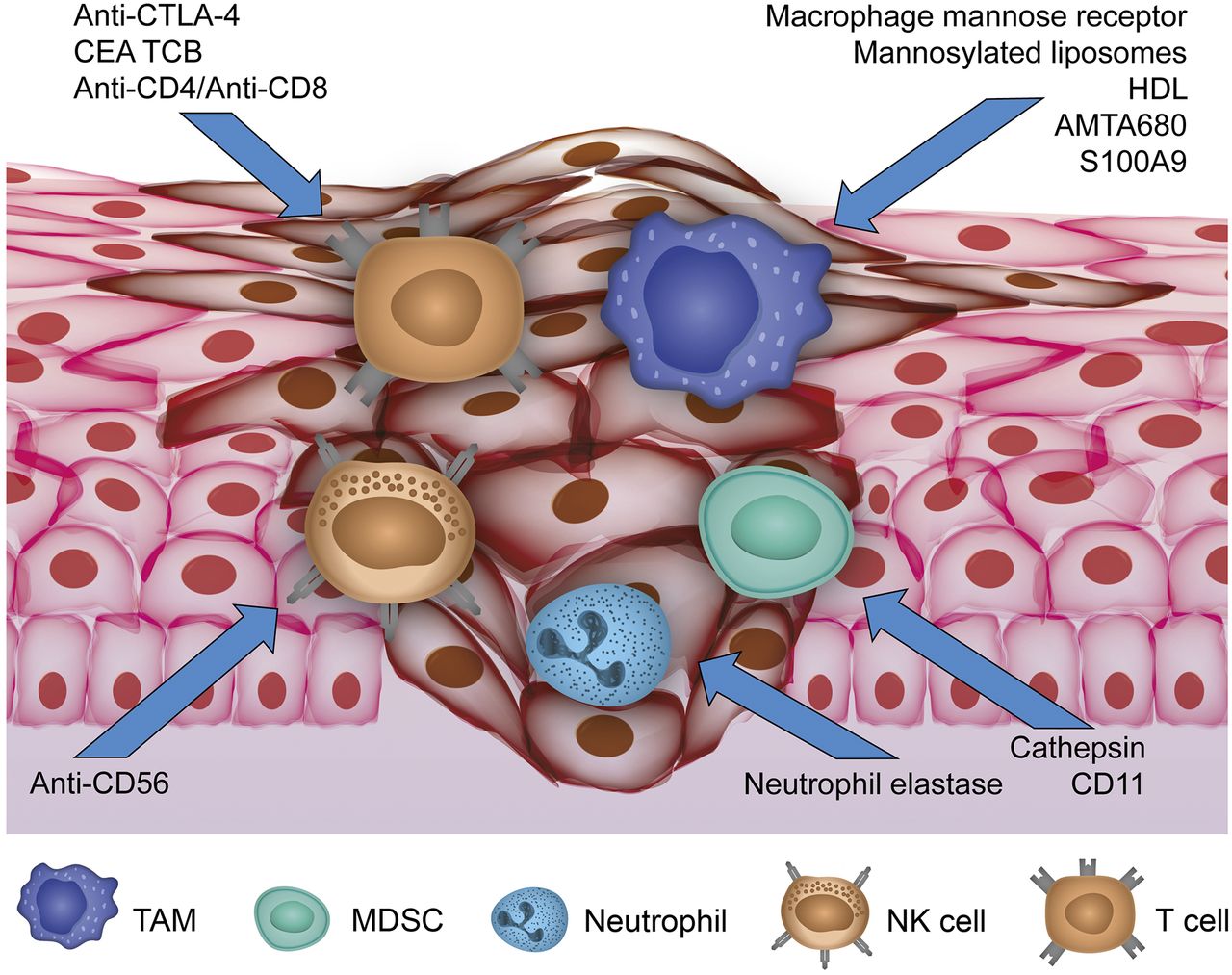
    Overview of current imaging approaches targeting cellular compounds of TME. Activity of TAMs, MDSCs, and neutrophils as protumoral immune cells infiltrating primary tumor is reflected by visualizing specific targets for current molecular imaging approaches. Antitumoral NK cells have been addressed by anti-CD56, whereas approaches targeting anti-CTLA-4, anti-CD4/CD8, and carcinoembryonic antigen T-cell–specific antibody (CEA TCB) for T-cell imaging have been reported.
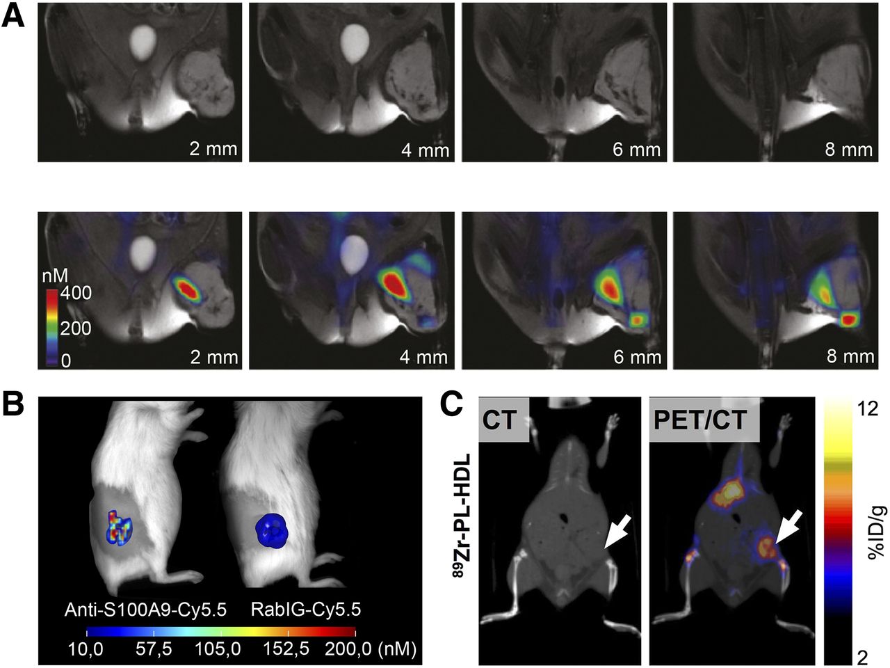
  • FIGURE 2.
    • Download figure
    • Open in new tab
    • Download powerpoint
    FIGURE 2.

    (A) Imaging of TAM distribution in mouse with soft-tissue sarcoma 24 h after intravenous injection of AMTA680, with naïve MR images shown at top and fused fluorescence-mediated tomography and MR images at bottom. (Adapted with permission of (21).) (B) Fluorescence imaging of TAM activity in murine 4T1 breast cancer. The specific tracer anti-S100A9-Cy5.5 shows high accumulation within tumor lesion, whereas homogeneous signal of nonspecifically binding rabIgG-Cy5.5 reflects tumor perfusion. (C) 89Zr-HDL–driven in vivo PET for imaging TAMs in murine 4T1 breast cancer 24 h after tracer injection. CT image is on left and PET/CT image on right. (Adapted with permission of (17).) %ID = percentage injected dose.
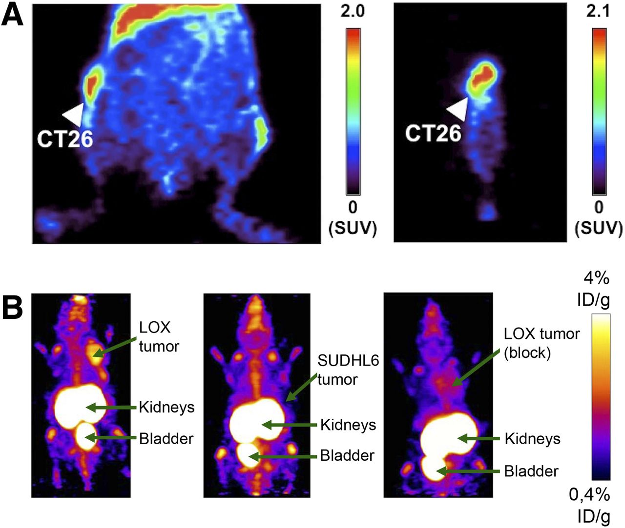
  • FIGURE 3.
    • Download figure
    • Open in new tab
    • Download powerpoint
    FIGURE 3.

    T-cell in vivo imaging within TME. (A) PET imaging using 64Cu-DOTA–labeled anti-CTLA-4 showing specific tracer accumulation in murine CT26 colon carcinoma in representative coronal (left) and sagittal (right) slices. Results suggest promise for evaluating targeted therapy by anti-CTLA-4 monoclonal antibodies. (Adapted with permission of (41).) (B) PET imaging after injection of 18F-labeled anti–PD-L1 Affibody molecule. Tracer allows for imaging of PD-L1 expressing LOX malignant melanoma (left) in comparison with negative controls of nonexpressing lymphoma SUDHL6 (middle) and blocked LOX tumor (right). (Adapted with permission of (48).) %ID = percentage injected dose.

Tables

  • Figures
    • View popup
    TABLE 1

    Characteristics of Immune Cells in TME

    Cell typeCell surface markersFunctions in TME
    TAMCD11b+ CD14+ CD31+ CD34+ CD45+ CD68+ CD117− CD133− CD146− CD204+ CD206+ CCR2+ CSF1R+ MHCII+ VEGFR1+ VEGFR2− (human/mouse); F4/80 (mouse); CD23+ CD16+ CXCR4+ (human)Enhancement of angiogenesis and remodeling; tumor promotion; association with poor prognosis
    MDSCCD11b+ CD14+ MHCI+ MHCIIlow (human/mouse); GR1+ CD11b+ (mouse); CD11b+/− CD33+ CD34+ CD68− (human)Increased in almost all patients/animals with cancer; ability to suppress T cells as defining trait
    NeutrophilCD11b+ CD14low CD31+ CD66B+ CXCR2+ (human/mouse); GR1+ VEGFR1+ CXCR1− (mouse); CD15+ CXCR1+ (human)Enhancement of angiogenesis and metastasis in animal models; increased levels in patients with colon, gastric and lung cancer; association with poor prognosis in bronchoalveolar carcinoma
    CD4+ T cellCD3+ CD4+ CD45+ (human/mouse)T-helper 1 cells: assistance to CD8+ cells in tumor rejection; T-helper 2 cells: polarization of immunity away from antitumor response
    CD8+ T cellCD3+ CD8+ CD45+ (human/mouse)Effector cells of adaptive immune system; specific recognition and destruction of cancer cells through perforin- and granzyme-mediated apoptosis
    Regulatory T cellsCD4+ CD25+ FOXP3+(human/mouse)Central role in tumor maintenance via suppression of antitumor immune response; blocking of CD8+ cell activation and NK cell killing; infiltration associated with poor prognosis (14)
    NK cellCD11b+ CD27+; CD3− CD16+/− CD56+; CD3− CD335+ NKp46+ (human/mouse)Effector lymphocytes; toxicity to cancer cells through perforin-granzyme–mediated apoptosis; contribution to immunosurveillance of cancer; low NK-like cytotoxicity in peripheral blood associated with increased risk of cancer
    • CCR = C-C chemokine receptor; CSF = colony-stimulating factor; CXCR = C-X-C chemokine receptor; FOXP = forkhead box protein; MHC = major histocompatibility complex; VEGFR = vascular endothelial growth factor.

    • Adapted with permission of (2).

PreviousNext
Back to top

In this issue

Journal of Nuclear Medicine: 59 (2)
Journal of Nuclear Medicine
Vol. 59, Issue 2
February 1, 2018
  • Table of Contents
  • Table of Contents (PDF)
  • About the Cover
  • Index by author
Print
Download PDF
Article Alerts
Sign In to Email Alerts with your Email Address
Email Article

Thank you for your interest in spreading the word on Journal of Nuclear Medicine.

NOTE: We only request your email address so that the person you are recommending the page to knows that you wanted them to see it, and that it is not junk mail. We do not capture any email address.

Enter multiple addresses on separate lines or separate them with commas.
In Vivo Imaging of Pro- and Antitumoral Cellular Components of the Tumor Microenvironment
(Your Name) has sent you a message from Journal of Nuclear Medicine
(Your Name) thought you would like to see the Journal of Nuclear Medicine web site.
Citation Tools
In Vivo Imaging of Pro- and Antitumoral Cellular Components of the Tumor Microenvironment
Anne Helfen, Johannes Roth, Tony Ng, Michel Eisenblaetter
Journal of Nuclear Medicine Feb 2018, 59 (2) 183-188; DOI: 10.2967/jnumed.117.198952

Citation Manager Formats

  • BibTeX
  • Bookends
  • EasyBib
  • EndNote (tagged)
  • EndNote 8 (xml)
  • Medlars
  • Mendeley
  • Papers
  • RefWorks Tagged
  • Ref Manager
  • RIS
  • Zotero
Share
In Vivo Imaging of Pro- and Antitumoral Cellular Components of the Tumor Microenvironment
Anne Helfen, Johannes Roth, Tony Ng, Michel Eisenblaetter
Journal of Nuclear Medicine Feb 2018, 59 (2) 183-188; DOI: 10.2967/jnumed.117.198952
Twitter logo Facebook logo LinkedIn logo Mendeley logo
  • Tweet Widget
  • Facebook Like
  • Google Plus One
Bookmark this article

Jump to section

  • Article
    • Abstract
    • ANTITUMORAL CELLULAR COMPONENTS OF TME
    • PROTUMORAL CELLULAR COMPONENTS OF TME
    • MOLECULAR IMAGING OF TME
    • TAMs
    • MDSCs
    • NEUTROPHILS
    • T CELLS
    • NK CELLS
    • CONCLUSION
    • DISCLOSURE
    • Footnotes
    • REFERENCES
  • Figures & Data
  • Info & Metrics
  • PDF

Related Articles

  • This Month in JNM
  • PubMed
  • Google Scholar

Cited By...

  • No citing articles found.
  • Google Scholar

More in this TOC Section

  • From Genome to Phenome: Opportunities and Challenges of Molecular Imaging
  • Imaging the Activity of Efflux Transporters at the Blood–Brain Barrier in Neurologic Diseases: Radiotracer Selection Criteria
  • Molecular Imaging of Pulmonary Fibrosis
Show more Focus on Molecular Imaging

Similar Articles

Keywords

  • tumor microenvironment
  • tumor-induced inflammation
  • tumor immunology
  • in vivo imaging
SNMMI

© 2025 SNMMI

Powered by HighWire