
Main menu

  • Home
  • Content
    • Current
    • Ahead of print
    • Past Issues
    • JNM Supplement
    • SNMMI Annual Meeting Abstracts
    • Continuing Education
    • JNM Podcasts
  • Subscriptions
    • Subscribers
    • Institutional and Non-member
    • Rates
    • Journal Claims
    • Corporate & Special Sales
  • Authors
    • Submit to JNM
    • Information for Authors
    • Assignment of Copyright
    • AQARA requirements
  • Info
    • Reviewers
    • Permissions
    • Advertisers
  • About
    • About Us
    • Editorial Board
    • Contact Information
  • More
    • Alerts
    • Feedback
    • Help
    • SNMMI Journals
  • SNMMI
    • JNM
    • JNMT
    • SNMMI Journals
    • SNMMI

User menu

  • Subscribe
  • My alerts
  • Log in
  • My Cart

Search

  • Advanced search
Journal of Nuclear Medicine
  • SNMMI
    • JNM
    • JNMT
    • SNMMI Journals
    • SNMMI
  • Subscribe
  • My alerts
  • Log in
  • My Cart
Journal of Nuclear Medicine

Advanced Search

  • Home
  • Content
    • Current
    • Ahead of print
    • Past Issues
    • JNM Supplement
    • SNMMI Annual Meeting Abstracts
    • Continuing Education
    • JNM Podcasts
  • Subscriptions
    • Subscribers
    • Institutional and Non-member
    • Rates
    • Journal Claims
    • Corporate & Special Sales
  • Authors
    • Submit to JNM
    • Information for Authors
    • Assignment of Copyright
    • AQARA requirements
  • Info
    • Reviewers
    • Permissions
    • Advertisers
  • About
    • About Us
    • Editorial Board
    • Contact Information
  • More
    • Alerts
    • Feedback
    • Help
    • SNMMI Journals
  • View or Listen to JNM Podcast
  • Visit JNM on Facebook
  • Join JNM on LinkedIn
  • Follow JNM on Twitter
  • Subscribe to our RSS feeds
Research ArticleOncology

A First-in-Human Study of 68Ga-Nanocolloid PET/CT Sentinel Lymph Node Imaging in Prostate Cancer Demonstrates Aberrant Lymphatic Drainage Pathways

Jacki A. Doughton, Michael S. Hofman, Peter Eu, Rodney J. Hicks and Scott Williams
Journal of Nuclear Medicine December 2018, 59 (12) 1837-1842; DOI: https://doi.org/10.2967/jnumed.118.209171
Jacki A. Doughton
1Department of Radiation Oncology and Cancer Imaging, Peter MacCallum Cancer Centre, Melbourne, Victoria, Australia; and
2University of Melbourne, Melbourne, Victoria, Australia
  • Find this author on Google Scholar
  • Find this author on PubMed
  • Search for this author on this site
Michael S. Hofman
1Department of Radiation Oncology and Cancer Imaging, Peter MacCallum Cancer Centre, Melbourne, Victoria, Australia; and
2University of Melbourne, Melbourne, Victoria, Australia
  • Find this author on Google Scholar
  • Find this author on PubMed
  • Search for this author on this site
Peter Eu
1Department of Radiation Oncology and Cancer Imaging, Peter MacCallum Cancer Centre, Melbourne, Victoria, Australia; and
  • Find this author on Google Scholar
  • Find this author on PubMed
  • Search for this author on this site
Rodney J. Hicks
1Department of Radiation Oncology and Cancer Imaging, Peter MacCallum Cancer Centre, Melbourne, Victoria, Australia; and
2University of Melbourne, Melbourne, Victoria, Australia
  • Find this author on Google Scholar
  • Find this author on PubMed
  • Search for this author on this site
Scott Williams
1Department of Radiation Oncology and Cancer Imaging, Peter MacCallum Cancer Centre, Melbourne, Victoria, Australia; and
2University of Melbourne, Melbourne, Victoria, Australia
  • Find this author on Google Scholar
  • Find this author on PubMed
  • Search for this author on this site
  • Article
  • Figures & Data
  • Info & Metrics
  • PDF
Loading

Article Figures & Data

Figures

  • Tables
  • FIGURE 1.
    • Download figure
    • Open in new tab
    • Download powerpoint
    FIGURE 1.

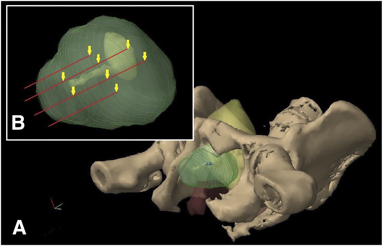
    (A) Diagram of pelvis from inferolateral view. Bony-anatomy cutaway shows prostate (green, not to scale), bladder and urethra (yellow), and rectum (orange). (B) Inset shows needle insertion tracts, with arrows indicating sites of injection along these tracts.

  • FIGURE 2.
    • Download figure
    • Open in new tab
    • Download powerpoint
    FIGURE 2.

    Eight syringes, each containing 0.2 mL of 68Ga-iron oxide. First syringe to be used is attached to 22-gauge 150-mm Sprotte cannula.

  • FIGURE 3.
    • Download figure
    • Open in new tab
    • Download powerpoint
    FIGURE 3.

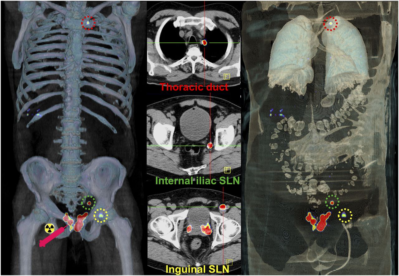
    Volume-rendered PET/CT (left and right) images and axial PET/CT (middle) images. After injection of tracer into prostate, imaging at 45 min demonstrated internal iliac SLN, inguinal SLN, and focal uptake at terminal thoracic duct near entry into left subclavian vein. No intervening SLN stations were seen in abdomen or chest. This intriguing finding correlates with increasing recognition of Virchow nodal metastases visualized with 68Ga-PSMA or 18F-choline PET.

  • FIGURE 4.
    • Download figure
    • Open in new tab
    • Download powerpoint
    FIGURE 4.

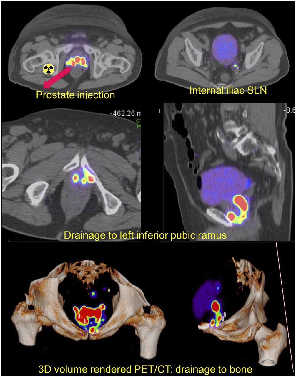
    After injection in prostate, left internal iliac SLN was identified. Additional drainage pathway from periprostatic region to inferior pubic ramus was observed and could represent drainage via periprostatic venous plexus rather than lymphatic pathway. Finding shows locoregional rather than hematogenous pathway of metastasis to pelvic bones and explains predominance of pelvic bony metastases in some patients.

Tables

  • Figures
    • View popup
    TABLE 1

    SLNs Identified in the Study Patients

    SLNsLymphatic drainage
    PatientDose* (MBq)Scan 1Scan 2Second-echelon nodesClassicAberrantNonlymphatic
    SPECT 182.6L external iliac, L common iliacL external iliac, L common iliacRetroperitoneal (paraaortic)YesNoNo
    SPECT 231.0R internal iliacR internal iliac, L internal iliac (probable)YesNoNo
    SPECT 319.9R external iliacR external iliacR common iliacYesNoNo
    PET 116.1R internal iliac, L perivesical, L internal iliac, L external iliac (probable)R internal iliac, L perivesicalYesYesNo
    PET 28.7L internal iliac, L anterior periprostatic into L inferior pubic ramusL internal iliac, L anterior periprostatic into L inferior pubic ramusL internal iliac 2YesNoYes
    PET 321.0L periprostaticL periprostatic, R periprostaticYesNoNo
    PET 411.2R mesorectal, L external iliacR mesorectal, L external iliacR presacralYesYesNo
    PET 53.1L internal iliac, L inguinalL internal iliac (disappears), L inguinalL common iliac, distal thoracic duct (Virchow node)YesYesNo
    • ↵* Injected dose of 99mTc-radiocolloid for SPECT and 68Ga-nanocolloid for PET.

    • SLNs were found both in classic sites of regional spread and in aberrant sites (boldface font). First 3 patients underwent 99mTc-radiocolloid SPECT/CT, and next 5 underwent 68Ga-nanocolloid PET/CT. Scans were obtained about 45 and 100 min after tracer injection.

PreviousNext
Back to top

In this issue

Journal of Nuclear Medicine: 59 (12)
Journal of Nuclear Medicine
Vol. 59, Issue 12
December 1, 2018
  • Table of Contents
  • Table of Contents (PDF)
  • About the Cover
  • Index by author
Print
Download PDF
Article Alerts
Sign In to Email Alerts with your Email Address
Email Article

Thank you for your interest in spreading the word on Journal of Nuclear Medicine.

NOTE: We only request your email address so that the person you are recommending the page to knows that you wanted them to see it, and that it is not junk mail. We do not capture any email address.

Enter multiple addresses on separate lines or separate them with commas.
A First-in-Human Study of 68Ga-Nanocolloid PET/CT Sentinel Lymph Node Imaging in Prostate Cancer Demonstrates Aberrant Lymphatic Drainage Pathways
(Your Name) has sent you a message from Journal of Nuclear Medicine
(Your Name) thought you would like to see the Journal of Nuclear Medicine web site.
Citation Tools
A First-in-Human Study of 68Ga-Nanocolloid PET/CT Sentinel Lymph Node Imaging in Prostate Cancer Demonstrates Aberrant Lymphatic Drainage Pathways
Jacki A. Doughton, Michael S. Hofman, Peter Eu, Rodney J. Hicks, Scott Williams
Journal of Nuclear Medicine Dec 2018, 59 (12) 1837-1842; DOI: 10.2967/jnumed.118.209171

Citation Manager Formats

  • BibTeX
  • Bookends
  • EasyBib
  • EndNote (tagged)
  • EndNote 8 (xml)
  • Medlars
  • Mendeley
  • Papers
  • RefWorks Tagged
  • Ref Manager
  • RIS
  • Zotero
Share
A First-in-Human Study of 68Ga-Nanocolloid PET/CT Sentinel Lymph Node Imaging in Prostate Cancer Demonstrates Aberrant Lymphatic Drainage Pathways
Jacki A. Doughton, Michael S. Hofman, Peter Eu, Rodney J. Hicks, Scott Williams
Journal of Nuclear Medicine Dec 2018, 59 (12) 1837-1842; DOI: 10.2967/jnumed.118.209171
Twitter logo Facebook logo LinkedIn logo Mendeley logo
  • Tweet Widget
  • Facebook Like
  • Google Plus One
Bookmark this article

Jump to section

  • Article
    • Abstract
    • MATERIALS AND METHODS
    • RESULTS
    • DISCUSSION
    • CONCLUSION
    • DISCLOSURE
    • Acknowledgments
    • Footnotes
    • REFERENCES
  • Figures & Data
  • Info & Metrics
  • PDF

Related Articles

  • This Month in JNM
  • PubMed
  • Google Scholar

Cited By...

  • No citing articles found.
  • Google Scholar

More in this TOC Section

Oncology

  • Comparative PSMA expression at early (1hour) vs late (2hour) in primary and secondary sites of involvement in prostate cancer.
  • Imaging spectrum of peritoneal carcinomatosis associated with various etiologies on 18F-FDG PET/CT
  • Hybrid Imaging Features of Musculoskeletal Tumors
Show more Oncology

Translational

  • Quantitative Total-Body Imaging of Blood Flow with High-Temporal-Resolution Early Dynamic 18F-FDG PET Kinetic Modeling
  • [99mTc]Tc-MY6349 Probe for Trop2-Targeted SPECT Imaging: From Preclinical to Pilot Clinical Study
  • Imaging Diverse Pathogenic Bacteria In Vivo with 18F-Fluoromannitol PET
Show more Translational

Similar Articles

Keywords

  • lymphoscintigraphy
  • lymph nodes
  • positron emission tomography
  • prostate neoplasms
  • sentinel lymph node
  • Magnetite nanoparticles
SNMMI

© 2025 SNMMI

Powered by HighWire