


Article Figures & Data
Figures
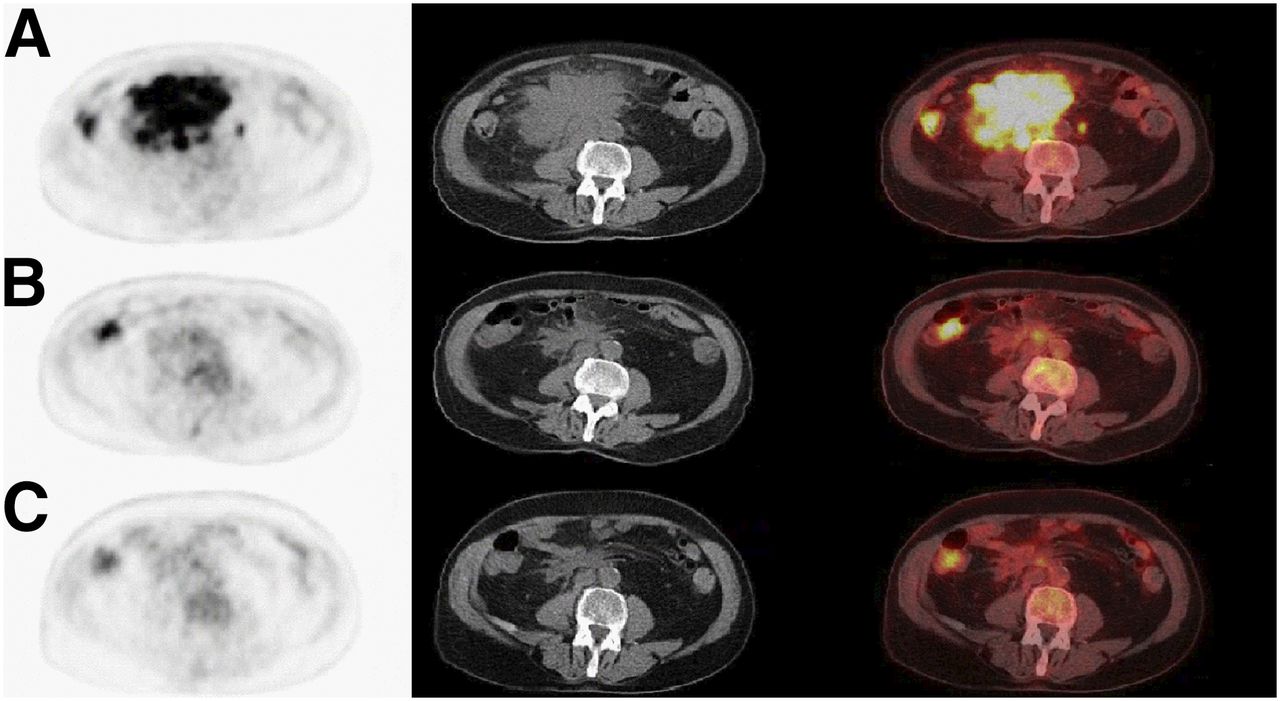
- FIGURE 1.
Example of discrepancy between reviewers’ assessment of mesenteric lymph nodes on, from left to right, axial attenuation-corrected PET, low-dose CT, and fused PET/CT images. (A) Baseline 18F-FDG PET/CT with mesenteric bulky mass. (B) I-PET/CT after 4 cycles of R-CHOP14. One reviewer scored scan negatively (DS 1) and the other reviewer scored DS 4 for residual uptake in mesenteric mass. (C) EoT-PET/CT after 6 cycles of R-CHOP14. Both reviewers scored scan negatively (DS 1 and DS 2, respectively).
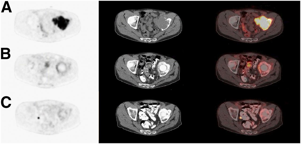
- FIGURE 2.
Example of discrepancy between reviewers’ assessment of skeletal lesion on, from left to right, axial attenuation-corrected PET, low-dose CT, and fused PET/CT images. (A) Baseline 18F-FDG PET/CT with skeletal lesion in left acetabulum. (B) I-PET/CT after 4 cycles of R-CHOP14 showing rim of uptake scored by one reviewer as DS 4 and by other reviewer as unclear. (C) EoT-PET/CT after 8 cycles of R-CHOP14 showing residual uptake scored by one reviewer as DS 4 and by other reviewer as unclear.
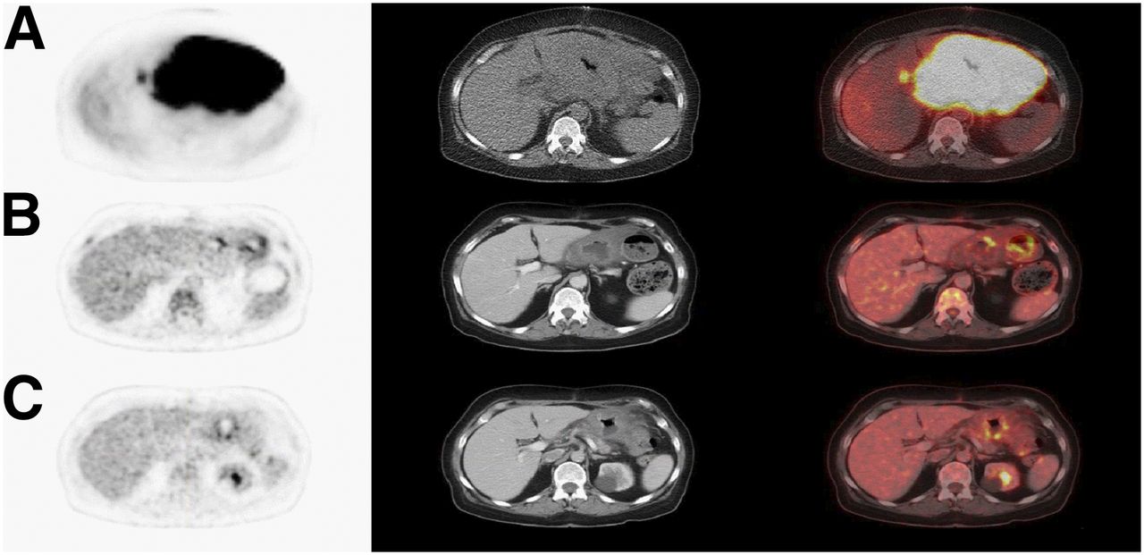
- FIGURE 3.
Example of discrepancy between reviewers’ assessment of stomach on, from left to right, axial attenuation-corrected PET, low-dose CT, and fused PET/CT images. (A) Baseline 18F-FDG PET/CT with clear localization of lymphoma in stomach. (B) I-PET/CT after 4 cycles of R-CHOP14. Reviewer 1 did not give final DS score and commented that stomach was “DS 4 but could be physiologic uptake.” Reviewer 2 scored this scan negatively (DS 2). (C) EoT-PET/CT after 6 cycles of R-CHOP14. Reviewer 1 still commented on stomach but now scored negatively. Reviewer 2 again scored scan negatively (DS 2).
Tables
I-PET/CT EoT-PET/CT Agreement Total (n = 465) Baseline CT only (n = 108) Baseline 18F-FDG PET or PET/CT (n = 357) P* Total (n = 457) Baseline CT only (n = 114) Baseline 18F-FDG PET or PET/CT (n = 343) P* Positivity† 23.3 18.5 24.8 17.5 18.0 17.3 Percentage OA 87.7 (84.7–90.8) 87.0 (80.2–93.8) 88.0 (84.4–91.5) 0.799 91.7 (89.0–94.3) 93.9 (89.0–98.7) 91.0 (87.8–94.1) 0.332 Percentage PA 73.7 (65.0–82.5) 65.0 (41.6–88.4) 75.7 (66.2–85.2) 0.347 76.3 (66.3–86.2) 82.9 (59.2–90.8) 73.9 (62.0–85.9) 0.486 Percentage NA 92.0 (89.1–95.0) 92.0 (85.8–98.3) 92.0 (88.6–95.4) 0.947 95.0 (92.6–97.3) 96.3 (89.5–97.9) 94.5 (91.7–97.4) 0.605 DS 1–3 = negative; DS 4–5 = positive.
↵* P values of χ2 test refer to comparison of baseline CT vs. baseline PET or PET/CT.
↵† Prevalence of positive scans was calculated as sum of number of scans in which both reviewers scored positively and half of scans with discrepancies divided by total number of scans.
Data are percentages, with 95%CIs in parentheses.
Location Number baseline positive Number of discrepancies on I-PET Agreement on negativity (absolute) Agreement on positivity (absolute) Percentage OA Related to baseline prevalence Nodal Paraaortic* 414 17 899 14 98.2 4.1% Cervical*† 302 8 915 6 99.1 2.6% Iliac* 272 6 917 7 99.4 2.2% Supraclavicular* 228 6 920 4 99.4 2.6% Axillary* 225 9 920 1 99.0 4.0% Mediastinal† 212 12 445 6 97.4 5.7% Inguinal* 210 3 926 1 99.7 1.4% Mesenteric 189 16 433 16 96.6 8.5% Hilar*† 147 7 918 3 99.2 4.8% Spleen† 115 11 442 6 97.6 9.6% Other 105 7 457 1 98.5 6.7% Waldeyer† 53 8 456 0 98.3 15.1% Extranodal Other extranodal† 124 17 436 8 96.3 13.7% Skeletal† 95 12 447 4 97.4 12.6% Gastrointestinal† 61 12 441 7 97.4 16.7% Lung† 55 3 455 6 99.4 5.5% Liver 37 3 461 1 99.4 8.1% Pleura 25 1 464 0 99.8 4.0% Skin 11 0 465 0 100.0 0.0% Central nervous system 0 0 465 0 100.0 0.0% ↵* Right and left are summed and presented together.
↵† Totals not 465 or 930, because of missing values or localization scored as unclear.
Percentage OA = (number of agreement on positivity + number of agreement on negativity)/(number of discrepancies + number of agreement on positivity + number of agreement on negativity) × 100%; related to baseline prevalence = (number of discrepancies/number baseline positive) × 100%.
Supplemental Data
Files in this Data Supplement:


Jump to section
Related Articles
Cited By...
- No citing articles found.