
Main menu

  • Home
  • Content
    • Current
    • Ahead of print
    • Past Issues
    • JNM Supplement
    • SNMMI Annual Meeting Abstracts
    • Continuing Education
    • JNM Podcasts
  • Subscriptions
    • Subscribers
    • Institutional and Non-member
    • Rates
    • Journal Claims
    • Corporate & Special Sales
  • Authors
    • Submit to JNM
    • Information for Authors
    • Assignment of Copyright
    • AQARA requirements
  • Info
    • Reviewers
    • Permissions
    • Advertisers
  • About
    • About Us
    • Editorial Board
    • Contact Information
  • More
    • Alerts
    • Feedback
    • Help
    • SNMMI Journals
  • SNMMI
    • JNM
    • JNMT
    • SNMMI Journals
    • SNMMI

User menu

  • Subscribe
  • My alerts
  • Log in
  • My Cart

Search

  • Advanced search
Journal of Nuclear Medicine
  • SNMMI
    • JNM
    • JNMT
    • SNMMI Journals
    • SNMMI
  • Subscribe
  • My alerts
  • Log in
  • My Cart
Journal of Nuclear Medicine

Advanced Search

  • Home
  • Content
    • Current
    • Ahead of print
    • Past Issues
    • JNM Supplement
    • SNMMI Annual Meeting Abstracts
    • Continuing Education
    • JNM Podcasts
  • Subscriptions
    • Subscribers
    • Institutional and Non-member
    • Rates
    • Journal Claims
    • Corporate & Special Sales
  • Authors
    • Submit to JNM
    • Information for Authors
    • Assignment of Copyright
    • AQARA requirements
  • Info
    • Reviewers
    • Permissions
    • Advertisers
  • About
    • About Us
    • Editorial Board
    • Contact Information
  • More
    • Alerts
    • Feedback
    • Help
    • SNMMI Journals
  • View or Listen to JNM Podcast
  • Visit JNM on Facebook
  • Join JNM on LinkedIn
  • Follow JNM on Twitter
  • Subscribe to our RSS feeds
Research ArticleOncology

Development of a SPECT Tracer to Image c-Met Expression in a Xenograft Model of Non–Small Cell Lung Cancer

Zhaoguo Han, Yadi Xiao, Kai Wang, Ji Yan, Zunyu Xiao, Fang Fang, Zhongnan Jin, Yang Liu, Xilin Sun and Baozhong Shen
Journal of Nuclear Medicine November 2018, 59 (11) 1686-1691; DOI: https://doi.org/10.2967/jnumed.117.206730
Zhaoguo Han
1Molecular Imaging Research Center, Harbin Medical University, Harbin, Heilongjiang, China
2TOF-PET/CT/MR Center, Fourth Hospital of Harbin Medical University, Harbin, Heilongjiang, China; and
  • Find this author on Google Scholar
  • Find this author on PubMed
  • Search for this author on this site
Yadi Xiao
1Molecular Imaging Research Center, Harbin Medical University, Harbin, Heilongjiang, China
2TOF-PET/CT/MR Center, Fourth Hospital of Harbin Medical University, Harbin, Heilongjiang, China; and
  • Find this author on Google Scholar
  • Find this author on PubMed
  • Search for this author on this site
Kai Wang
1Molecular Imaging Research Center, Harbin Medical University, Harbin, Heilongjiang, China
2TOF-PET/CT/MR Center, Fourth Hospital of Harbin Medical University, Harbin, Heilongjiang, China; and
  • Find this author on Google Scholar
  • Find this author on PubMed
  • Search for this author on this site
Ji Yan
1Molecular Imaging Research Center, Harbin Medical University, Harbin, Heilongjiang, China
2TOF-PET/CT/MR Center, Fourth Hospital of Harbin Medical University, Harbin, Heilongjiang, China; and
  • Find this author on Google Scholar
  • Find this author on PubMed
  • Search for this author on this site
Zunyu Xiao
1Molecular Imaging Research Center, Harbin Medical University, Harbin, Heilongjiang, China
2TOF-PET/CT/MR Center, Fourth Hospital of Harbin Medical University, Harbin, Heilongjiang, China; and
  • Find this author on Google Scholar
  • Find this author on PubMed
  • Search for this author on this site
Fang Fang
1Molecular Imaging Research Center, Harbin Medical University, Harbin, Heilongjiang, China
2TOF-PET/CT/MR Center, Fourth Hospital of Harbin Medical University, Harbin, Heilongjiang, China; and
  • Find this author on Google Scholar
  • Find this author on PubMed
  • Search for this author on this site
Zhongnan Jin
1Molecular Imaging Research Center, Harbin Medical University, Harbin, Heilongjiang, China
2TOF-PET/CT/MR Center, Fourth Hospital of Harbin Medical University, Harbin, Heilongjiang, China; and
  • Find this author on Google Scholar
  • Find this author on PubMed
  • Search for this author on this site
Yang Liu
1Molecular Imaging Research Center, Harbin Medical University, Harbin, Heilongjiang, China
2TOF-PET/CT/MR Center, Fourth Hospital of Harbin Medical University, Harbin, Heilongjiang, China; and
  • Find this author on Google Scholar
  • Find this author on PubMed
  • Search for this author on this site
Xilin Sun
1Molecular Imaging Research Center, Harbin Medical University, Harbin, Heilongjiang, China
2TOF-PET/CT/MR Center, Fourth Hospital of Harbin Medical University, Harbin, Heilongjiang, China; and
3Molecular Imaging Program at Stanford, Department of Radiology, Stanford University School of Medicine, Stanford, California
  • Find this author on Google Scholar
  • Find this author on PubMed
  • Search for this author on this site
Baozhong Shen
1Molecular Imaging Research Center, Harbin Medical University, Harbin, Heilongjiang, China
2TOF-PET/CT/MR Center, Fourth Hospital of Harbin Medical University, Harbin, Heilongjiang, China; and
  • Find this author on Google Scholar
  • Find this author on PubMed
  • Search for this author on this site
  • Article
  • Figures & Data
  • Supplemental
  • Info & Metrics
  • PDF
Loading

Article Figures & Data

Figures

  • Additional Files
  • FIGURE 1.
    • Download figure
    • Open in new tab
    • Download powerpoint
    FIGURE 1.

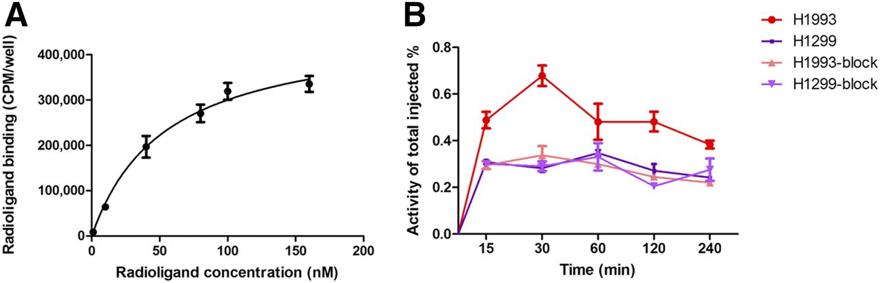
    (A) Saturation curve for binding of 99mTc-HYNIC-cMBP to H1993 cells with Kd of 56.30 ± 2.11. (B) Cell uptake and block-uptake of 99mTc-HYNIC-cMBP in NSCLC cells. CPM = counts/min.

  • FIGURE 2.
    • Download figure
    • Open in new tab
    • Download powerpoint
    FIGURE 2.

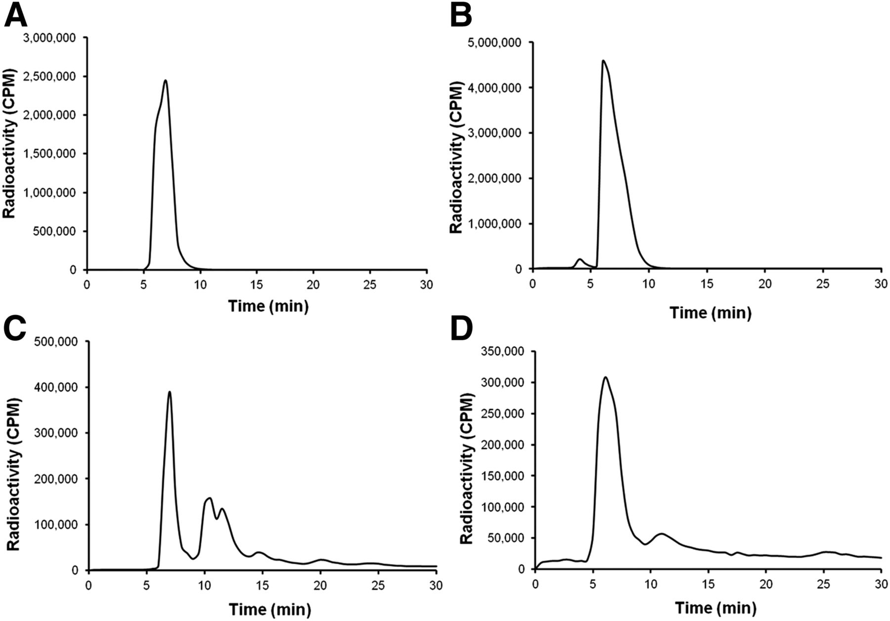
    In vivo stability of 99mTc-HYNIC-cMBP as indicated by radiochemical purity in samples of plasma (A), tumor (B), liver (C), and urine (D) at 1 h. CPM = counts/min.
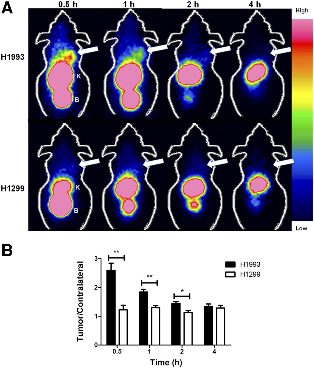
  • FIGURE 3.
    • Download figure
    • Open in new tab
    • Download powerpoint
    FIGURE 3.

    (A) Representative coronal SPECT images of H1993 and H1299 xenografts 0–4 h after injection of 99mTc-HYNIC-cMBP. Arrows = tumor; B = bladder; K = kidney. (B) Quantitative analysis of images through counts in region of interest.*P < 0.05.**P < 0.01.

  • FIGURE 4.
    • Download figure
    • Open in new tab
    • Download powerpoint
    FIGURE 4.

    (A) Representative coronal SPECT images for H1993 and block-H1993 xenografts at 0.5 h. Arrows = tumor; B = bladder; K = kidney. (B) Quantitative analysis of images through counts in region of interest. **P < 0.01.

  • FIGURE 5.
    • Download figure
    • Open in new tab
    • Download powerpoint
    FIGURE 5.

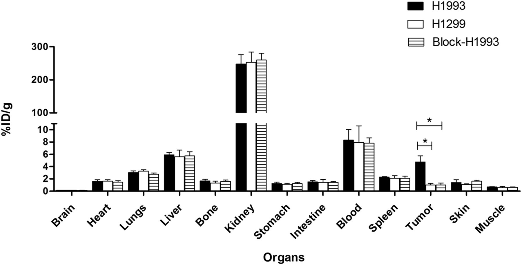
    Biodistribution of 99mTc-HYNIC-cMBP in H1993, H1299, and block-H1993 groups at 0.5 h after injection. *P < 0.05.
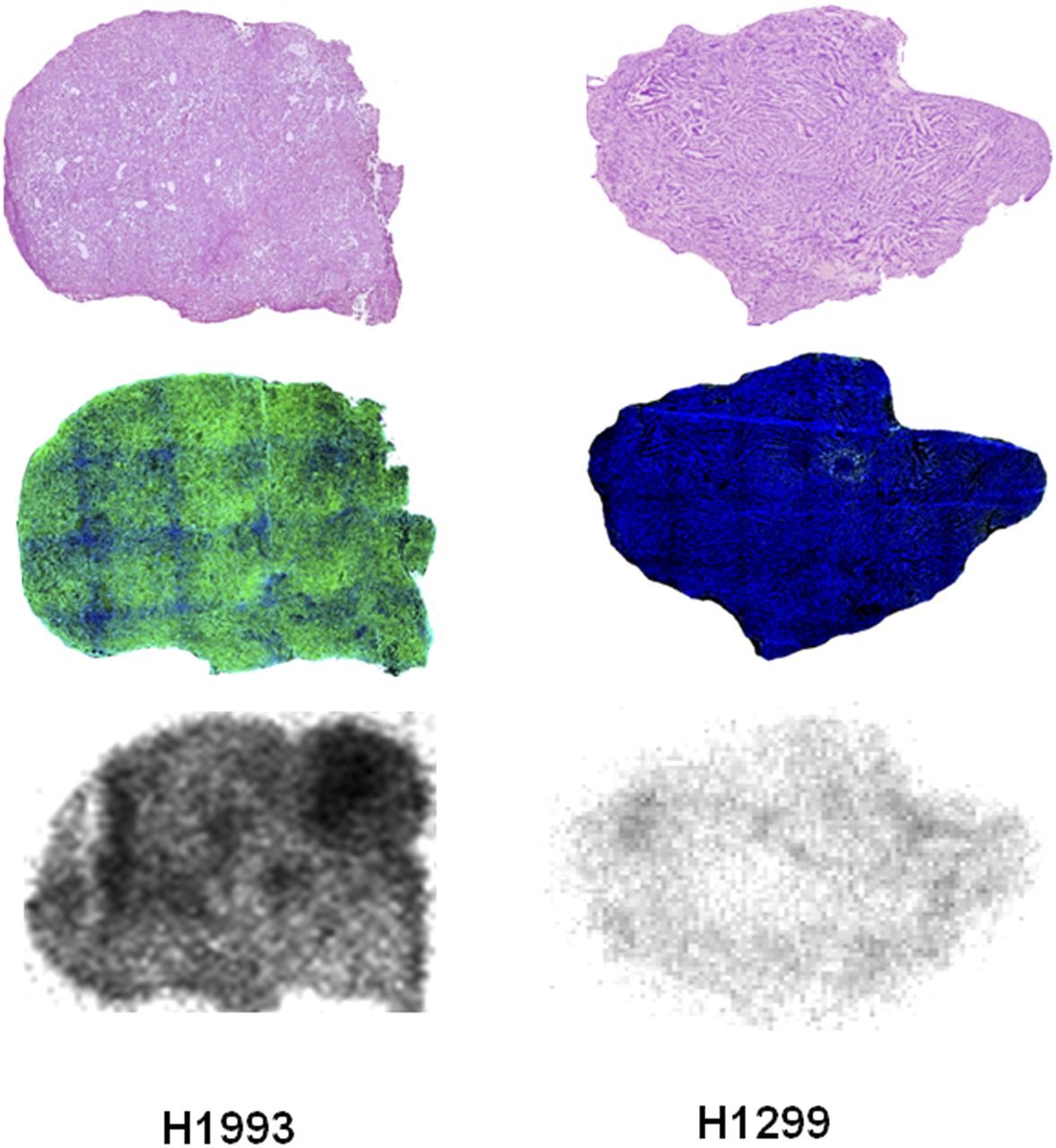
  • FIGURE 6.
    • Download figure
    • Open in new tab
    • Download powerpoint
    FIGURE 6.

    Comparison of hematoxylin and eosin staining (top), immunofluorescence staining (middle), and autoradiography images (bottom) of H1993 and H1299 tumor sections at 0.5 h after injection of 99mTc-HYNIC-cMBP. Green fluorescence indicates high expression of c-Met.

Additional Files

  • Figures
  • Supplemental Data

    Files in this Data Supplement:

    • Supplemental Data
PreviousNext
Back to top

In this issue

Journal of Nuclear Medicine: 59 (11)
Journal of Nuclear Medicine
Vol. 59, Issue 11
November 1, 2018
  • Table of Contents
  • Table of Contents (PDF)
  • About the Cover
  • Index by author
Print
Download PDF
Article Alerts
Sign In to Email Alerts with your Email Address
Email Article

Thank you for your interest in spreading the word on Journal of Nuclear Medicine.

NOTE: We only request your email address so that the person you are recommending the page to knows that you wanted them to see it, and that it is not junk mail. We do not capture any email address.

Enter multiple addresses on separate lines or separate them with commas.
Development of a SPECT Tracer to Image c-Met Expression in a Xenograft Model of Non–Small Cell Lung Cancer
(Your Name) has sent you a message from Journal of Nuclear Medicine
(Your Name) thought you would like to see the Journal of Nuclear Medicine web site.
Citation Tools
Development of a SPECT Tracer to Image c-Met Expression in a Xenograft Model of Non–Small Cell Lung Cancer
Zhaoguo Han, Yadi Xiao, Kai Wang, Ji Yan, Zunyu Xiao, Fang Fang, Zhongnan Jin, Yang Liu, Xilin Sun, Baozhong Shen
Journal of Nuclear Medicine Nov 2018, 59 (11) 1686-1691; DOI: 10.2967/jnumed.117.206730

Citation Manager Formats

  • BibTeX
  • Bookends
  • EasyBib
  • EndNote (tagged)
  • EndNote 8 (xml)
  • Medlars
  • Mendeley
  • Papers
  • RefWorks Tagged
  • Ref Manager
  • RIS
  • Zotero
Share
Development of a SPECT Tracer to Image c-Met Expression in a Xenograft Model of Non–Small Cell Lung Cancer
Zhaoguo Han, Yadi Xiao, Kai Wang, Ji Yan, Zunyu Xiao, Fang Fang, Zhongnan Jin, Yang Liu, Xilin Sun, Baozhong Shen
Journal of Nuclear Medicine Nov 2018, 59 (11) 1686-1691; DOI: 10.2967/jnumed.117.206730
Twitter logo Facebook logo LinkedIn logo Mendeley logo
  • Tweet Widget
  • Facebook Like
  • Google Plus One
Bookmark this article

Jump to section

  • Article
    • Abstract
    • MATERIALS AND METHODS
    • RESULTS
    • DISCUSSION
    • CONCLUSION
    • DISCLOSURE
    • Acknowledgments
    • Footnotes
    • REFERENCES
  • Figures & Data
  • Supplemental
  • Info & Metrics
  • PDF

Related Articles

  • This Month in JNM
  • PubMed
  • Google Scholar

Cited By...

  • SPECT/CT: Standing on the Shoulders of Giants, It Is Time to Reach for the Sky!
  • Light-Induced Radiosynthesis of 89Zr-DFO-Azepin-Onartuzumab for Imaging the Hepatocyte Growth Factor Receptor
  • Google Scholar

More in this TOC Section

Oncology

  • 18F-FDG PET/CT manifestations of three Cases of Female Desmoplastic Small Round Cell Tumor
  • 18F-FDG PET/CT imaging features of parotid lesions; Case based pictorial review and its multi-modality correlation.
  • Utility of Fluorine-18-fluorodeoxyglucose PET/CT in Uterine sarcoma patients.
Show more Oncology

Basic

  • Dopamine D1 Receptor Agonist PET Tracer Development: Assessment in Nonhuman Primates
  • Optical Navigation of the Drop-In γ-Probe as a Means to Strengthen the Connection Between Robot-Assisted and Radioguided Surgery
  • Synthesis and Preclinical Evaluation of a 68Ga-Labeled Adnectin, 68Ga-BMS-986192, as a PET Agent for Imaging PD-L1 Expression
Show more Basic

Similar Articles

Keywords

  • c-MET
  • targeted peptide
  • NSCLC
  • SPECT
SNMMI

© 2025 SNMMI

Powered by HighWire