
Main menu

  • Home
  • Content
    • Current
    • Ahead of print
    • Past Issues
    • JNM Supplement
    • SNMMI Annual Meeting Abstracts
    • Continuing Education
    • JNM Podcasts
  • Subscriptions
    • Subscribers
    • Institutional and Non-member
    • Rates
    • Journal Claims
    • Corporate & Special Sales
  • Authors
    • Submit to JNM
    • Information for Authors
    • Assignment of Copyright
    • AQARA requirements
  • Info
    • Reviewers
    • Permissions
    • Advertisers
  • About
    • About Us
    • Editorial Board
    • Contact Information
  • More
    • Alerts
    • Feedback
    • Help
    • SNMMI Journals
  • SNMMI
    • JNM
    • JNMT
    • SNMMI Journals
    • SNMMI

User menu

  • Subscribe
  • My alerts
  • Log in
  • My Cart

Search

  • Advanced search
Journal of Nuclear Medicine
  • SNMMI
    • JNM
    • JNMT
    • SNMMI Journals
    • SNMMI
  • Subscribe
  • My alerts
  • Log in
  • My Cart
Journal of Nuclear Medicine

Advanced Search

  • Home
  • Content
    • Current
    • Ahead of print
    • Past Issues
    • JNM Supplement
    • SNMMI Annual Meeting Abstracts
    • Continuing Education
    • JNM Podcasts
  • Subscriptions
    • Subscribers
    • Institutional and Non-member
    • Rates
    • Journal Claims
    • Corporate & Special Sales
  • Authors
    • Submit to JNM
    • Information for Authors
    • Assignment of Copyright
    • AQARA requirements
  • Info
    • Reviewers
    • Permissions
    • Advertisers
  • About
    • About Us
    • Editorial Board
    • Contact Information
  • More
    • Alerts
    • Feedback
    • Help
    • SNMMI Journals
  • View or Listen to JNM Podcast
  • Visit JNM on Facebook
  • Join JNM on LinkedIn
  • Follow JNM on Twitter
  • Subscribe to our RSS feeds
Meeting ReportInstrumentation & Data Analysis Track

Artificial neural network based regularization for brain PET image reconstruction

Bao Yang and Jing Tang
Journal of Nuclear Medicine May 2017, 58 (supplement 1) 95;
Bao Yang
1Oakland University Rochester MI United States
  • Find this author on Google Scholar
  • Find this author on PubMed
  • Search for this author on this site
Jing Tang
1Oakland University Rochester MI United States
  • Find this author on Google Scholar
  • Find this author on PubMed
  • Search for this author on this site
  • Article
  • Figures & Data
  • Info & Metrics
Loading

Abstract

95

Objectives: PET imaging provides quantitative measurement of physiological and biochemical processes. As the inverse problem to solve for the emission distribution from the acquired data is ill-posed, maximum a posteriori (MAP) algorithms have been developed to regularize the iterative process of image formation. The performance of MAP algorithms strongly depends on the weighting applied to the regularization, which results in different tradeoffs between variance and spatial resolution. Instead of trying to tune the regularization weight for a MAP algorithm, the goal of this study is to take advantage of all the information in images reconstructed with different weights. Artificial neural network (ANN) solves regression or discrimination problems by learning from examples and it has successful applications in the medical image analysis and image interpretation fields. We aim to build a local fusion scheme using an ANN to extract and fuse the information from a collection of MAP reconstructed image versions.

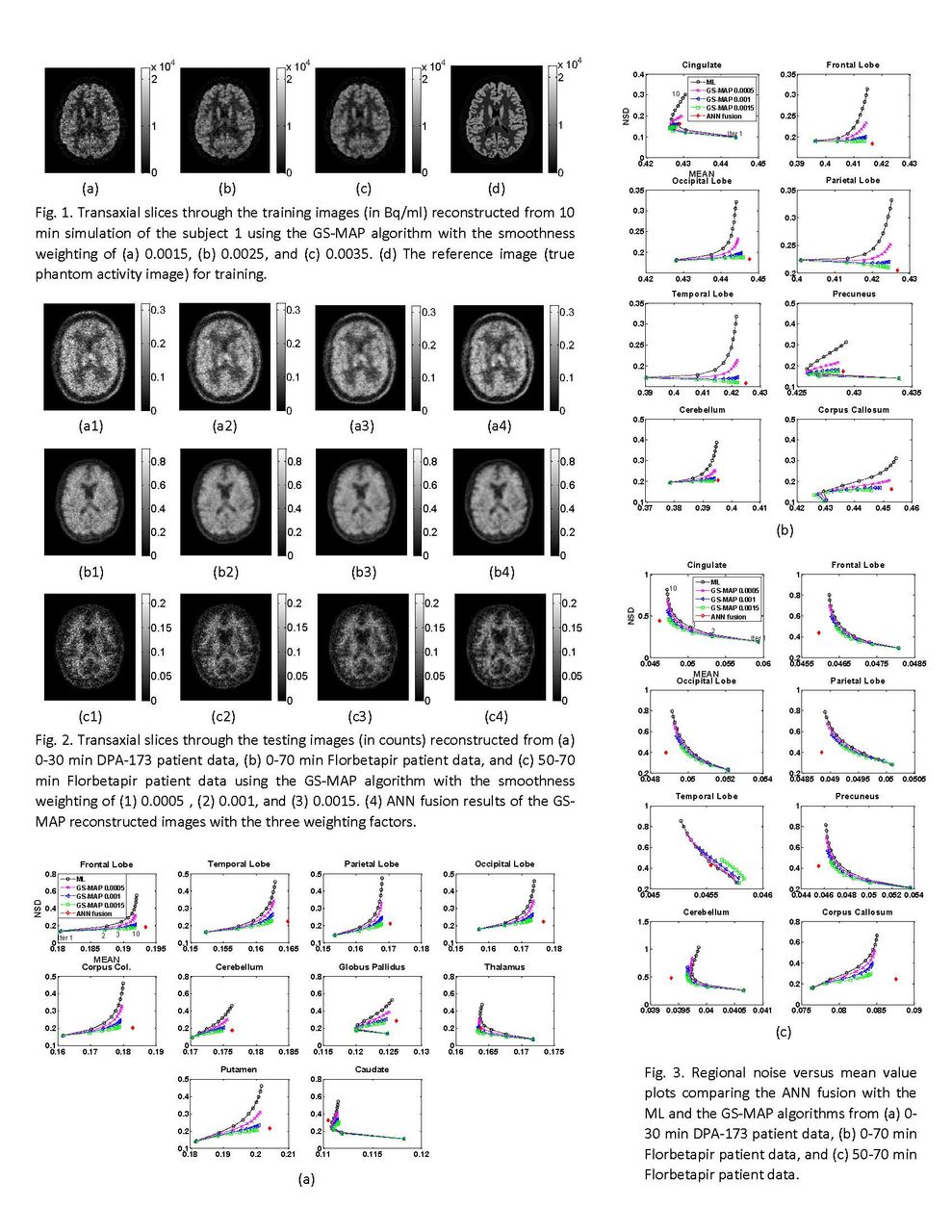
Methods: We designed an ANN serving as a nonlinear multivariate regression function with four hidden layers: two fully-connected lagers, a Hyperbolic Tangent (TanH) activation layer, and a Euclidean loss layer. Using simulated PET data from one subject of the BrainWeb phantom at a high-count level, we obtained the training images from reconstruction with the Green smoothness (GS) MAP algorithm applying three different regularization weights. A set of small patches extracted from the training images at the same location were the inputs fed to the ANN. Using the back-propagation algorithm, the ANN was trained to compute a patch with values as close as possible to those in the patch of the reference image, i.e., the true phantom activity image. We validated the trained ANN by fusing the reconstructed images from simulations at lower count levels of the same subject and various count levels of other subjects.We tested the developed ANN fusion scheme using patient brain PET imaging datasets taken on a second-generation Siemens HRRT scanner with the tracers DPA-173 and Florbetapir. The DPA-173 data corresponded to a 30 min acquisition and the Florbetapir data had two count levels corresponding to 0-70 min and 50-70 min acquisitions. We reconstructed each set of patient data using the GS-MAP algorithms with three different weighting factors and then performed the ANN fusion on the reconstructed images. To ensure the adaptability of the trained ANN model on the patient PET images, we normalized both training and testing images to the range of [0, 1] separately using their respective maximum intensity values. To evaluate the performance of the proposed ANN fusion technique, we calculated the tradeoff between the mean value and the normalized standard deviation on regions of interest (ROIs) of each brain image.

Results: For the DPA-173 study, the ANN fused image reaches the noise measure as low as the GS-MAP reconstruction achieves at heavy regularization while approaches the regional mean value the maximum likelihood (ML) algorithm obtained in most of the ROIs. Similarly, at both count levels of the Florbetapir studies, the ANN fusion suppresses noise as well as the GS-MAP algorithm with large weighting factor while arriving at mean regional values close to what the ML algorithm achieves in most of the ROIs.

Conclusion: An ANN fusion scheme was developed and evaluated to extract and fuse information from images reconstructed using a MAP algorithm of different smoothness weights. The proposed technique reached the lowest variance and best spatial resolution achieved by the GS-MAP algorithm at different weightings in patient PET studies of different tracers at different count levels. With robust performance, the ANN-based fusion contributes to PET image reconstruction by removing the need for tuning the regularization weight of MAP algorithms. Research Support: National Science Foundation ECCS-1454552

Figure
  • Download figure
  • Open in new tab
  • Download powerpoint
Previous
Back to top

In this issue

Journal of Nuclear Medicine
Vol. 58, Issue supplement 1
May 1, 2017
  • Table of Contents
  • Index by author
Article Alerts
Sign In to Email Alerts with your Email Address
Email Article

Thank you for your interest in spreading the word on Journal of Nuclear Medicine.

NOTE: We only request your email address so that the person you are recommending the page to knows that you wanted them to see it, and that it is not junk mail. We do not capture any email address.

Enter multiple addresses on separate lines or separate them with commas.
Artificial neural network based regularization for brain PET image reconstruction
(Your Name) has sent you a message from Journal of Nuclear Medicine
(Your Name) thought you would like to see the Journal of Nuclear Medicine web site.
Citation Tools
Artificial neural network based regularization for brain PET image reconstruction
Bao Yang, Jing Tang
Journal of Nuclear Medicine May 2017, 58 (supplement 1) 95;

Citation Manager Formats

  • BibTeX
  • Bookends
  • EasyBib
  • EndNote (tagged)
  • EndNote 8 (xml)
  • Medlars
  • Mendeley
  • Papers
  • RefWorks Tagged
  • Ref Manager
  • RIS
  • Zotero
Share
Artificial neural network based regularization for brain PET image reconstruction
Bao Yang, Jing Tang
Journal of Nuclear Medicine May 2017, 58 (supplement 1) 95;
Twitter logo Facebook logo LinkedIn logo Mendeley logo
  • Tweet Widget
  • Facebook Like
  • Google Plus One
Bookmark this article

Jump to section

  • Article
  • Figures & Data
  • Info & Metrics

Related Articles

  • No related articles found.
  • Google Scholar

Cited By...

  • No citing articles found.
  • Google Scholar

More in this TOC Section

Instrumentation & Data Analysis Track

  • Deep Learning Based Kidney Segmentation for Glomerular Filtration Rate Measurement Using Quantitative SPECT/CT
  • The Benefit of Time-of-Flight in Digital Photon Counting PET Imaging: Physics and Clinical Evaluation
  • Preclinical validation of a single-scan rest/stress imaging technique for 13NH3 cardiac perfusion studies
Show more Instrumentation & Data Analysis Track

Computer and Instrumentation Council Young Investigator Award Symposium

  • Improved Image Quality with Locally Adaptive Gating in 4D PET Image Reconstruction
  • Studies of a Next Generation Silicon-Photomultiplier-Based Time-of-Flight PET/CT System
  • Ultrahigh Resolution Coplanar PET Imaging of Microfluidic Devices for Synthesizing PET Radiopharmaceuticals
Show more Computer and Instrumentation Council Young Investigator Award Symposium

Similar Articles

SNMMI

© 2025 SNMMI

Powered by HighWire