
Main menu

  • Home
  • Content
    • Current
    • Ahead of print
    • Past Issues
    • JNM Supplement
    • SNMMI Annual Meeting Abstracts
    • Continuing Education
    • JNM Podcasts
  • Subscriptions
    • Subscribers
    • Institutional and Non-member
    • Rates
    • Journal Claims
    • Corporate & Special Sales
  • Authors
    • Submit to JNM
    • Information for Authors
    • Assignment of Copyright
    • AQARA requirements
  • Info
    • Reviewers
    • Permissions
    • Advertisers
  • About
    • About Us
    • Editorial Board
    • Contact Information
  • More
    • Alerts
    • Feedback
    • Help
    • SNMMI Journals
  • SNMMI
    • JNM
    • JNMT
    • SNMMI Journals
    • SNMMI

User menu

  • Subscribe
  • My alerts
  • Log in
  • Log out
  • My Cart

Search

  • Advanced search
Journal of Nuclear Medicine
  • SNMMI
    • JNM
    • JNMT
    • SNMMI Journals
    • SNMMI
  • Subscribe
  • My alerts
  • Log in
  • Log out
  • My Cart
Journal of Nuclear Medicine

Advanced Search

  • Home
  • Content
    • Current
    • Ahead of print
    • Past Issues
    • JNM Supplement
    • SNMMI Annual Meeting Abstracts
    • Continuing Education
    • JNM Podcasts
  • Subscriptions
    • Subscribers
    • Institutional and Non-member
    • Rates
    • Journal Claims
    • Corporate & Special Sales
  • Authors
    • Submit to JNM
    • Information for Authors
    • Assignment of Copyright
    • AQARA requirements
  • Info
    • Reviewers
    • Permissions
    • Advertisers
  • About
    • About Us
    • Editorial Board
    • Contact Information
  • More
    • Alerts
    • Feedback
    • Help
    • SNMMI Journals
  • View or Listen to JNM Podcast
  • Visit JNM on Facebook
  • Join JNM on LinkedIn
  • Follow JNM on Twitter
  • Subscribe to our RSS feeds
Meeting ReportInstrumentation & Data Analysis Track

Model-based 3D reconstruction of myocardial perfusion SPECT images

Yinfeng Dong, Eric Frey and Yong Du
Journal of Nuclear Medicine May 2017, 58 (supplement 1) 26;
Yinfeng Dong
1The Johns Hopkins University Baltimore MD United States
  • Find this author on Google Scholar
  • Find this author on PubMed
  • Search for this author on this site
Eric Frey
1The Johns Hopkins University Baltimore MD United States
  • Find this author on Google Scholar
  • Find this author on PubMed
  • Search for this author on this site
Yong Du
1The Johns Hopkins University Baltimore MD United States
  • Find this author on Google Scholar
  • Find this author on PubMed
  • Search for this author on this site
  • Article
  • Figures & Data
  • Info & Metrics
Loading

Abstract

26

Objectives: Partial volume (PV) effects resulting from finite spatial resolution significantly degrade the accuracy of activity quantification and left ventricle (LV) segmentation in the analysis of myocardial perfusion SPECT (MPS) images. High-resolution anatomical images, such as contrast CT or MRI, can be used for segmentation and PV compensation, but they are typically not available in routine clinical practice. The objective of this study was to develop and evaluate a model-based 3D reconstruction of MPS images to improve quantification and LV segmentation accuracy without the use of anatomical images.

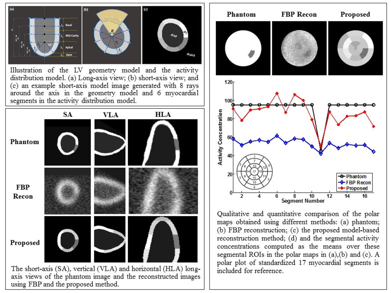
Methods: The proposed model-based reconstruction method describes the LV using 3D geometry and activity distribution models instead of voxels. The geometry model parameterizes the endo- and epicardial surfaces using a set of rays originating from the long axis of the LV. The rays are in planes perpendicular to the axis and at equiangular increments in the basal and mid-ventricular regions. In the apex, the rays radiate from a point with equal polar and azimuthal angular increments. The surfaces are obtained by interpolating the intersection points of each surface and the rays using cubic-spline functions. The activity distribution model divides the myocardium into segments similar to those used in standard quantitative MPS analysis and assumes uniform activity concentrations in each segment and the blood pool and body background. The method estimates the model parameters of voxel intensities, greatly reducing the number of unknowns. The parameters are estimated that give the best match between the image generated from the geometry and activity models and the measured data. The metric for the goodness of fit was a maximum likelihood criterion based on the modeling of noise statistics in the reconstructed image. The effects of finite resolution were modeled with a spatially invariant Gaussian function. A penalty encouraging uniform wall thickness was incorporated into the objective function to regularize the reconstruction problem and to increase robustness to perfusion defects and noise. The parameters were optimized by seeking the maximum of the objective function. The proposed method was evaluated using simulated MPS images based on the XCAT phantom. The myocardium contained a perfusion defect with 50° angular and 20mm axial extent, and 50% severity. The relative activity concentrations were: blood pool=15, background=5, normal myocardium=95, perfusion defect=45. The noise level matched that in clinical MPS images. For simplicity, attenuation and scatter were not modeled in the projection data.

Results: The resulting parameters provided an accurately segmented LV myocardium and PV compensated measurements of the true activities. The overlap between the resulting segmented LV myocardium and the truth was about 91%. The proposed model-based reconstruction method reduced the RMSE of estimated LV myocardial activity concentrations from 40.37 to 11.92 in comparison to FBP reconstruction.

Conclusion: We have developed a model-based 3D reconstruction method that incorporates prior knowledge about the targeted object via a model and models the image formation process. The method can simultaneously produce accurate LV segmentation and PV compensated images without using anatomical images. Research Support: AHA 16GRNT3109007

Figure
  • Download figure
  • Open in new tab
  • Download powerpoint
Previous
Back to top

In this issue

Journal of Nuclear Medicine
Vol. 58, Issue supplement 1
May 1, 2017
  • Table of Contents
  • Index by author
Article Alerts
Sign In to Email Alerts with your Email Address
Email Article

Thank you for your interest in spreading the word on Journal of Nuclear Medicine.

NOTE: We only request your email address so that the person you are recommending the page to knows that you wanted them to see it, and that it is not junk mail. We do not capture any email address.

Enter multiple addresses on separate lines or separate them with commas.
Model-based 3D reconstruction of myocardial perfusion SPECT images
(Your Name) has sent you a message from Journal of Nuclear Medicine
(Your Name) thought you would like to see the Journal of Nuclear Medicine web site.
Citation Tools
Model-based 3D reconstruction of myocardial perfusion SPECT images
Yinfeng Dong, Eric Frey, Yong Du
Journal of Nuclear Medicine May 2017, 58 (supplement 1) 26;

Citation Manager Formats

  • BibTeX
  • Bookends
  • EasyBib
  • EndNote (tagged)
  • EndNote 8 (xml)
  • Medlars
  • Mendeley
  • Papers
  • RefWorks Tagged
  • Ref Manager
  • RIS
  • Zotero
Share
Model-based 3D reconstruction of myocardial perfusion SPECT images
Yinfeng Dong, Eric Frey, Yong Du
Journal of Nuclear Medicine May 2017, 58 (supplement 1) 26;
Twitter logo Facebook logo LinkedIn logo Mendeley logo
  • Tweet Widget
  • Facebook Like
  • Google Plus One
Bookmark this article

Jump to section

  • Article
  • Figures & Data
  • Info & Metrics

Related Articles

  • No related articles found.
  • Google Scholar

Cited By...

  • No citing articles found.
  • Google Scholar

More in this TOC Section

Instrumentation & Data Analysis Track

  • Deep Learning Based Kidney Segmentation for Glomerular Filtration Rate Measurement Using Quantitative SPECT/CT
  • The Benefit of Time-of-Flight in Digital Photon Counting PET Imaging: Physics and Clinical Evaluation
  • Preclinical validation of a single-scan rest/stress imaging technique for 13NH3 cardiac perfusion studies
Show more Instrumentation & Data Analysis Track

INTEGRATED SESSION: Cardiac SPECT

  • Collimator Design for a Dedicated Human Cardiac SPECT System with High Resolution Detectors
  • Comparison of Left Ventricular Ejection Fraction Calculation Methods between CT and SPECT with a Dynamic Cardiac Phantom
Show more INTEGRATED SESSION: Cardiac SPECT

Similar Articles

SNMMI

© 2025 SNMMI

Powered by HighWire