


Abstract
1053
Objectives: We compared 99mTc-sestamibi breast specific gamma imaging (BSGI) and fluorine-18 fluorodeoxyglucose positron emission tomography/computed tomography (18F-FDG PET/CT) to determine their clinical value in differentiating malignant breast lesions and discriminating between two malignant subtypes, invasive ductal carcinoma (IDC) and ductal carcinoma in situ (DCIS).
Methods: A total of 224 patients who underwent preoperative DTP BSGI and 18F-FDG PET/CT between November 2010 and April 2014 were studied. All primary lesions were histopathologically confirmed as IDC or DCIS. BSGI and PET/CT results were compared with the final histopathologic diagnoses based on surgical reports. The benign histopathologic analysis was performed on specimens acquired by ultrasonography (US) guided needle biopsy or surgical procedure (e.g., excision). Tumor-to-normal background ratio (TNR) of early (TNR1) and delayed (TNR2) BSGI were acquired by measuring the number of uptake counts of the primary lesion and normal background breast tissue, respectively. The percent changes in TNR (Δ% TNR) were computed for the images by subtracting TNR1 from TNR2 and dividing by TNR1. For quantitative assessment of PET/CT data, the maximal standardized uptake value (SUVmax) was measured. The SUVmax of early images (SUVmax1) and delayed images (SUVmax2) were obtained. Thereafter, the percent changes in SUVmax (Δ% SUVmax) were calculated by subtracting SUVmax1 from SUVmax2 and dividing by SUVmax1. To estimate an optimal cutoff value of quantitative indices of BSGI and PET/CT parameters for each group, receiver-operating characteristic (ROC) analysis was performed. The comparison of diagnostic performance of individual quantitative parameters were investigated through the area under curve (AUC). After that, significant differences between individual parameters were assessed using the McNemar test.
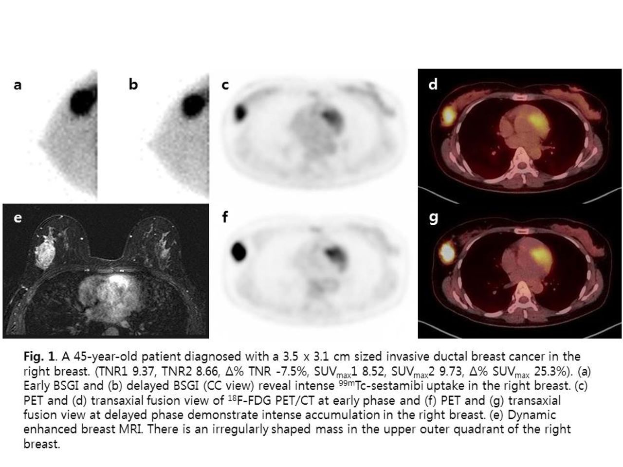
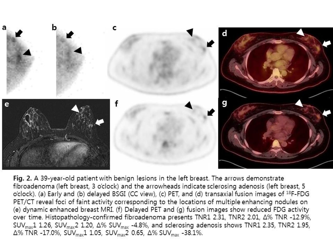
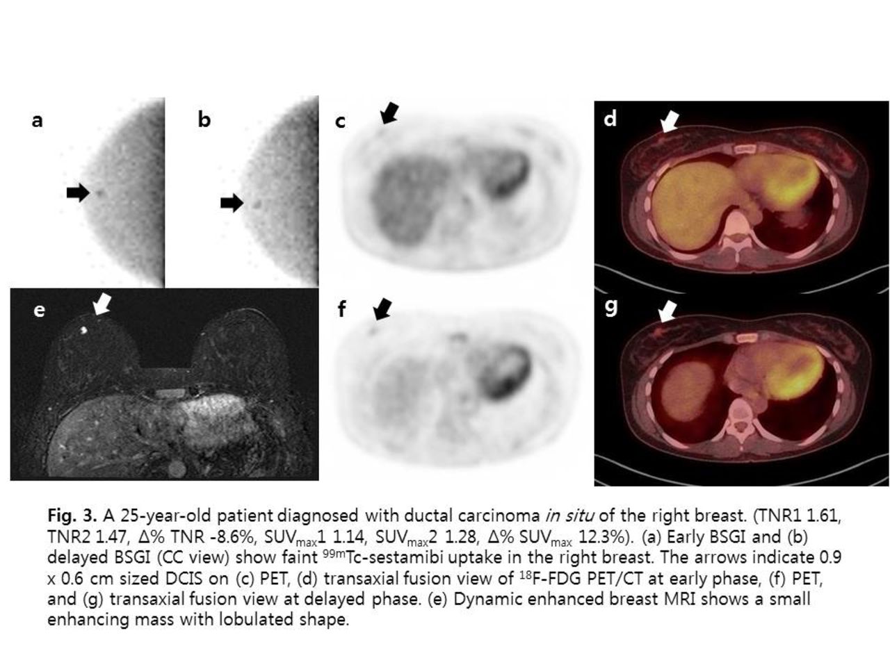
Results: Of the 281 breast lesions, 194 were IDC, 36 were DCIS, and 51 were benign. The sensitivity and accuracy of delayed TNR > 2.05 (TNR2) were higher than those of delayed SUVmax > 2.20 (SUVmax2; 86.1 vs. 78.3% and 84.7 vs. 81.5%, respectively; p = 0.0055 and 0.0003, respectively). However, the specificity of TNR2 was lower than that of SUVmax2 (78.4 vs. 96.1%, p = 0.0126) in discriminating malignancy from benign lesions. For differentiation of malignant subtypes, the sensitivity and accuracy for detecting IDC were lower with TNR2 than with SUVmax2 (65.5 vs. 85.1% and 67.8 vs. 85.7%, respectively; p < 0.0001). Representative cases are illustrated in Fig. 1, 2 and 3.
Conclusion: In differentiating between malignant and benign breast lesions, DTP BSGI parameters showed significantly higher sensitivity and accuracy than DTP PET/CT parameters. However, DTP PET/CT parameters had a tendency for improved specificity, while DTP PET/CT parameters had significantly higher sensitivity and accuracy when discriminating between IDC and DCIS. In conclusion, combined DTP BSGI and PET/CT have good diagnostic performance and are effective imaging techniques for determining malignant breast diseases and differentiating IDC from DCIS. Research Support: This research was supported by grants (2015R1C1A1A02037051, 2012M3A9B6055379, 2015R1C1A2A01054113) from the National Research Foundation of South Korea.


In this issue


Jump to section
Related Articles
Cited By...
- No citing articles found.