
Main menu

  • Home
  • Content
    • Current
    • Ahead of print
    • Past Issues
    • JNM Supplement
    • SNMMI Annual Meeting Abstracts
    • Continuing Education
    • JNM Podcasts
  • Subscriptions
    • Subscribers
    • Institutional and Non-member
    • Rates
    • Journal Claims
    • Corporate & Special Sales
  • Authors
    • Submit to JNM
    • Information for Authors
    • Assignment of Copyright
    • AQARA requirements
  • Info
    • Reviewers
    • Permissions
    • Advertisers
  • About
    • About Us
    • Editorial Board
    • Contact Information
  • More
    • Alerts
    • Feedback
    • Help
    • SNMMI Journals
  • SNMMI
    • JNM
    • JNMT
    • SNMMI Journals
    • SNMMI

User menu

  • Subscribe
  • My alerts
  • Log in
  • My Cart

Search

  • Advanced search
Journal of Nuclear Medicine
  • SNMMI
    • JNM
    • JNMT
    • SNMMI Journals
    • SNMMI
  • Subscribe
  • My alerts
  • Log in
  • My Cart
Journal of Nuclear Medicine

Advanced Search

  • Home
  • Content
    • Current
    • Ahead of print
    • Past Issues
    • JNM Supplement
    • SNMMI Annual Meeting Abstracts
    • Continuing Education
    • JNM Podcasts
  • Subscriptions
    • Subscribers
    • Institutional and Non-member
    • Rates
    • Journal Claims
    • Corporate & Special Sales
  • Authors
    • Submit to JNM
    • Information for Authors
    • Assignment of Copyright
    • AQARA requirements
  • Info
    • Reviewers
    • Permissions
    • Advertisers
  • About
    • About Us
    • Editorial Board
    • Contact Information
  • More
    • Alerts
    • Feedback
    • Help
    • SNMMI Journals
  • View or Listen to JNM Podcast
  • Visit JNM on Facebook
  • Join JNM on LinkedIn
  • Follow JNM on Twitter
  • Subscribe to our RSS feeds
Meeting ReportOncology, Clinical Diagnosis Track

Relationship between FDG PETCT imaging and CA 125 levels in treated patients with Ovarian cancers - Can FDG PETCT define and predict the disease burden in clinically suspected recurrence ?

Palaniswamy Shanmuga Sundaram and Subramanyam Padma
Journal of Nuclear Medicine May 2018, 59 (supplement 1) 398;
Palaniswamy Shanmuga Sundaram
1Nuclear Medicine & PETCT, Amrita Institute of Medical Sciences, Amrita Vishwavidyapeetham Cochin India
  • Find this author on Google Scholar
  • Find this author on PubMed
  • Search for this author on this site
Subramanyam Padma
1Nuclear Medicine & PETCT, Amrita Institute of Medical Sciences, Amrita Vishwavidyapeetham Cochin India
  • Find this author on Google Scholar
  • Find this author on PubMed
  • Search for this author on this site
  • Article
  • Figures & Data
  • Info & Metrics
Loading

Abstract

398

Introduction: Cancer antigen 125 (CA-125) is used for monitoring disease status in post treatment patients of ovarian cancer as rising concentrations predict recurrence in most cases. Many studies have demonstrated that 18F-FDG PET/CT imaging is sensitive & a better modality when compared to conventional anatomical imaging in detection of recurrent disease in patients with raised CA-125 levels.

Objectives: The retrospective study was designed to determine: 1) If FDG PETCT can serve as an accurate maker to define and predict disease burden 2) If there exists any linear relationship between raised CA-125 levels and degree of disease burden 3) A possible CA-125 cutoff value which could predict whether the active disease predominantly involves lymph nodal stations or intra abdominal soft tissue (omental/peritoneal/mesenteric) sites.

Methods: 42 patients (35-74 years, Median 52 years) with suspected disease recurrence based on raised CA-125 levels referred for PET/CT examination were studied. Patients were classified into three groups based on range of CA-125 levels: Group A (35-175), Group B (175-350) and Group C (more than 350). Number of lesions per patient was counted in the following manner: Each lymph nodal station was given a score of 1; Each segment of liver was given a score of 1 irrespective to of number of lesions present in that segment.; Peritoneal soft tissue deposit along the surface of an organ was given a score of 1 and omental, mesenteric and serosal deposits were assigned a score of 1 individually. Data analysis was done using appropriate statistical tests. To arrive at a possible cutoff value for CA-125, various levels of CA-125 were analysed using Chi square test.

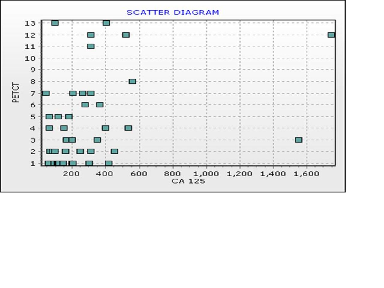
Results: PET/CT detected a total of 57 lesions in Group A (n=18), 68 in Group B (n=14) and 65 lesions in Group C (n=10) respectively. No significant statistical difference was found in total no of lesions detected in all the three groups with different levels of CA-125 (One Way ANOVA: p value 0.071). When comparing total no of lesions detected on PET/CT in groups with marked difference in CA-125 levels - Group A (CA-125: 35-175) and Group C (CA-125: >350), no significant statistical difference was seen (Proportional test: p value 0.380). Also, when number of lesions detected on PET/CT in each patient was correlated with the respective CA-125 levels and data analysed using Spearman Correlation coefficient, no correlation (r = 0.342) between the CA-125 levels and disease burden was observed. Scatter diagram given below depicts X axis: number of PET/CT lesions and Y axis: Level of CA-125. Using Chi square test, a cut off CA-125 value of 350 was established to be significant (p value <0.001) in suggesting whether the active disease predominantly involves lymph nodal or intra abdominal soft tissue sites. In our study, lymph nodal metastases were more common in patients with CA-125 value less than/equal to 350 (n=32/42, 76.2%) & soft tissue deposits were more common in patients with CA-125 levels more than 350 (n=10/42, 23.8%).

Conclusions: Whole body FDG PETCT imaging serves as a reliable, accurate marker of disease recurrence in treated cases of Ca Ovary with raised CA 125 levels. There exists no relationship between CA-125 levels and degree of disease burden. This suggests that PETCT can detect higher degree of disease burden even in patients with low levels of raised CA-125 and vice versa. A cutoff of 350 of CA-125 may be useful to predict the predominant disease distribution pattern i.e. lymph nodal stations versus soft tissue deposits in these patients.

Figure
  • Download figure
  • Open in new tab
  • Download powerpoint
Previous
Back to top

In this issue

Journal of Nuclear Medicine
Vol. 59, Issue supplement 1
May 1, 2018
  • Table of Contents
  • Index by author
Article Alerts
Sign In to Email Alerts with your Email Address
Email Article

Thank you for your interest in spreading the word on Journal of Nuclear Medicine.

NOTE: We only request your email address so that the person you are recommending the page to knows that you wanted them to see it, and that it is not junk mail. We do not capture any email address.

Enter multiple addresses on separate lines or separate them with commas.
Relationship between FDG PETCT imaging and CA 125 levels in treated patients with Ovarian cancers - Can FDG PETCT define and predict the disease burden in clinically suspected recurrence ?
(Your Name) has sent you a message from Journal of Nuclear Medicine
(Your Name) thought you would like to see the Journal of Nuclear Medicine web site.
Citation Tools
Relationship between FDG PETCT imaging and CA 125 levels in treated patients with Ovarian cancers - Can FDG PETCT define and predict the disease burden in clinically suspected recurrence ?
Palaniswamy Shanmuga Sundaram, Subramanyam Padma
Journal of Nuclear Medicine May 2018, 59 (supplement 1) 398;

Citation Manager Formats

  • BibTeX
  • Bookends
  • EasyBib
  • EndNote (tagged)
  • EndNote 8 (xml)
  • Medlars
  • Mendeley
  • Papers
  • RefWorks Tagged
  • Ref Manager
  • RIS
  • Zotero
Share
Relationship between FDG PETCT imaging and CA 125 levels in treated patients with Ovarian cancers - Can FDG PETCT define and predict the disease burden in clinically suspected recurrence ?
Palaniswamy Shanmuga Sundaram, Subramanyam Padma
Journal of Nuclear Medicine May 2018, 59 (supplement 1) 398;
Twitter logo Facebook logo LinkedIn logo Mendeley logo
  • Tweet Widget
  • Facebook Like
  • Google Plus One
Bookmark this article

Jump to section

  • Article
  • Figures & Data
  • Info & Metrics

Related Articles

  • No related articles found.
  • Google Scholar

Cited By...

  • No citing articles found.
  • Google Scholar

More in this TOC Section

Oncology, Clinical Diagnosis Track

  • Immunohistochemical analysis of Gastrin-Releasing-Peptide receptor (GRPr) and Prostate-Specific- Membrane Antigen (PSMA) in primary prostate cancer: comparison with radiolabeled GRPr antagonist (68Ga-RM2) PET/CT
  • Cancer-associated fibroblasts enhance tumor 18F-FDG uptake and contribute to the intratumor heterogeneity of SUVmax
Show more Oncology, Clinical Diagnosis Track

Gynecological

  • Ms
  • Multi-parametric FDG PET/MRI as an Early Predictor of Response to Neoadjuvant Chemotherapy in Patients wit Epithelial Ovarian Cancer
Show more Gynecological

Similar Articles

SNMMI

© 2025 SNMMI

Powered by HighWire