


Article Figures & Data
Figures
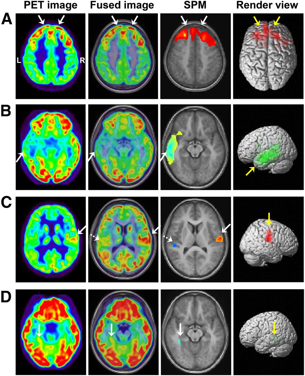
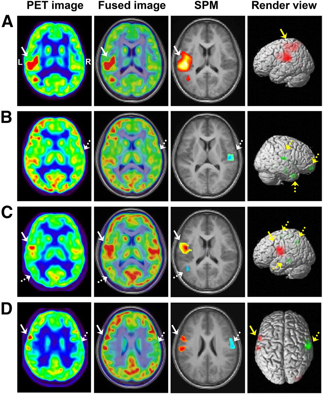
- FIGURE 1.
Comparison between visual and SPM analysis of 18F-FDG PET images. (A) Hypermetabolism in bilateral frontal lobes (solid arrow) was detected by both visual and SPM analysis. (B) Hypometabolism in left temporal lobe (solid arrow) was detected by both visual and SPM analysis. (C) Hypometabolism was found in left rolandic area (dashed arrow) by visual assessment, and hypermetabolic region was further identified in right rolandic area (solid arrow) by SPM analysis. (D) Hypometabolic region undetected by visual assessment was identified in left mesial temporal lobe (solid arrow) by SPM analysis.
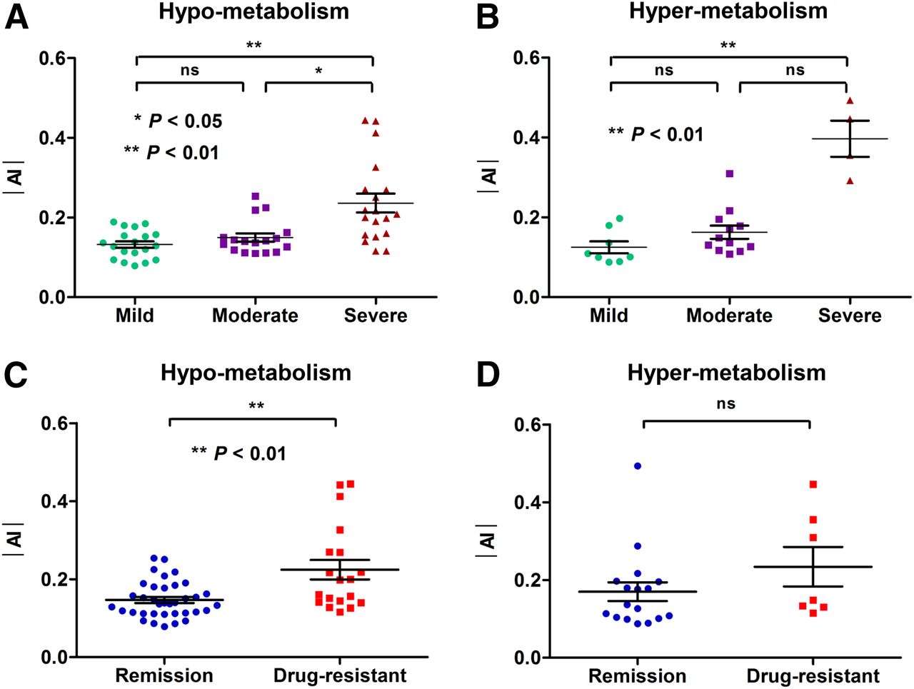
- FIGURE 2.
Clinical value of 18F-FDG PET in severity and outcome of pediatric epilepsy. (A) In patients with hypometabolism (n = 55), severe group had higher value of |AI| than mild and moderate groups (P = 0.001 and 0.030, respectively). (B) In patients with hypermetabolism (n = 24), severe group had higher value of |AI| than mild group (P = 0.002) and slightly higher value than moderate group (P = 0.051). (C) On the basis of follow-up, in patients with hypometabolism, drug-resistant patients had higher values of |AI| than seizure-free patients (P = 0.002). (D) In patients with hypermetabolism, drug-resistant patients had slightly higher values of |AI| than seizure-free patients (P = 0.209).
- FIGURE 3.
Metabolic features of rolandic epilepsy. (A) Hypermetabolism was found in left rolandic area (solid arrow). (B) Hypometabolic regions were found in right frontal and temporal lobes and rolandic areas (dashed arrow). (C) Hypermetabolism (solid arrow) with associated surrounding hypometabolism (dashed arrow) was observed in left rolandic area. (D) Hypermetabolism and its associated remote hypometabolism were found in left (solid arrow) and right (dashed arrow) rolandic areas, respectively.
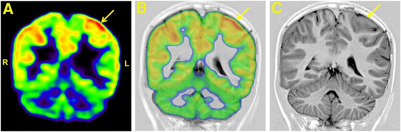
- FIGURE 4.
Interictal 18F-FDG PET and MRI studies of 14-y-old girl who experienced 6 times of right hemifacial contraction with drooling during a 4-mo time. 18F-FDG PET image (A) and PET/MR image (B) coregistration guided second reading of MR images, in which previously missed cortical thickening in the left parietal lobe (solid arrow) on T1-weighted image (C) was detected.
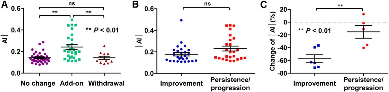
- FIGURE 5.
Diagnostic value of 18F-FDG PET imaging during clinical course of pediatric epilepsy. (A) Patients with add-on therapy after 18F-FDG PET study had higher values of |AI| than those with unchanged therapy or withdrawal (P = 0.006 and <0.001, respectively), but no significant difference in |AI| was found between patients with unchanged therapy and those at withdrawal (P = 1.0). (B) Improvement course was defined as remaining seizure-free throughout follow-up of patients in moderate or severe groups; and persistence or progression course was defined as being drug-resistant throughout follow-up. Patients with persistence or progression course had slightly higher values of |AI| than those with improvement (P = 0.051). (C) Of 11 patients who also underwent post-1-y follow-up PET scans, significant change of |AI| (%) was found in patients with clinical improvement compared than with persistence or progression (P = 0.005). n.s. = not significant.


Tables
- TABLE 2
Comparison between Visual Assessment and SPM Analysis of 18F-FDG PET Studies in Pediatric Epilepsy Patients (n = 100)
SPM analysis Visual assessment Normal Hypometabolism Hypermetabolism Total Normal 21 6 3 30 Hypometabolism 8 41 14 63 Hypermetabolism 0 0 7 7 Total 29 47 24 100 κ = 0.483, P < 0.001.
- TABLE 3
Clinical Characteristics and 18F-FDG PET Findings in Pediatric Epilepsy Patients (n = 100)
Clinical characteristic Abnormal (n = 79) Normal (n = 21) P Sex 0.745 Female/male 37/42 9/12 Age (y) 10.3 ± 2.0 10.8 ± 2.0 0.293 Age of onset (y) 6.7 ± 2.6 7.7 ± 2.5 0.149 Duration of epilepsy (y) 3.6 ± 2.4 3.2 ± 2.1 0.713 Seizure frequency (times/y) 6.0 ± 13.4 1.0 ± 1.4 0.034 Seizure severity 0.026 Mild 27 (19/8) 12 Moderate 28 (16/12) 7 Severe 24 (20/4) 2 Time since last seizure 0.010 1 d to 1 m 38 (25/13) 3 1 m to 1 y 14 (11/3) 6 1 y 27 (19/8) 12 Antiepileptic drug 0.127 Drug naive 11 (7/4) 3 Single drug 35 (25/10) 14 Combined drugs 33 (23/10) 4 *Data in parentheses refer to number of patients with hypo- vs. hypermetabolism.
- TABLE 4
Correlation Between |AI| and Clinical Severity in Pediatric Epilepsy Patients with Abnormal Glucose Metabolism (n = 79)
Seizure frequency Time since last seizure Abnormal metabolism rs P rs P Hypometabolism (n = 55) 0.516 <0.001 −0.477 <0.001 Hypermetabolism (n = 24) 0.653 0.001 −0.621 0.001
Supplemental Data
Files in this Data Supplement:


