
Main menu

  • Home
  • Content
    • Current
    • Ahead of print
    • Past Issues
    • JNM Supplement
    • SNMMI Annual Meeting Abstracts
    • Continuing Education
    • JNM Podcasts
  • Subscriptions
    • Subscribers
    • Institutional and Non-member
    • Rates
    • Journal Claims
    • Corporate & Special Sales
  • Authors
    • Submit to JNM
    • Information for Authors
    • Assignment of Copyright
    • AQARA requirements
  • Info
    • Reviewers
    • Permissions
    • Advertisers
  • About
    • About Us
    • Editorial Board
    • Contact Information
  • More
    • Alerts
    • Feedback
    • Help
    • SNMMI Journals
  • SNMMI
    • JNM
    • JNMT
    • SNMMI Journals
    • SNMMI

User menu

  • Subscribe
  • My alerts
  • Log in
  • My Cart

Search

  • Advanced search
Journal of Nuclear Medicine
  • SNMMI
    • JNM
    • JNMT
    • SNMMI Journals
    • SNMMI
  • Subscribe
  • My alerts
  • Log in
  • My Cart
Journal of Nuclear Medicine

Advanced Search

  • Home
  • Content
    • Current
    • Ahead of print
    • Past Issues
    • JNM Supplement
    • SNMMI Annual Meeting Abstracts
    • Continuing Education
    • JNM Podcasts
  • Subscriptions
    • Subscribers
    • Institutional and Non-member
    • Rates
    • Journal Claims
    • Corporate & Special Sales
  • Authors
    • Submit to JNM
    • Information for Authors
    • Assignment of Copyright
    • AQARA requirements
  • Info
    • Reviewers
    • Permissions
    • Advertisers
  • About
    • About Us
    • Editorial Board
    • Contact Information
  • More
    • Alerts
    • Feedback
    • Help
    • SNMMI Journals
  • View or Listen to JNM Podcast
  • Visit JNM on Facebook
  • Join JNM on LinkedIn
  • Follow JNM on Twitter
  • Subscribe to our RSS feeds
Research ArticleOncology

Prospective Evaluation of the Clinical Implications of the Tumor Metabolism and Chemotherapy-Related Changes in Advanced Biliary Tract Cancer

Jaemin Jo, Hyun Woo Kwon, Seongyeol Park, Do-Youn Oh, Gi Jeong Cheon and Yung-Jue Bang
Journal of Nuclear Medicine August 2017, 58 (8) 1255-1261; DOI: https://doi.org/10.2967/jnumed.116.186239
Jaemin Jo
1Department of Internal Medicine, Seoul National University College of Medicine, Seoul, Korea
  • Find this author on Google Scholar
  • Find this author on PubMed
  • Search for this author on this site
Hyun Woo Kwon
2Department of Nuclear Medicine, Seoul National University Hospital, Seoul, Korea; and
  • Find this author on Google Scholar
  • Find this author on PubMed
  • Search for this author on this site
Seongyeol Park
1Department of Internal Medicine, Seoul National University College of Medicine, Seoul, Korea
  • Find this author on Google Scholar
  • Find this author on PubMed
  • Search for this author on this site
Do-Youn Oh
1Department of Internal Medicine, Seoul National University College of Medicine, Seoul, Korea
3Cancer Research Institute, Seoul National University College of Medicine, Seoul, Korea
  • Find this author on Google Scholar
  • Find this author on PubMed
  • Search for this author on this site
Gi Jeong Cheon
2Department of Nuclear Medicine, Seoul National University Hospital, Seoul, Korea; and
3Cancer Research Institute, Seoul National University College of Medicine, Seoul, Korea
  • Find this author on Google Scholar
  • Find this author on PubMed
  • Search for this author on this site
Yung-Jue Bang
1Department of Internal Medicine, Seoul National University College of Medicine, Seoul, Korea
3Cancer Research Institute, Seoul National University College of Medicine, Seoul, Korea
  • Find this author on Google Scholar
  • Find this author on PubMed
  • Search for this author on this site
  • Article
  • Figures & Data
  • Supplemental
  • Info & Metrics
  • PDF
Loading

Article Figures & Data

Figures

  • Tables
  • Additional Files
  • FIGURE 1.
    • Download figure
    • Open in new tab
    • Download powerpoint
    FIGURE 1.

    (A) Distribution of initial SUVmax. (B) SUVmax reduction at best metabolic response and at first 18F-FDG PET evaluation. (C) Distribution of initial SUVmax according to primary tumor origin.

  • FIGURE 2.
    • Download figure
    • Open in new tab
    • Download powerpoint
    FIGURE 2.

    PFS according to initial SUVmax (A) and SUVmax reduction at best metabolic response (B). OS according to initial SUVmax (C) and SUVmax reduction at best metabolic response (D).
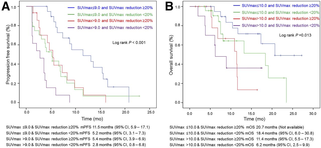
  • FIGURE 3.
    • Download figure
    • Open in new tab
    • Download powerpoint
    FIGURE 3.

    PFS (A) and OS (B) after patients were divided into 4 groups by initial SUVmax and its response.

Tables

  • Figures
  • Additional Files
    • View popup
    TABLE 1

    Baseline Characteristics of Patients

    CharacteristicValue (n = 75)
    Women (n)32 (42.7%)
    Median age (y)64.0 (range, 46.0–83.0)
    Median BMI (kg/m2)23.4 (range, 15.8–30.0)
    ECOG performance-status score (n)
     0/1 to 220 (26.7%)/55 (73.3%)
    Primary tumor site (n)
     Gallbladder cancer28 (37.3%)
     IHCC22 (29.3%)
     EHCC19 (25.3%)
     AoV cancer6 (8.0%)
    Pathologic differentiation (n)
     WD/MD/PD3 (4.0%)/38 (50.7%)/12 (16.0%)
    HER 2 immunohistochemistry (n)
     Negative to 1+33 (44.0%)
     2+ to 3+10 (13.3%)
    HER 2 FISH (n)
     Negative/positive5 (6.7%)/3 (4.0%)
    c-MET cytoplasm immunohistochemistry (n)
     Negative to 1+36 (48.0%)
     2+ to 3+7 (9.3%)
    c-MET membrane immunohistochemistry (n)
     Negative to 1+16 (21.3%)
     2+ to 3+27 (36.0%)
    c-Myc
     Negative/positive28 (37.3%)/12 (16.0%)
    Median CEA (ng/mL)2.6 (range, 0.5–182.9)
    Median CA 19-9 (U/mL)133 (range, 2.0–36,000.0)
    Median WBC (μL)6,300 (range, 2,890–16,330)
    Median total bilirubin (mg/dL)0.6 (range, 0.3–3.3)
    Median albumin (mg/dL)3.9 (range, 3.3–4.7)
    Curative/palliative operation (n)40 (78.4%)/11 (21.6%)
    Unresectable/recurrent disease (n)35 (46.7%)/40 (53.3%)
    Best response (n)
     PR/SD/PD12 (16.7%)/45 (62.5%)/15 (20.8%)
    Median follow-up duration (mo)6.8 (1.0–27.2)
    Median PFS (mo)5.6 (95% CI, 4.4–6.8)
    Median OS (mo)13.2 (95% CI, 7.1–19.3)
    • BMI = body mass index; ECOG = Eastern Cooperative Oncology Group; WD = well differentiated; MD = moderately differentiated; PD = poorly differentiated; HER 2 = human epidermal growth factor receptor 2; FISH = fluorescent in situ hybridization; CEA = carcinoembryonic antigen; CA 19-9 = carbohydrate antigen 19-9; WBC = white blood cell; CR = complete response; PR = partial response; SD = stable disease; PD = progressive disease.

    • View popup
    TABLE 2

    Comparison of Patient Characteristics Between High-/Low-Metabolism Groups

    CharacteristicLow-metabolism group, SUVmax ≤ 9.0 (n = 46)High-metabolism group, SUVmax > 9.0 (n = 29)P
    Primary tumor site (n)0.013
     Gallbladder cancer12 (26.1%)16 (55.2%)
     IHCC14 (30.4%)8 (27.6%)
     EHCC17 (37.0%)2 (6.9%)
     AoV cancer3 (6.5%)3 (10.3%)
    Histologic differentiation (n)0.034
     WD1 (2.6%)2 (13.3%)
     MD31 (81.6%)7 (46.7%)
     PD6 (15.8%)6 (40.0%)
    c-Myc, positive3 (12,5%)9 (56.2%)0.005
    Initial presentation at enrollment (n)<0.001
     Metastatic disease14 (30.4%)21 (72.4%)
     Recurrent disease32 (69.6%)8 (27.6%)
    Mean WBC (μL)5,980.4 ± 1,857.47,473.1 ± 3,032.80.010
    Mean no. of organs with 18F-FDG uptake1.5 ± 1.12.3 ± 1.00.004
    Mean no. of lesions with 18F-FDG uptake2.9 ± 3.57.0 ± 7.90.012
    • WD = well differentiated; MD = moderately differentiated; PD = poorly differentiate; WBC = white blood cell.

    • View popup
    TABLE 3

    Analysis of Prognostic Factors of PFS

    Univariate analysisMultivariate analysis
    VariableMedian PFS (mo)95% CIPHR95% CIP
    Primary tumor origin0.0130.003
     Gallbladder cancer5.32.8–7.80.4760.17–1.32
     IHCC8.35.0–11.6Ref
     EHCC5.02.2–7.82.310.83–6.50
     AoV cancer1.30.7–1.93.260.83–12.85
    c-Myc0.044
     Negative7.02.5–11.5
     Positive3.80.4–7.2
    Initial SUVmax0.0024.091.73–9.660.001
     ≤9.07.04.8–9.2
     >9.03.82.2–5.4
    SUVmax reduction (at best 18F-FDG PET response)<0.0013.351.55–7.200.002
     ≥20.0%8.85.8–11.8
     <20.0%3.93.3–4.5
    Organs with 18F-FDG uptake (n)0.134
     0–26.34.8–7.9
     ≥33.92.2–5.6
    • View popup
    TABLE 4

    Analysis of Prognostic Factors of OS

    Univariate analysisMultivariate analysis
    VariableMedian OS (mo)95% CIPHR95% CIP
    Age (y)0.094
     ≤6519.19.8–28.4
     >658.96.0–11.8
    CEA (ng/mL)0.062
     ≤5.018.415.8–21.0
     >5.08.94.7–13.1
    Initial SUVmax0.0032.611.18–5.810.019
     ≤10.019.116.1–22.1
     10.010.93.8–18.1
    SUVmax reduction (at best 18F-FDG PET response)0.0741.960.91–4.200.082
     ≥20.0%20.75.8–35.6
     20.0%13.22.8–23.6
    Organs with 18F-FDG uptake (n)0.0392.080.95–4.570.068
     0–218.410.3–26.5
     ≥38.92.3–15.5
    • CEA = carcinoembryonic antigen.

Additional Files

  • Figures
  • Tables
  • Supplemental Data

    Files in this Data Supplement:

    • Supplemental Data
PreviousNext
Back to top

In this issue

Journal of Nuclear Medicine: 58 (8)
Journal of Nuclear Medicine
Vol. 58, Issue 8
August 1, 2017
  • Table of Contents
  • Table of Contents (PDF)
  • About the Cover
  • Index by author
Print
Download PDF
Article Alerts
Sign In to Email Alerts with your Email Address
Email Article

Thank you for your interest in spreading the word on Journal of Nuclear Medicine.

NOTE: We only request your email address so that the person you are recommending the page to knows that you wanted them to see it, and that it is not junk mail. We do not capture any email address.

Enter multiple addresses on separate lines or separate them with commas.
Prospective Evaluation of the Clinical Implications of the Tumor Metabolism and Chemotherapy-Related Changes in Advanced Biliary Tract Cancer
(Your Name) has sent you a message from Journal of Nuclear Medicine
(Your Name) thought you would like to see the Journal of Nuclear Medicine web site.
Citation Tools
Prospective Evaluation of the Clinical Implications of the Tumor Metabolism and Chemotherapy-Related Changes in Advanced Biliary Tract Cancer
Jaemin Jo, Hyun Woo Kwon, Seongyeol Park, Do-Youn Oh, Gi Jeong Cheon, Yung-Jue Bang
Journal of Nuclear Medicine Aug 2017, 58 (8) 1255-1261; DOI: 10.2967/jnumed.116.186239

Citation Manager Formats

  • BibTeX
  • Bookends
  • EasyBib
  • EndNote (tagged)
  • EndNote 8 (xml)
  • Medlars
  • Mendeley
  • Papers
  • RefWorks Tagged
  • Ref Manager
  • RIS
  • Zotero
Share
Prospective Evaluation of the Clinical Implications of the Tumor Metabolism and Chemotherapy-Related Changes in Advanced Biliary Tract Cancer
Jaemin Jo, Hyun Woo Kwon, Seongyeol Park, Do-Youn Oh, Gi Jeong Cheon, Yung-Jue Bang
Journal of Nuclear Medicine Aug 2017, 58 (8) 1255-1261; DOI: 10.2967/jnumed.116.186239
Twitter logo Facebook logo LinkedIn logo Mendeley logo
  • Tweet Widget
  • Facebook Like
  • Google Plus One
Bookmark this article

Jump to section

  • Article
    • Abstract
    • MATERIALS AND METHODS
    • RESULTS
    • DISCUSSION
    • CONCLUSION
    • DISCLOSURE
    • Footnotes
    • REFERENCES
  • Figures & Data
  • Supplemental
  • Info & Metrics
  • PDF

Related Articles

  • This Month in JNM
  • PubMed
  • Google Scholar

Cited By...

  • Predictive Role of Temporal Changes in Intratumoral Metabolic Heterogeneity During Palliative Chemotherapy in Patients with Advanced Pancreatic Cancer: A Prospective Cohort Study
  • Google Scholar

More in this TOC Section

Oncology

  • Rediscovering the indispensable role of FDG PET in kidney cancer imaging
  • The role of FDG PET/CT in clinical evaluation and management of nasopharyngeal carcinoma
  • Immune-related adverse events on 18F-FDG PET/CT in patients undergoing immune checkpoint inhibitor therapy
Show more Oncology

Clinical

  • TauIQ: A Canonical Image Based Algorithm to Quantify Tau PET Scans
  • Dual PET Imaging in Bronchial Neuroendocrine Neoplasms: The NETPET Score as a Prognostic Biomarker
  • Addition of 131I-MIBG to PRRT (90Y-DOTATOC) for Personalized Treatment of Selected Patients with Neuroendocrine Tumors
Show more Clinical

Similar Articles

Keywords

  • Biliary Tract Neoplasm
  • carcinoma
  • positron-emission tomography
  • metabolism
  • prognosis
SNMMI

© 2025 SNMMI

Powered by HighWire