


Article Figures & Data
Figures
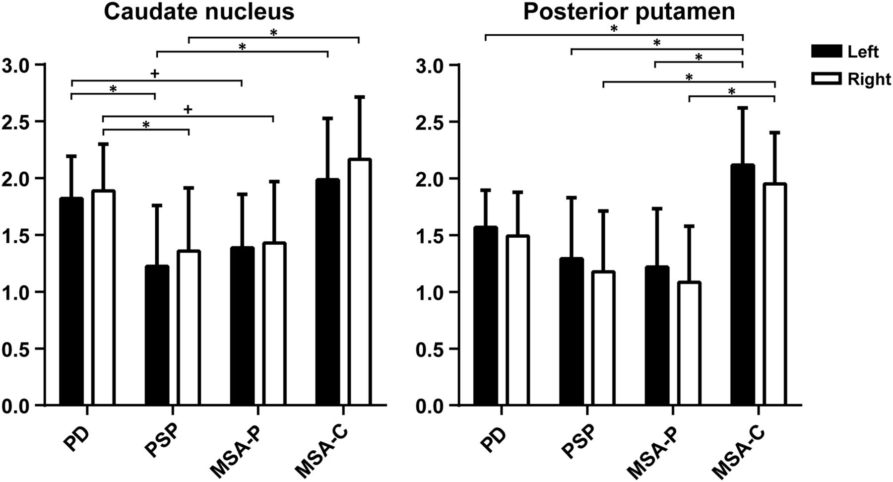
- FIGURE 1.
Binding ratios per ROI. * = statistically significant difference; + = trend. Striatal regions, including patients using SSRIs.
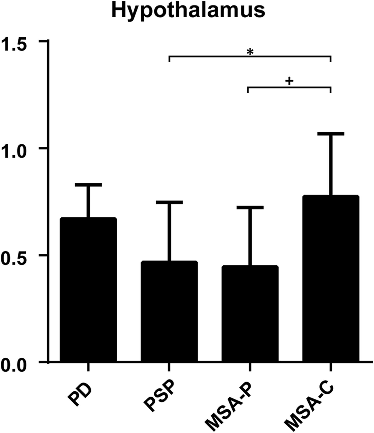
- FIGURE 2.
Binding ratios in hypothalamus, without patients using SSRIs. Data on thalamus and pons are provided in supplemental materials.
- FIGURE 3.
Voxel-by-voxel analysis of hypothalamus at (x,y,z), −2,0,−8. (Lower) Quantification per diagnosis.


Tables
Characteristic PD MSA-P MSA-C PSP Test statistic/df/P No. of patients 30 9 7 13 Sex χ2 = 2.986/3/0.394 Female 14 7 4 6 Male 16 2 3 7 Age at DAT SPECT 66.39 ± 7.55 61.37 ± 9.61 67.72 ± 10.63 70.46 ± 6.29 F = 2.322/3,55/0.085 Disease duration (y) 3.59 ± 2.95 3.15 ± 2.59 3.57 ± 1.43 5.69 ± 4.71 Kruskal–Wallis test = 4.98/3/0.173 UPDRS III 26.8 ± 12.4 41.38 ± 22.83 36.50 ± 7.78 33.17 ± 12.13 Kruskal–Wallis test = 6.58/3/0.087 SCOPA-AUT 35.57 ± 8.45 44.88 ± 12.05 53,00 ± 11.31 36.62 ± 6.62 F = 12.662/2,55/0.001* SSRI (n) 5 (16.7) 3 (33.3) 0 (0) 3 (23.1) ↵* For this analysis MSA-P and MSA-C were pooled into 1 group.
df = degree of freedom; UPDRS III = Unified Parkinson's Disease Rating Scale, motor evaluation; SCOPA-AUT = SCale for Outcomes of PArkinson’s Disease–AUTonomic Symptoms.
Values are mean ± SD, unless otherwise specified. Data in parentheses are percentages.
ROIs PD MSA-P MSA-C PSP F/df/P Striatal Caudate left 1.82 ± 0.37 1.39 ± 0.47 1.99 ± 0.54 1.22 ± 0.53 7.619/3,55/<0.001 Caudate right 1.89 ± 0.41 1.43 ± 0.54 2.16 ± 0.55 1.36 ± 0.56 6.621/3,55/0.001 Putamen left 1.57 ± 0.33 1.22 ± 0.51 2.12 ± 0.51 1.28 ± 0.52 7.559/3,55/<0.001 Putamen right 1.49 ± 0.39 1.08 ± 0.50 1.95 ± 0.46 1.18 ± 0.54 6.588/3,55/0.001 Extrastriatal Hypothalamus 0.67 ± 0.16 0.45 ± 0.28 0.78 ± 0.30 0.47 ± 0.28 4.307/3,43/0.012 Thalamus left 0.79 ± 0.18 0.58 ± 0.27 0.80 ± 0.29 0.70 ± 0.29 1.576/3,43/0.236 Thalamus right 0.81 ± 0.18 0.68 ± 0.24 0.90 ± 0.20 0.68 ± 0.26 2.332/3,43/0.087 Pons 0.55 ± 0.03 0.41 ± 0.09 0.50 ± 0.05 0.48 ± 0.07 0.803/3,43/0.375 Extrastriatal groups depicted for patients without SSRI; F, df, and P values given are between-group analysis of covariance results, corrected for age.
Values given are mean ± SD, unless otherwise specified.
df = degrees of freedom.
Region Group 1 > group 2 Ke PFWE peak-voxel T x/y/z (mm) Caudate nucleus left (df, 1,54) PD PSP 13 0.005 4.80 −14/4/20 MSA-C PSP 4 0.008 4.67 −12/18/12 Caudate nucleus right (df, 1,54) PD PSP 22 0.005 4.85 12/4/20 MSA-P 2 0.024 4.30 16/0/24 MSA-C PSP 2 0.018 4.41 18/0/18 Posterior putamen left (df, 1,54) MSA-C PSP 41 0.004 4.69 −26/−2/4 MSA-P 16 0.007 4.51 −26/−2/4 PD 33 0.003 4.83 −26/−14/6 Posterior putamen right (df, 1,54) PD PSP 2 0.018 4.17 36/−16/−8 MSA-C PSP 30 0.004 4.73 28/−10/2 MSA-P 28 0.001 5.22 28/−10/2 13 0.002 4.88 32/−12/8 PD 20 <0.001 5.73 28/−10/2 Hypothalamus* (df, 1,43) PD PSP 17 <0.001 5.93 −4/0/−10 MSA-P 6 0.002 4.79 −6/0/−10 MSA-C PSP 8 <0.001 5.47 −2/0/−8 MSA-P 4 0.002 4.79 −6/0/−10 ↵* For this analysis, patients not on SSRIs were included.
Difference between 2 diagnoses for specific cluster.
df =degrees of freedom; Ke = cluster extent in no. of voxels; PFWE = familywise error–corrected P values; T = T statistic; x/y/z = location of significantly most different between-groups cluster from midpoint in millimeter.
Supplemental Data
Files in this Data Supplement:


