


Article Figures & Data
Figures
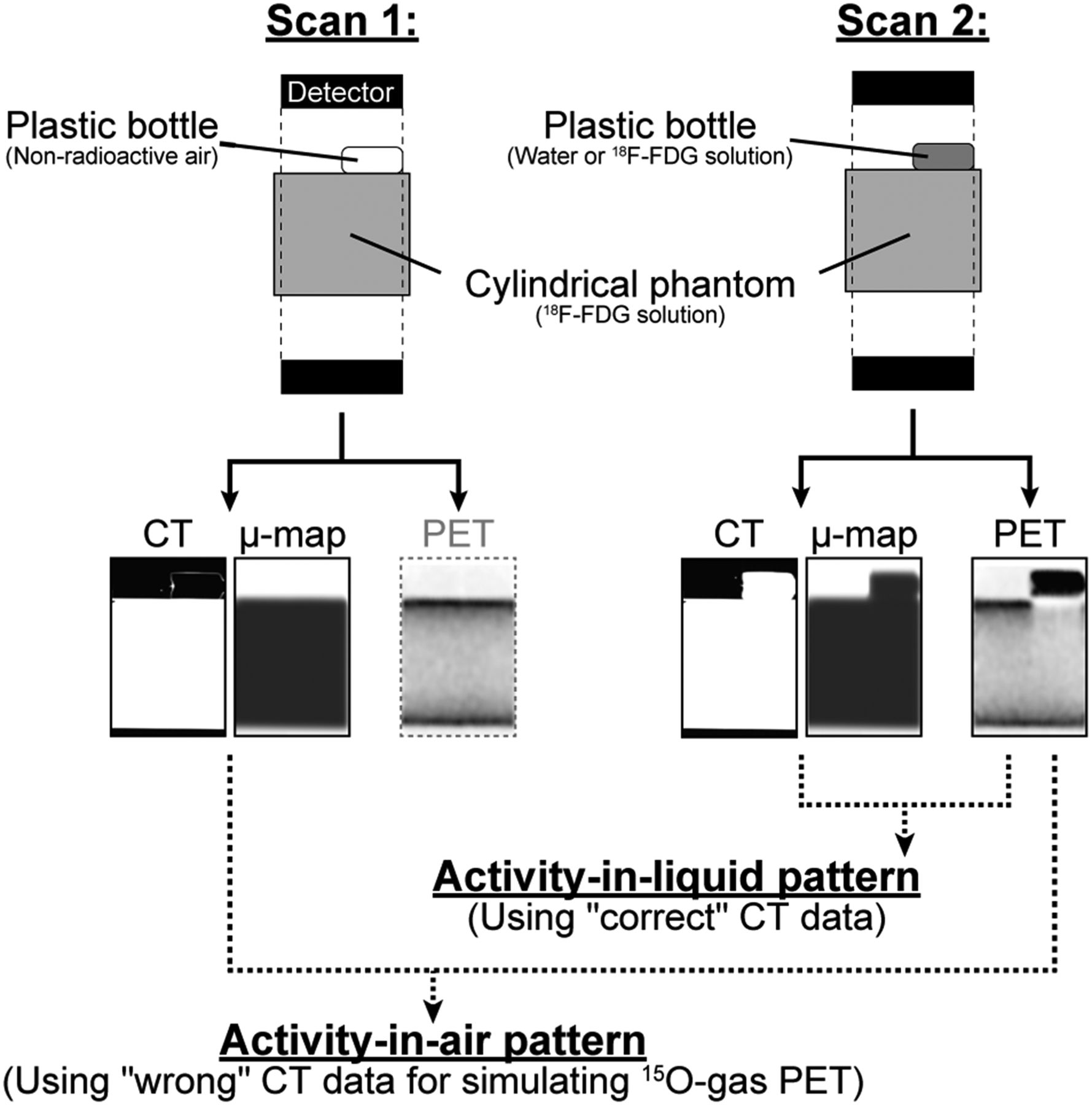
- FIGURE 1.
Procedures used for data acquisition and image reconstruction in phantom experiments. Plastic bottle filled with nonradioactive air (scan 1) and water or an 18F-FDG solution (scan 2) was attached to cylindric phantom. Attenuation and scatter corrections were performed for PET data in scan 2 using the CT data obtained in scan 1 (activity-in-air pattern) and using CT and PET data in scan 2 (activity-in-liquid pattern).
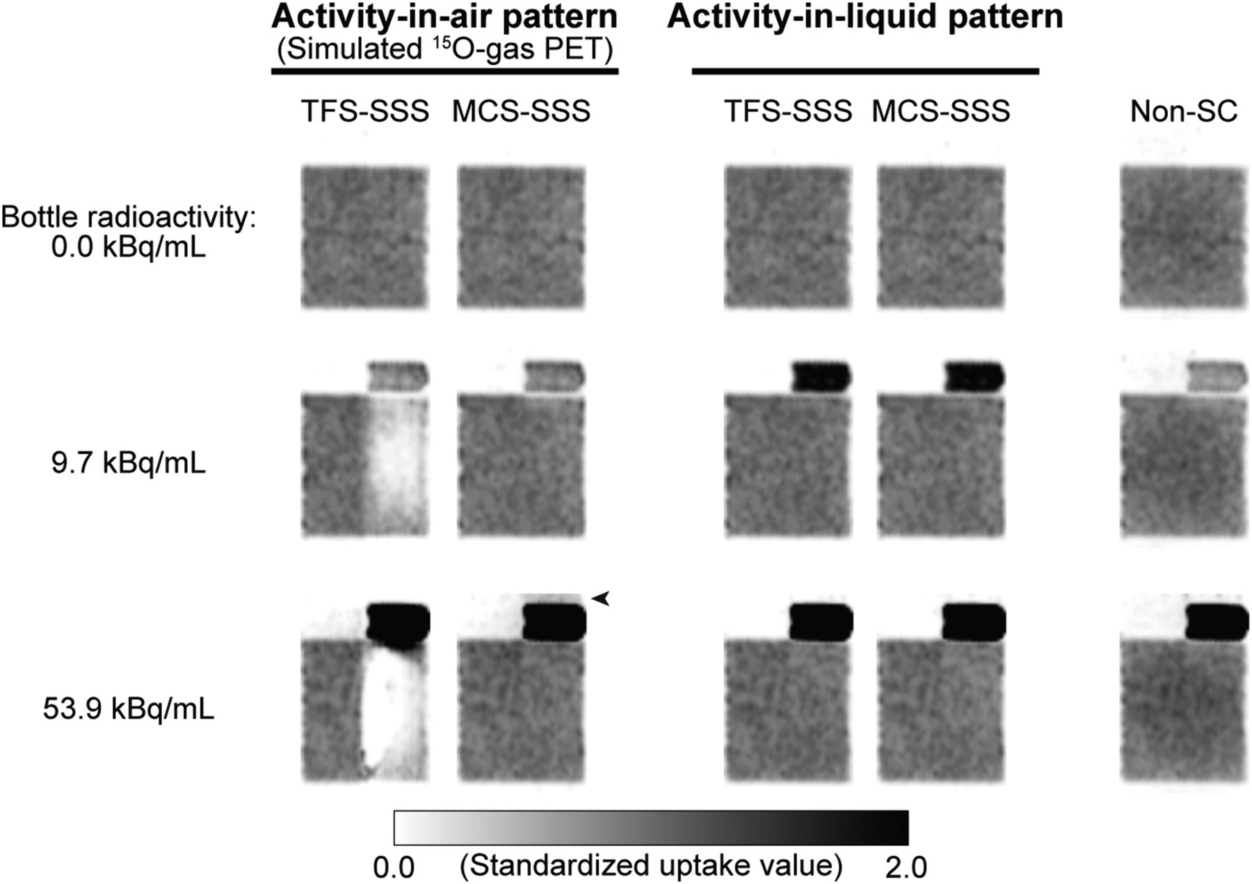
- FIGURE 2.
Typical sagittal images of phantom for activity-in-air pattern, activity-in-liquid pattern, and case without scatter correction, when bottle radioactivity levels were 0.0, 9.7, and 53.9 kBq/mL. Black arrowhead = slight high-activity artifact.
- FIGURE 3.
(A and B) Percentage errors of activity concentration in cylindric phantom and true activity concentration with respect to bottle radioactivity. Dotted line = TFS-SSS; solid line = MCS-SSS. Open and closed symbols represent slices with and without bottle, respectively. Scatter fractions in A and B are shown in C and D, respectively. Activity-in-air pattern (A and C); activity-in-liquid pattern (B and D).
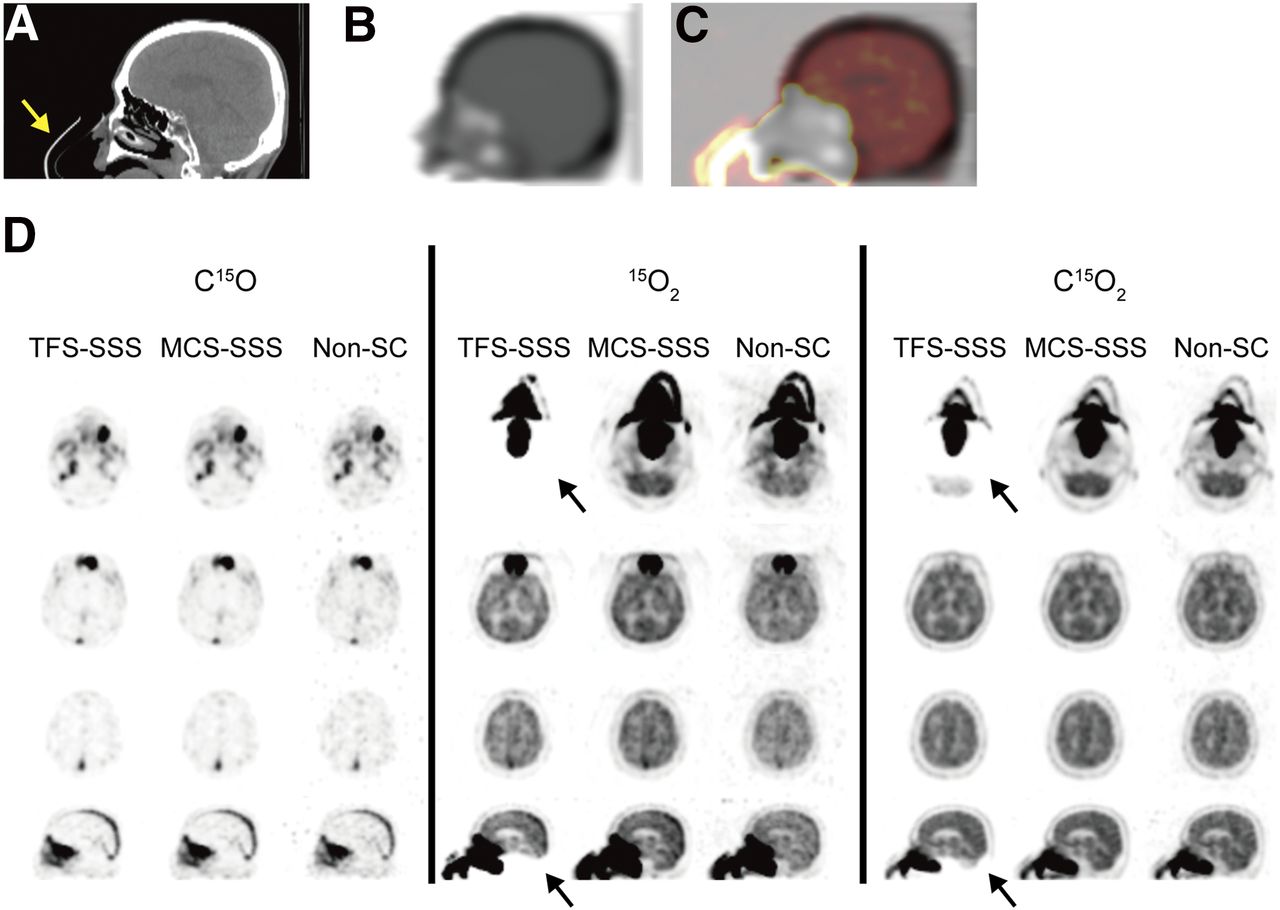
- FIGURE 4.
CT image (A), μ-map (B), image obtained by superimposing μ-map and C15O2 PET image (C), and PET images (D) of patient in case 1 in sagittal view. (D) Axial (rows 1–3) and sagittal (row 4) images of C15O, 15O2, and C15O2 PET. Yellow arrow = face mask; black arrows = cold artifacts.
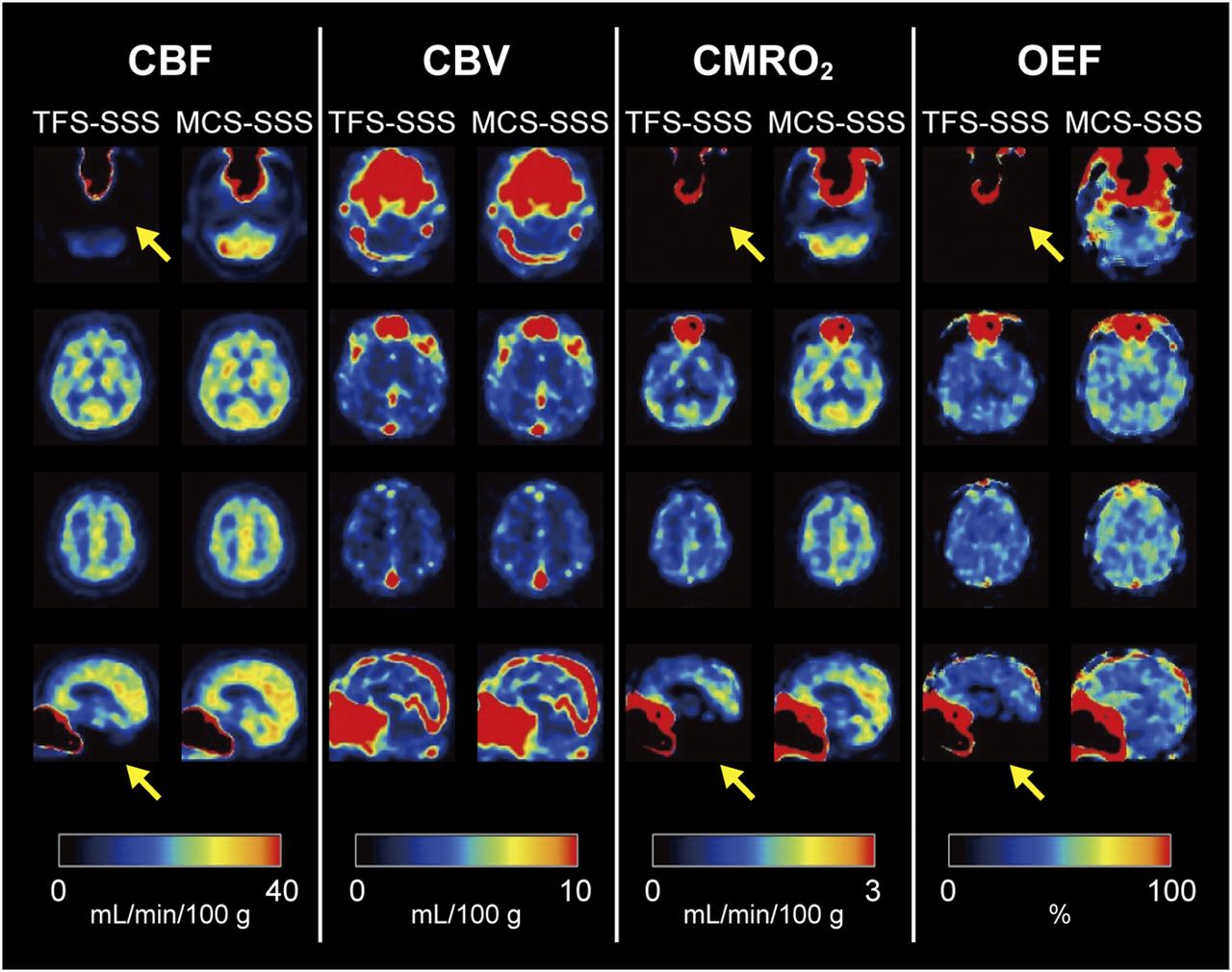
- FIGURE 5.
Axial (rows 1–3) and sagittal (row 4) images for assessment of CBF, CBV, CMRO2, and OEF of patient case 1. Yellow arrows = cold artifacts.


Additional Files
Supplemental Data
Files in this Data Supplement:


Jump to section
Related Articles
Cited By...
- No citing articles found.