


Article Figures & Data
Figures
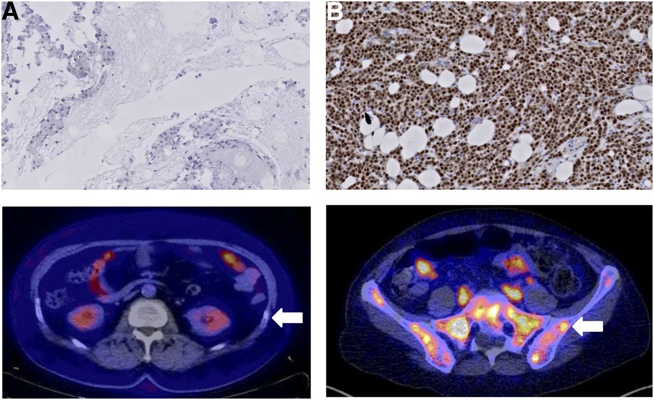
- FIGURE 1.
Comparison of immunohistochemistry staining of AR between an AR-negative (0% AR staining) lesion (A, top) and AR-positive (100% staining) lesion (B, top). (Bottom) Horizontal 18F-FDHT PET/CT fusion images. (A, bottom) Physiologic uptake in small intestines and kidneys. Arrow indicates biopsied lesion (rib) with no visual enhanced uptake. (B, bottom) Physiologic uptake in small intestines and high uptake throughout pelvic bones. Arrow indicates biopsied lesion in iliac bone with visually enhanced uptake.
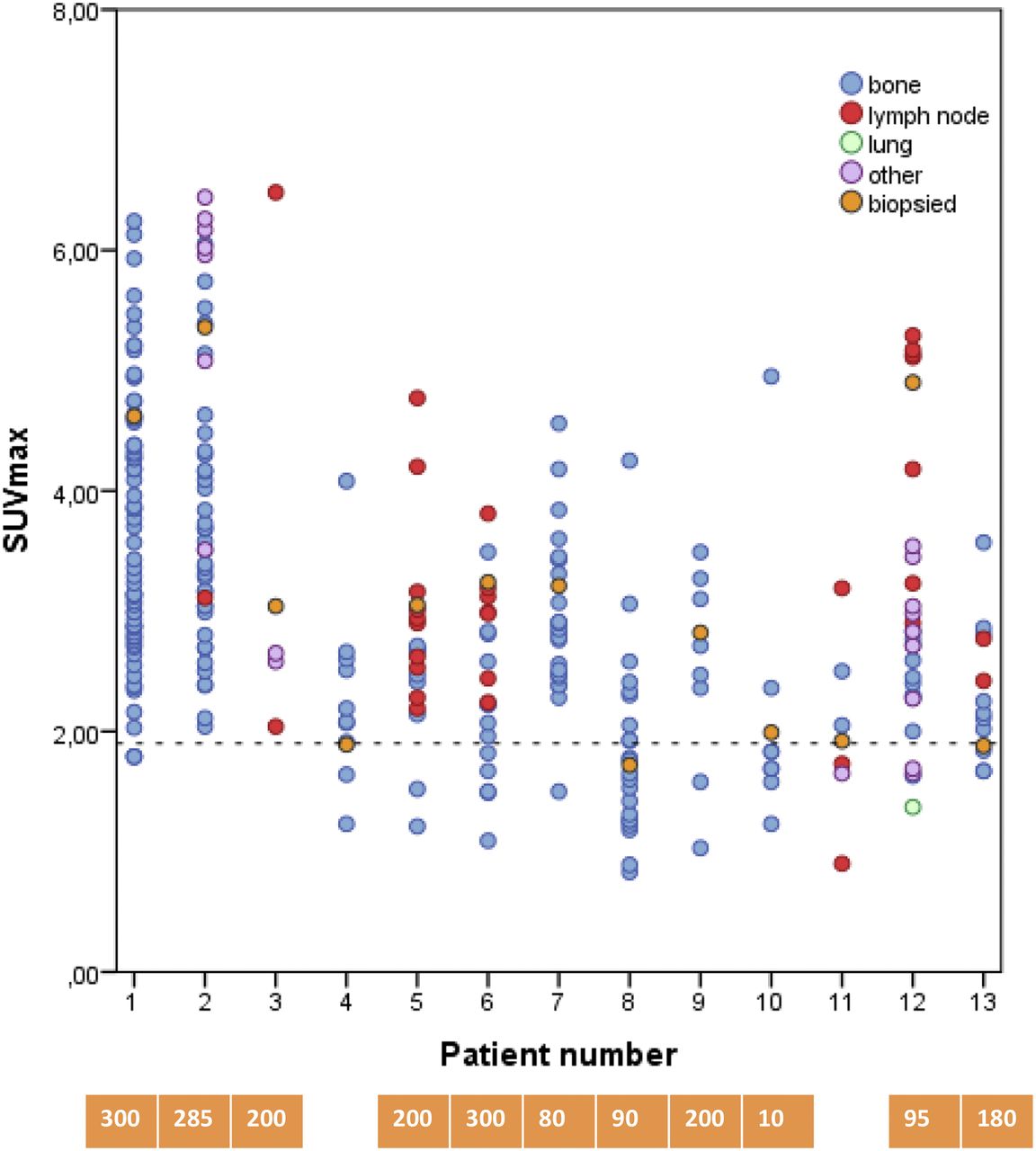
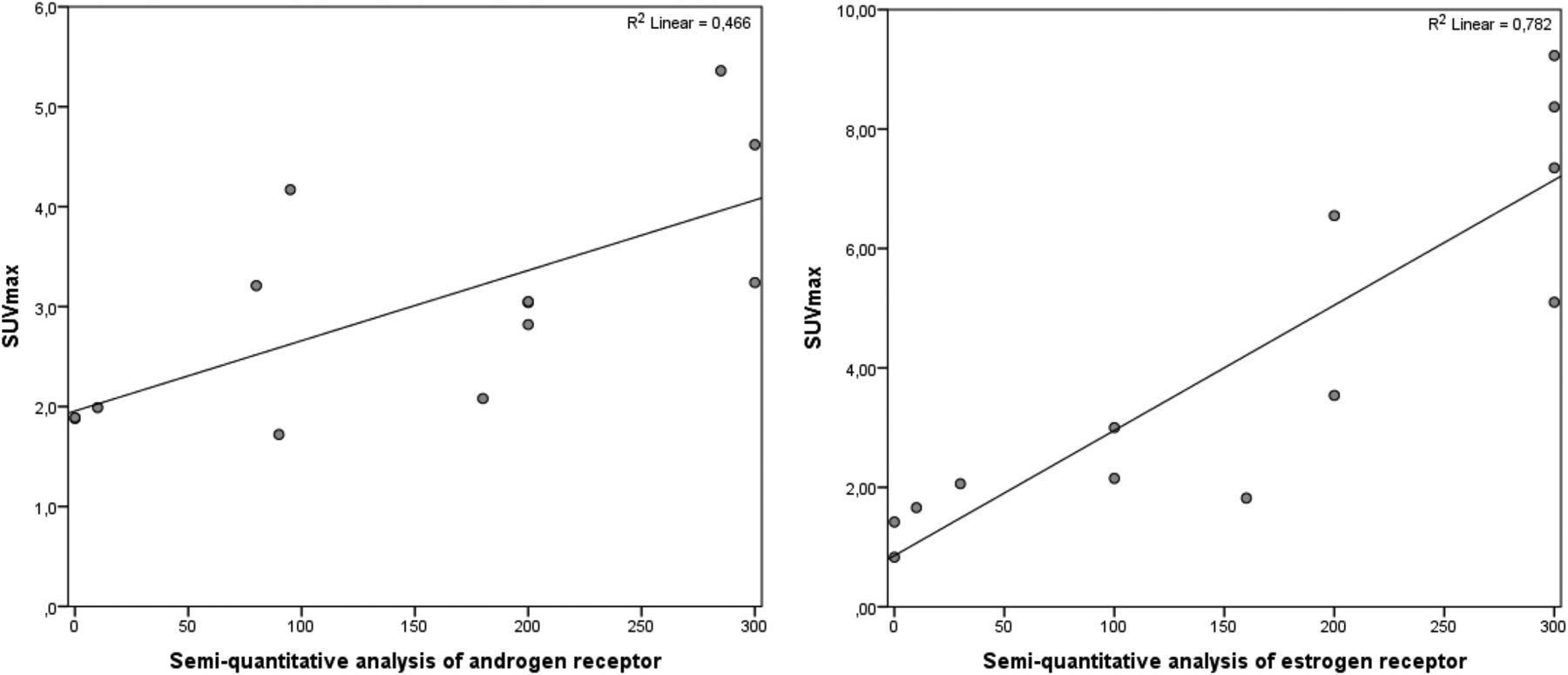
- FIGURE 2.
Correlation plot of semiquantitative analysis of receptor status and SUVmax as measured by PET scan for AR (left) and ER (right).
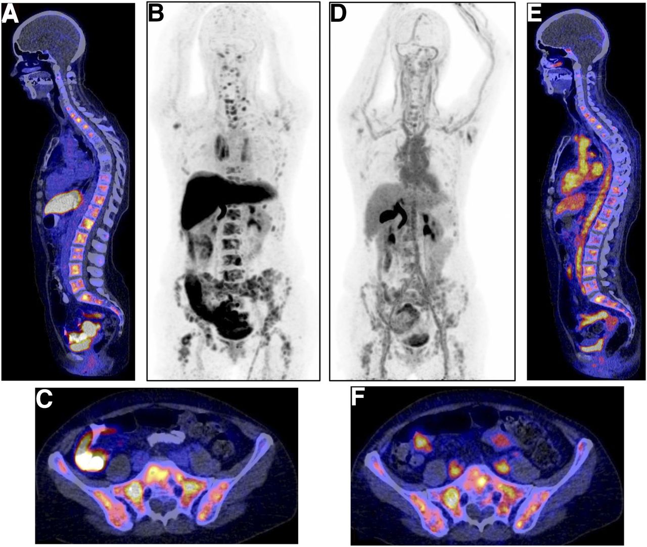
- FIGURE 3.
Example of typical 18F-FES (A–C) and 18F-FDHT (D–F) distribution in same patient with multiple bone metastases. (A) Sagittal 18F-FES PET/CT fusion image with physiologic uptake in liver, small intestine, and urinary tract and pathologic uptake in multiple vertebra. (B) 18F-FES PET maximum-intensity-pixel format to allow visualization of biodistribution of 18F-FES tracer. (C) Horizontal 18F-FES PET/CT fusion image with physiologic uptake in small intestine and pathologic uptake throughout pelvic bones. (D) Maximum-intensity-pixel format of 18F-FDHT PET scan, with physiologic uptake in blood pool of heart and liver and excretion via bile to small intestine, and urinary tract. (E) Sagittal 18F-FDHT PET/CT fusion image with physiologic uptake and pathologic uptake in multiple vertebrae. (F) Horizontal 18F-FES PET/CT fusion image with physiologic uptake in large vessels and small intestines and pathologic uptake throughout pelvic bones.
- FIGURE 4.
Distribution of SUVmax per lesion per patient measured by 18F-FES PET. Lesions are divided into bone (blue), lymph nodes (red), lung (green), and others (purple). Orange circles are biopsied lesions. Blue boxes indicate ER-positive biopsies (>1% staining); numbers indicate score of biopsy (i.e., intensity times percentage positive cells). Dashed line indicates threshold set based on receiver-operating-characteristic analysis. White boxes indicate negative biopsies (<1% staining).
- FIGURE 5.
Distribution of SUVmax per lesion per patient measured by 18F-FDHT PET. Lesions are divided into bone (blue), lymph nodes (red), lung (green), and others (purple). Orange circles are biopsied lesions. Orange boxes indicate AR-positive biopsies (i.e., >10% staining); white boxes indicate negative biopsies. Numbers in boxes indicate score of biopsy (i.e., intensity times percentage positive cells). Dashed line indicates threshold set based on receiver-operating-characteristic analysis.


Tables
Characteristic n % Mean age (y) 64 Sex Female 11 85 Male 2 15 Primary tumor characteristics IHC ER+/AR+ 13 100 ER+/AR− 0 ER−/AR+ 0 ER−/AR− 0 Primary tumor stage T1N0M0 4 31 T1N1M0 1 8 T2N0M0 4 31 T2N1M0 1 8 T3N2M0 3 23 Metastatic tumor characteristics IHC ER+/AR+ 10 77 ER+/AR− 1 8 ER−/AR+ 1 8 ER−/AR− 1 8 Treatment at time of 18F-FES and 18F-FDHT PET scans Aromatase inhibitor 5 38 Chemotherapy 4 31 None 4 31 ER+ = ER-positive; AR+ = AR-positive; ER− = ER-negative; AR− = AR-negative; IHC = immunohistochemistry.
Supplemental Data
Files in this Data Supplement:


Jump to section
Related Articles
Cited By...
- Report on the PET/CT Image-Based Radiation Dosimetry of [18F]FDHT in Women, a Validated Imaging Agent with New Applications for Evaluation of Androgen Receptor Status in Women with Metastatic Breast Cancer
- Report on the PET/CT Image-Based Radiation Dosimetry of [18F]FDHT in Women, a Validated Imaging Agent with New Applications for Evaluation of Androgen Receptor Status in Women with Metastatic Breast Cancer
- Summary: Appropriate Use Criteria for Estrogen Receptor-Targeted PET Imaging with 16{alpha}-18F-Fluoro-17{beta}-Fluoroestradiol
- Imaging Androgen Receptors in Breast Cancer with 18F-Fluoro-5{alpha}-Dihydrotestosterone PET: A Pilot Study
- Breast Cancer: Evaluating Tumor Estrogen Receptor Status with Molecular Imaging to Increase Response to Therapy and Improve Patient Outcomes
- Recent Advances in Imaging Steroid Hormone Receptors in Breast Cancer