
Main menu

  • Home
  • Content
    • Current
    • Ahead of print
    • Past Issues
    • JNM Supplement
    • SNMMI Annual Meeting Abstracts
    • Continuing Education
    • JNM Podcasts
  • Subscriptions
    • Subscribers
    • Institutional and Non-member
    • Rates
    • Journal Claims
    • Corporate & Special Sales
  • Authors
    • Submit to JNM
    • Information for Authors
    • Assignment of Copyright
    • AQARA requirements
  • Info
    • Reviewers
    • Permissions
    • Advertisers
  • About
    • About Us
    • Editorial Board
    • Contact Information
  • More
    • Alerts
    • Feedback
    • Help
    • SNMMI Journals
  • SNMMI
    • JNM
    • JNMT
    • SNMMI Journals
    • SNMMI

User menu

  • Subscribe
  • My alerts
  • Log in
  • My Cart

Search

  • Advanced search
Journal of Nuclear Medicine
  • SNMMI
    • JNM
    • JNMT
    • SNMMI Journals
    • SNMMI
  • Subscribe
  • My alerts
  • Log in
  • My Cart
Journal of Nuclear Medicine

Advanced Search

  • Home
  • Content
    • Current
    • Ahead of print
    • Past Issues
    • JNM Supplement
    • SNMMI Annual Meeting Abstracts
    • Continuing Education
    • JNM Podcasts
  • Subscriptions
    • Subscribers
    • Institutional and Non-member
    • Rates
    • Journal Claims
    • Corporate & Special Sales
  • Authors
    • Submit to JNM
    • Information for Authors
    • Assignment of Copyright
    • AQARA requirements
  • Info
    • Reviewers
    • Permissions
    • Advertisers
  • About
    • About Us
    • Editorial Board
    • Contact Information
  • More
    • Alerts
    • Feedback
    • Help
    • SNMMI Journals
  • View or Listen to JNM Podcast
  • Visit JNM on Facebook
  • Join JNM on LinkedIn
  • Follow JNM on Twitter
  • Subscribe to our RSS feeds
Research ArticleImaging/Infection

Imaging Macrophage Accumulation in a Murine Model of Chronic Pancreatitis with 125I-Iodo-DPA-713 SPECT/CT

Catherine A. Foss, Liansheng Liu, Ronnie C. Mease, Haofan Wang, Pankaj Pasricha and Martin G. Pomper
Journal of Nuclear Medicine October 2017, 58 (10) 1685-1690; DOI: https://doi.org/10.2967/jnumed.117.189571
Catherine A. Foss
1Russell H. Morgan Department of Radiology and Radiological Science, Johns Hopkins Medical Institutions, Baltimore, Maryland; and
  • Find this author on Google Scholar
  • Find this author on PubMed
  • Search for this author on this site
Liansheng Liu
2Center for Neurogastroenterology, Department of Medicine, Johns Hopkins Medical Institutions, Baltimore, Maryland
  • Find this author on Google Scholar
  • Find this author on PubMed
  • Search for this author on this site
Ronnie C. Mease
1Russell H. Morgan Department of Radiology and Radiological Science, Johns Hopkins Medical Institutions, Baltimore, Maryland; and
  • Find this author on Google Scholar
  • Find this author on PubMed
  • Search for this author on this site
Haofan Wang
1Russell H. Morgan Department of Radiology and Radiological Science, Johns Hopkins Medical Institutions, Baltimore, Maryland; and
  • Find this author on Google Scholar
  • Find this author on PubMed
  • Search for this author on this site
Pankaj Pasricha
2Center for Neurogastroenterology, Department of Medicine, Johns Hopkins Medical Institutions, Baltimore, Maryland
  • Find this author on Google Scholar
  • Find this author on PubMed
  • Search for this author on this site
Martin G. Pomper
1Russell H. Morgan Department of Radiology and Radiological Science, Johns Hopkins Medical Institutions, Baltimore, Maryland; and
  • Find this author on Google Scholar
  • Find this author on PubMed
  • Search for this author on this site
  • Article
  • Figures & Data
  • Supplemental
  • Info & Metrics
  • PDF
Loading

Article Figures & Data

Figures

  • Additional Files
  • FIGURE 1.
    • Download figure
    • Open in new tab
    • Download powerpoint
    FIGURE 1.

    125I-iodo-DPA-713 SPECT/CT of treated (Cer) and control (vehicle) mice. Coregistered images show radiotracer uptake in inflamed pancreas (arrows) and duodenum of treated mouse but not in either tissue of control mouse. Stomach is delineated by dotted circles. Gastrointestinal uptake of radiotracer is specific for peritoneal macrophages. Scale unit is %ID/g. D = duodenum; GI = gastrointestinal tract; K = kidney.

  • FIGURE 2.
    • Download figure
    • Open in new tab
    • Download powerpoint
    FIGURE 2.

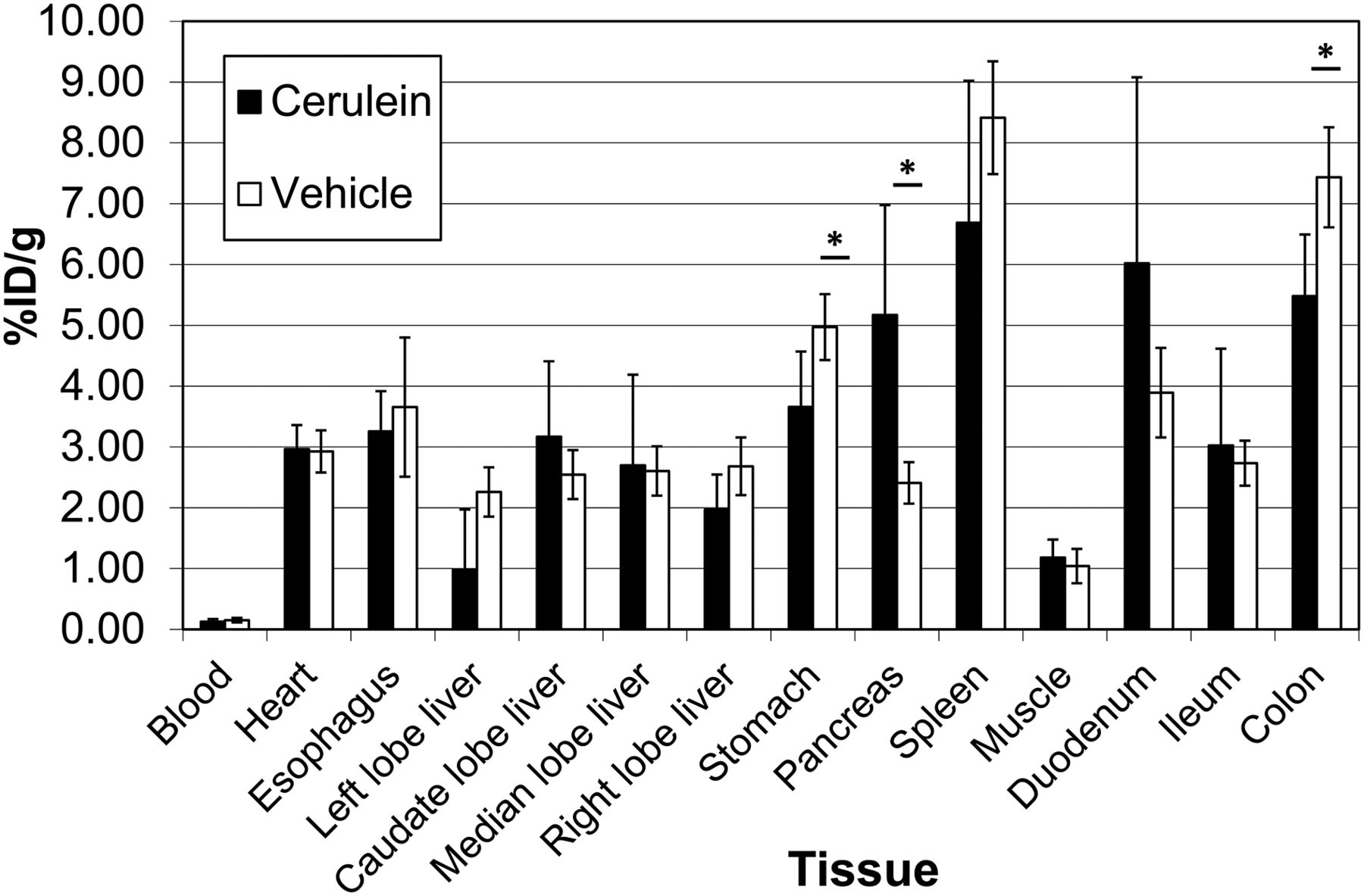
    Ex vivo biodistribution in 4 treated (cerulein) and 4 control (vehicle) mice injected intravenously with 259 kBq (7 μCi) of 125I-iodo-DPA-713. The indicated tissues were collected and weighed, and their radioactivity was counted along with a standard dose for comparison. Data are mean ± SD. *P < 0.05.
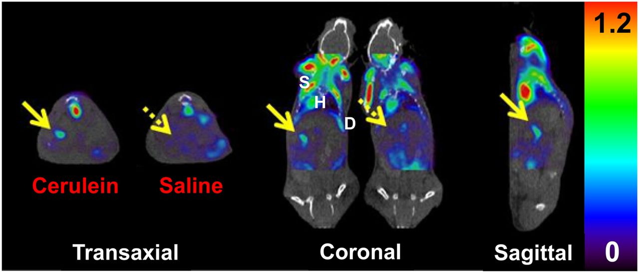
  • FIGURE 3.
    • Download figure
    • Open in new tab
    • Download powerpoint
    FIGURE 3.

    18F-FDG PET/CT of treated (cerulein) and control (saline) mice. Coregistered images show radiotracer uptake in inflamed pancreas (solid arrow) of cerulein-treated mouse, with reduced background uptake (dashed arrow) seen in control mouse. Prominent uptake is also apparent in skeletal muscle, heart, and diaphragm. Scale unit is SUV (%ID/g/body weight). D = diaphragm; H = heart; S = skeletal muscle.
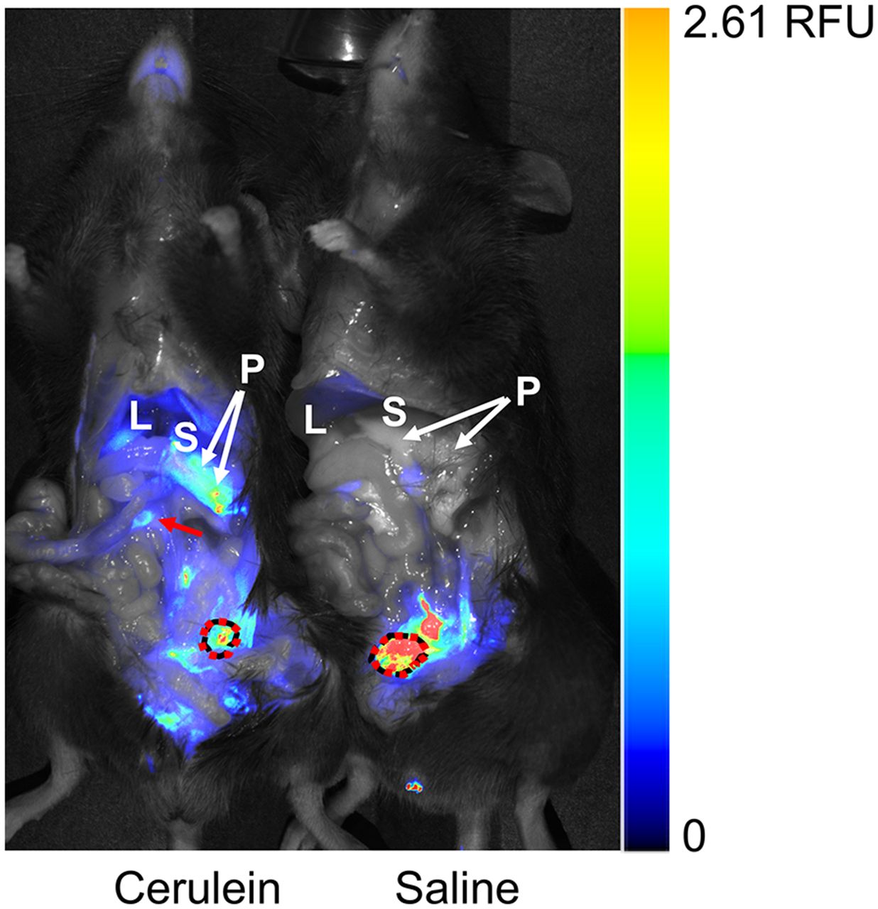
  • FIGURE 4.
    • Download figure
    • Open in new tab
    • Download powerpoint
    FIGURE 4.

    Ex vivo near-infrared fluorescence imaging of DPA-713-IRDye800CW in treated (cerulein) and control (saline) mice. Fluorescent tracer uptake is seen in pancreas, gastrointestinal tract (arrow), and urinary bladder (dotted circle) of treated mouse but in only urinary bladder of control mouse. Adjacent stomach is labeled for context. Color bar is relative fluorescence units (RFU). L = liver; P = pancreas; S = stomach.
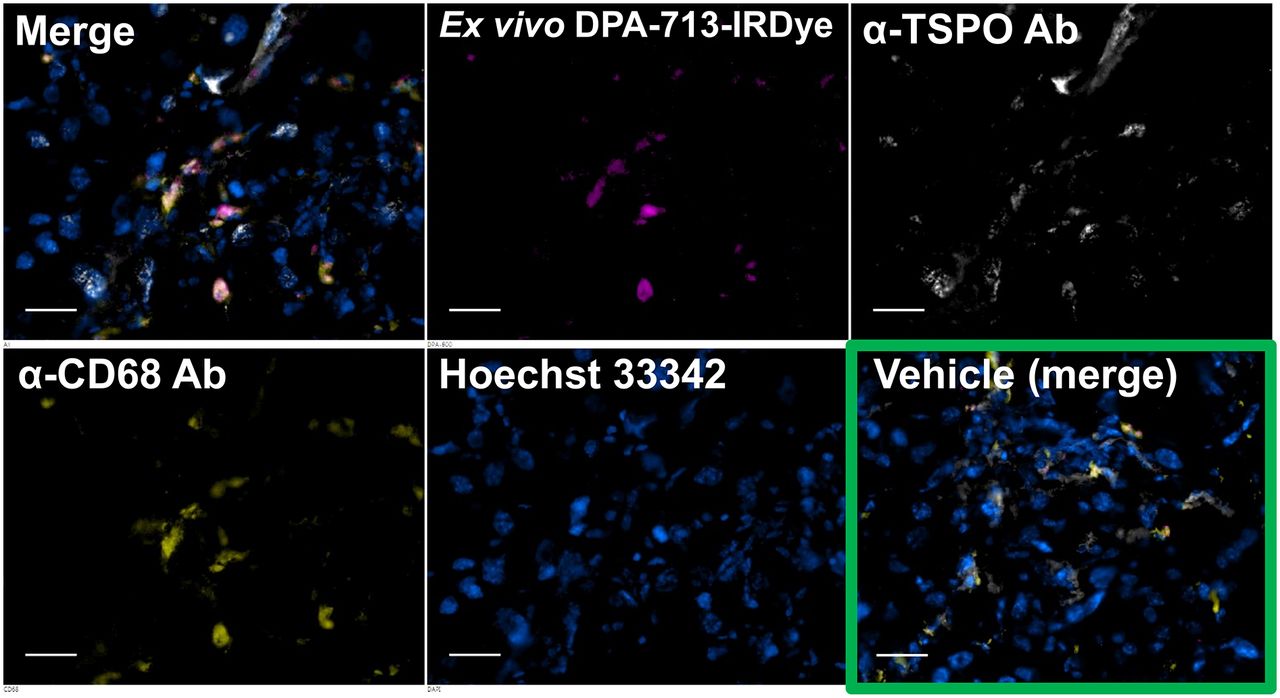
  • FIGURE 5.
    • Download figure
    • Open in new tab
    • Download powerpoint
    FIGURE 5.

    Ex vivo near-infrared fluorescence microscopy of DPA-713-IRDye800CW in pancreatic sections from mice in Figure 4. Separate and merged images are shown for treated mouse, whereas only merged image is shown for control mouse (vehicle). Treated pancreas has abundant fluorescent tracer uptake (magenta) in CD68-positive phagocytic cells (yellow) along with abundant TSPO expression (white), whereas control pancreas has almost no phagocytic cells present, small amounts of TSPO expression, and no visible fluorescent probe uptake. Scale bar = 50 μm. Ab = antibody.

Additional Files

  • Figures
  • Supplemental Data

    Files in this Data Supplement:

    • Supplemental Data
PreviousNext
Back to top

In this issue

Journal of Nuclear Medicine: 58 (10)
Journal of Nuclear Medicine
Vol. 58, Issue 10
October 1, 2017
  • Table of Contents
  • Table of Contents (PDF)
  • About the Cover
  • Index by author
Print
Download PDF
Article Alerts
Sign In to Email Alerts with your Email Address
Email Article

Thank you for your interest in spreading the word on Journal of Nuclear Medicine.

NOTE: We only request your email address so that the person you are recommending the page to knows that you wanted them to see it, and that it is not junk mail. We do not capture any email address.

Enter multiple addresses on separate lines or separate them with commas.
Imaging Macrophage Accumulation in a Murine Model of Chronic Pancreatitis with 125I-Iodo-DPA-713 SPECT/CT
(Your Name) has sent you a message from Journal of Nuclear Medicine
(Your Name) thought you would like to see the Journal of Nuclear Medicine web site.
Citation Tools
Imaging Macrophage Accumulation in a Murine Model of Chronic Pancreatitis with 125I-Iodo-DPA-713 SPECT/CT
Catherine A. Foss, Liansheng Liu, Ronnie C. Mease, Haofan Wang, Pankaj Pasricha, Martin G. Pomper
Journal of Nuclear Medicine Oct 2017, 58 (10) 1685-1690; DOI: 10.2967/jnumed.117.189571

Citation Manager Formats

  • BibTeX
  • Bookends
  • EasyBib
  • EndNote (tagged)
  • EndNote 8 (xml)
  • Medlars
  • Mendeley
  • Papers
  • RefWorks Tagged
  • Ref Manager
  • RIS
  • Zotero
Share
Imaging Macrophage Accumulation in a Murine Model of Chronic Pancreatitis with 125I-Iodo-DPA-713 SPECT/CT
Catherine A. Foss, Liansheng Liu, Ronnie C. Mease, Haofan Wang, Pankaj Pasricha, Martin G. Pomper
Journal of Nuclear Medicine Oct 2017, 58 (10) 1685-1690; DOI: 10.2967/jnumed.117.189571
Twitter logo Facebook logo LinkedIn logo Mendeley logo
  • Tweet Widget
  • Facebook Like
  • Google Plus One
Bookmark this article

Jump to section

  • Article
    • Abstract
    • MATERIALS AND METHODS
    • RESULTS
    • DISCUSSION
    • CONCLUSION
    • DISCLOSURE
    • Acknowledgments
    • Footnotes
    • REFERENCES
  • Figures & Data
  • Supplemental
  • Info & Metrics
  • PDF

Related Articles

  • This Month in JNM
  • PubMed
  • Google Scholar

Cited By...

  • No citing articles found.
  • Google Scholar

More in this TOC Section

Imaging/Infection

  • Specific Imaging of Bacterial Infection Using 6″-18F-Fluoromaltotriose: A Second-Generation PET Tracer Targeting the Maltodextrin Transporter in Bacteria
  • Evaluation of Spleen Glucose Metabolism Using 18F-FDG PET/CT in Patients with Febrile Autoimmune Disease
Show more Imaging/Infection

Basic

  • Specific Imaging of Bacterial Infection Using 6″-18F-Fluoromaltotriose: A Second-Generation PET Tracer Targeting the Maltodextrin Transporter in Bacteria
  • Evaluation of Spleen Glucose Metabolism Using 18F-FDG PET/CT in Patients with Febrile Autoimmune Disease
  • Dopamine D1 Receptor Agonist PET Tracer Development: Assessment in Nonhuman Primates
Show more Basic

Similar Articles

Keywords

  • macrophages
  • DPA-713
  • pancreatitis
  • SPECT-CT
SNMMI

© 2025 SNMMI

Powered by HighWire