
Main menu

  • Home
  • Content
    • Current
    • Ahead of print
    • Past Issues
    • JNM Supplement
    • SNMMI Annual Meeting Abstracts
    • Continuing Education
    • JNM Podcasts
  • Subscriptions
    • Subscribers
    • Institutional and Non-member
    • Rates
    • Journal Claims
    • Corporate & Special Sales
  • Authors
    • Submit to JNM
    • Information for Authors
    • Assignment of Copyright
    • AQARA requirements
  • Info
    • Reviewers
    • Permissions
    • Advertisers
  • About
    • About Us
    • Editorial Board
    • Contact Information
  • More
    • Alerts
    • Feedback
    • Help
    • SNMMI Journals
  • SNMMI
    • JNM
    • JNMT
    • SNMMI Journals
    • SNMMI

User menu

  • Subscribe
  • My alerts
  • Log in
  • My Cart

Search

  • Advanced search
Journal of Nuclear Medicine
  • SNMMI
    • JNM
    • JNMT
    • SNMMI Journals
    • SNMMI
  • Subscribe
  • My alerts
  • Log in
  • My Cart
Journal of Nuclear Medicine

Advanced Search

  • Home
  • Content
    • Current
    • Ahead of print
    • Past Issues
    • JNM Supplement
    • SNMMI Annual Meeting Abstracts
    • Continuing Education
    • JNM Podcasts
  • Subscriptions
    • Subscribers
    • Institutional and Non-member
    • Rates
    • Journal Claims
    • Corporate & Special Sales
  • Authors
    • Submit to JNM
    • Information for Authors
    • Assignment of Copyright
    • AQARA requirements
  • Info
    • Reviewers
    • Permissions
    • Advertisers
  • About
    • About Us
    • Editorial Board
    • Contact Information
  • More
    • Alerts
    • Feedback
    • Help
    • SNMMI Journals
  • View or Listen to JNM Podcast
  • Visit JNM on Facebook
  • Join JNM on LinkedIn
  • Follow JNM on Twitter
  • Subscribe to our RSS feeds
Meeting ReportInstrumentation & Data Analysis Track

A pre-clinical PET scanner based on a single piece of annular scintillator: a simulation study

Alexander Stolin, Peter Martone, Gangadhar Jaliparthi and Raymond Raylman
Journal of Nuclear Medicine May 2016, 57 (supplement 2) 1952;
Alexander Stolin
2Radiology West Virginia University Morgantown WV United States
  • Find this author on Google Scholar
  • Find this author on PubMed
  • Search for this author on this site
Peter Martone
2Radiology West Virginia University Morgantown WV United States
  • Find this author on Google Scholar
  • Find this author on PubMed
  • Search for this author on this site
Gangadhar Jaliparthi
2Radiology West Virginia University Morgantown WV United States
  • Find this author on Google Scholar
  • Find this author on PubMed
  • Search for this author on this site
Raymond Raylman
1Accounts Payable Morgantown WV United States
  • Find this author on Google Scholar
  • Find this author on PubMed
  • Search for this author on this site
  • Article
  • Figures & Data
  • Info & Metrics
Loading

Abstract

1952

Objectives One challenge of pre-clinical PET imaging is the amount of scan time required to produce high quality, high-resolution images. This issue is important because many pre-clinical studies require the rapid scanning of numerous animals. To create a very high sensitivity and high resolution pre-clinical PET scanner, one based on a single scintillator crystal formed into an annulus is proposed. The objective of this study was to evaluate, via simulation, the potential performance of this system.

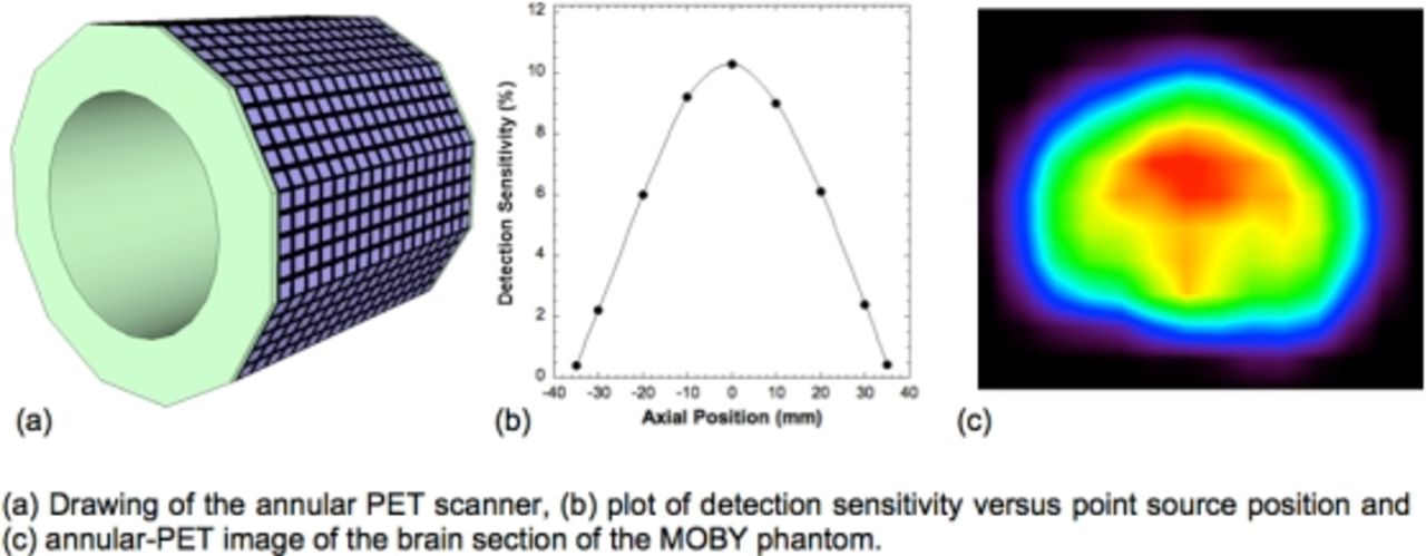
Methods The simulated scanner consisted of a 72.7mm long cylindrical piece of LYSO crystal with a diameter of 75 mm. The cylinder was formed into an annulus by machining a hole (diameter of 50mm) in the center of the scintillator. Twelve flat surfaces were formed on the outer surface of the annulus to accommodate light detection devices. Specifically, an array of 5 by 17 discrete SiPMs with a size of 3mmx3mm (pitch of 4.1mm, similar to the SensL C-series SiPMs) was attached to each facet. Evaluation of scanner performance was carried out in three steps. First, GATE simulation software was used to obtain a list mode distribution of detected 511keV photons. Next, a trial GATE data set was used in conjunction with the DETECT2000 optical simulation software to explore light transport in the detector. From these results, the three-dimensional positions of the simulated positron annihilations in the scintillator were calculated from the detected optical photons distribution detected at the surfaces of the SiPMs (depth of the events were estimated by the shape of the light distribution). Calculated positions were compared to original emission points to determine spatial accuracy of the event reconstruction. Finally, the data were histogrammed into 100 radial and 180 angular bins and reconstructed using the SSRB-FBP algorithm. Images from a point source and the brain section of the MOBY phantom were created. Data from the point source were used to measure spatial resolution and detection sensitivity.

Results Event positioning was accurate to within 1.9mm in the tangential and radial directions. Interaction depth resolution was ~4.4mm. Detection sensitivity at the center of the scanner was 10.3% which is more than twice of any pre-clinical scanner of comparable axial size and diameter. Spatial resolution at the center of the scanner was 1.04mm FWHM. Finally, images of the MOBY mouse brain phantom resulted in good results for FBP reconstruction.

Conclusions The results from this initial study demonstrated the promise of a solid, annular pre-clinical PET scanner.

Figure
  • Download figure
  • Open in new tab
  • Download powerpoint
Previous
Back to top

In this issue

Journal of Nuclear Medicine
Vol. 57, Issue supplement 2
May 1, 2016
  • Table of Contents
  • Index by author
Article Alerts
Sign In to Email Alerts with your Email Address
Email Article

Thank you for your interest in spreading the word on Journal of Nuclear Medicine.

NOTE: We only request your email address so that the person you are recommending the page to knows that you wanted them to see it, and that it is not junk mail. We do not capture any email address.

Enter multiple addresses on separate lines or separate them with commas.
A pre-clinical PET scanner based on a single piece of annular scintillator: a simulation study
(Your Name) has sent you a message from Journal of Nuclear Medicine
(Your Name) thought you would like to see the Journal of Nuclear Medicine web site.
Citation Tools
A pre-clinical PET scanner based on a single piece of annular scintillator: a simulation study
Alexander Stolin, Peter Martone, Gangadhar Jaliparthi, Raymond Raylman
Journal of Nuclear Medicine May 2016, 57 (supplement 2) 1952;

Citation Manager Formats

  • BibTeX
  • Bookends
  • EasyBib
  • EndNote (tagged)
  • EndNote 8 (xml)
  • Medlars
  • Mendeley
  • Papers
  • RefWorks Tagged
  • Ref Manager
  • RIS
  • Zotero
Share
A pre-clinical PET scanner based on a single piece of annular scintillator: a simulation study
Alexander Stolin, Peter Martone, Gangadhar Jaliparthi, Raymond Raylman
Journal of Nuclear Medicine May 2016, 57 (supplement 2) 1952;
Twitter logo Facebook logo LinkedIn logo Mendeley logo
  • Tweet Widget
  • Facebook Like
  • Google Plus One
Bookmark this article

Jump to section

  • Article
  • Figures & Data
  • Info & Metrics

Related Articles

  • No related articles found.
  • Google Scholar

Cited By...

  • No citing articles found.
  • Google Scholar

More in this TOC Section

Instrumentation & Data Analysis Track

  • Deep Learning Based Kidney Segmentation for Glomerular Filtration Rate Measurement Using Quantitative SPECT/CT
  • The Benefit of Time-of-Flight in Digital Photon Counting PET Imaging: Physics and Clinical Evaluation
  • Preclinical validation of a single-scan rest/stress imaging technique for 13NH3 cardiac perfusion studies
Show more Instrumentation & Data Analysis Track

MTA II: Instrumentation Posters

  • Dedicated cardiac SPECT imaging with multi-pinhole collimators on a clinical scanner
  • Hybrid X-ray Fluorescence, Luminescence and Transmission Computed Tomography for Image-Guided Nanoparticle Mediated X-ray Micro-Beam Therapy
  • An analysis of side readouts of monolithic scintillation crystals
Show more MTA II: Instrumentation Posters

Similar Articles

SNMMI

© 2025 SNMMI

Powered by HighWire