


Abstract
1346
Objectives 3-[18F]fluoro-α-methyl-L-tyrosine (FAMT) and 4-borono-2-[18F]fluoro-L-phenylalanine (FBPA) were both reported as tumor selective tracer, which accumulate through L-type amino acid transporter 1 (LAT1). We performed the dynamic PET of FAMT, FBPA, and 15O-water to estimate the effect of tumor blood flow on the accumulation of these tracers.
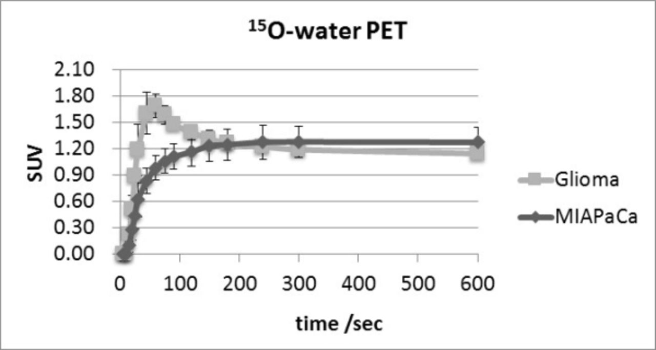
Methods Rats (F344 rats, male, 8 weeks old, 213.1±27.0 g) were prepared as xenograft model of C6 glioma (n=12, 14 days after the implantation) and MIAPaCa (human pancreatic cancer cell line) (n=4, 28 days after the implantation). Dynamic PET scans were performed just after injection of FAMT (33.4±7.7 MBq) or FBPA (27.3±5.1 MBq) up to 70min, 15O-water (63.1±13.3 MBq) up to 10min under isoflurane anesthesia by small animal PET. Regions of interest (ROIs) were placed on the tumor of each PET image and the time activity curves (TACs) were compared between glioma and MIAPaCa. We calculated K1 and k2 from the 15O-water PET using one tissue compartment model analysis by PMOD 3.4, in which ROIs of left ventricle cavity were placed as image derived input function. K1 (an index of tumoral blood flow) and k2 were compared by t-test.
Results TAC of FAMT showed a rapid increase and continuous wash-out pattern in glioma, while low uptake and slow wash-out pattern in MIAPaCa. Tumoral uptake showed marked difference in the early phase (SUV at 10min = 3.20±0.42 in glioma and 1.11±0.15 in MIAPaCa), whereas no significant difference was observed in the late phase (SUV at 70min = 1.16±0.16 in glioma and 1.09±0.07 in MIAPaCa). Similar uptake patterns were observed in TAC of FBPA for both glioma and MIAPaCa (SUV at 10min = 3.17±0.39 in glioma and 1.11±0.16 in MIAPaCa) although kinetic of FBPA was slow compared to FAMT. K1 of glioma (0.93±0.45 ml/ccm/min) was significantly higher than that of MIAPaCa (0.31±0.07) (p<0.01), and k2 of glioma (1.51±0.75 /min) was also significantly higher than that of MIAPaCa (0.42±0.13) (p<0.01). It was suggested that difference in tumoral blood flow and wash-out of the tumor affected the kinetic patterns of glioma and MIAPaCa.
Conclusions The SUV ratio of glioma to MIAPaCa during first 10min was close to the K1 ratio of glioma to MIAPaCa, suggesting the accumulations of FAMT and FBPA into the malignant tumors were mainly influenced by the tumoral blood flow in the early phase. $$graphic_011D658E-B56B-4AA0-B31B-9348FE626064$$ $$graphic_3BB62279-A3CA-41E5-BA8C-ED1D2B4FBA44$$ $$graphic_93152865-8E84-41A7-9A7C-F2C48FF6EAA6$$
Tumoral blood flow volume
SUV at 10 min in malignant tumors


In this issue


Jump to section
Related Articles
Cited By...
- No citing articles found.