


Article Figures & Data
Figures
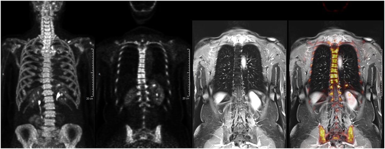
- FIGURE 1.
Normal coronal 18F-NaF PET/MR images representing (from left to right) PET maximum-intensity projection, PET coronal slice, T1-weighted MR image, and fusion of PET with MRI.
- FIGURE 2.
73-y-old man (serum PSA, 38 ng/mL) with history of 2 negative TRUS-guided prostate biopsies. (A) Axial T2-weighted MRI shows hypointense lesion in midline anterior transition zone (A) (long arrows); short arrow indicates benign hyperplastic lesion. (B and C) Lesion had restricted diffusion on apparent-diffusion-coefficient maps (B) and DW MRI (b = 2,000 s/mm2) (C) (arrows). (D) On permeability map derived from dynamic contrast-enhanced MRI, lesion—in comparison with remainder of prostate—has increased vascularity (arrows). (E and F) 18F-DCFBC PET (E) and software-based PET/MRI fusion (F) images show specific uptake within midline anterior transition zone lesion (long arrows); benign hyperplastic nodule in left transition zone did not show any 18F-DCFBC uptake (short arrow). TRUS/MRI fusion–guided biopsy of this lesion revealed prostate adenocarcinoma with Gleason scores of 4 and 5.
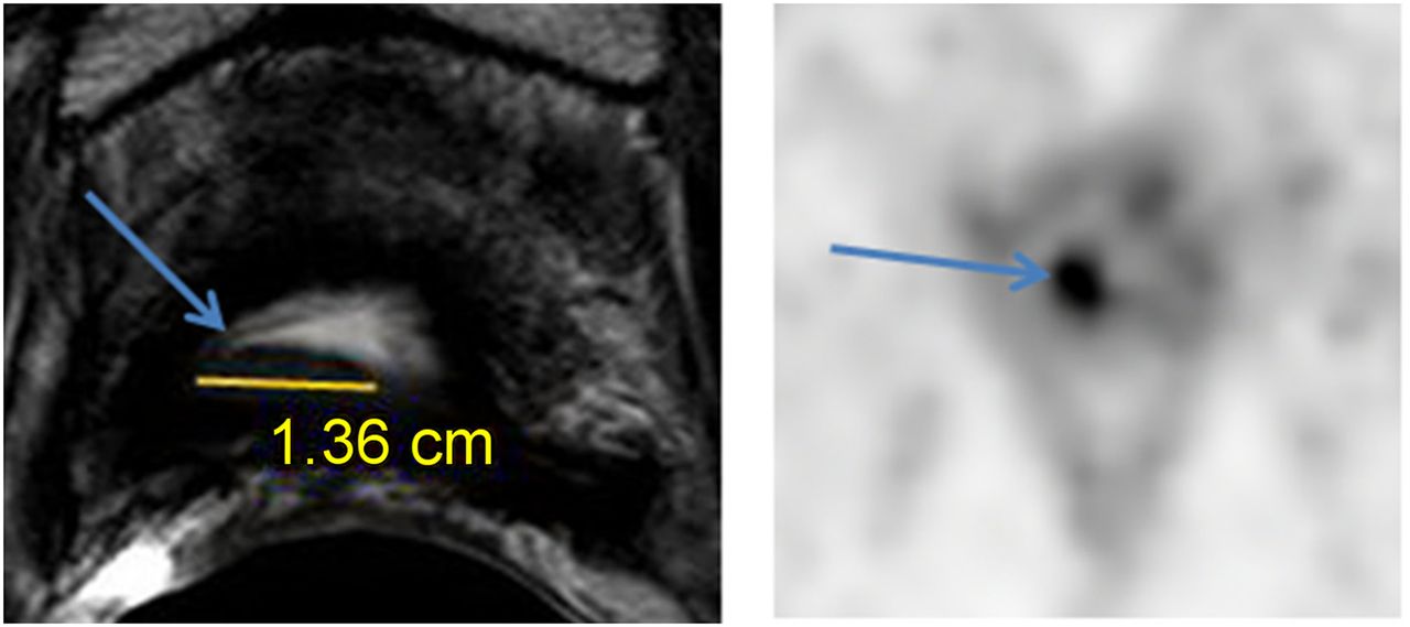
- FIGURE 3.
64-y-old man with history of PCa treated by radical prostatectomy 4 y earlier and current biochemical recurrence (PSA, 0.61 ng/mL). Suggestive findings in prostate fossa on MRI (left) matched uptake on 18F-DCFBC PET (right) (arrows).

Tables
PET tracer Mechanism 18F-FDG Glucose metabolism 11C- or 18F-acetate Lipid metabolism 11C- or 18F-choline Lipid metabolism 11C-methionine Amino acid transport 18F-FACBC Amino acid transport 18F-DCFBC, 18F-DCFPyL, 64Cu- or 89Zr-J591, and 68Ga-PSMA compounds: 68Ga-DKFZ-PSMA-1 (same as 68Ga-PSMA-HBED-CC), 68Ga-PSMA-DKFZ-617, and 68Ga-PSMA I&T PSMA inhibitors/antibodies 18F-FDHT Androgen receptor 18F-FLT Cell proliferation 18F-FMAU Cell proliferation 18F-NaF Calcium analog 68Ga-bombesin Gastrin-releasing peptide receptor 18F-FACBC = 1-amino-3-18F-fluorocyclobutane-1-carboxylic acid; 18F-DCFPyL = 2-(3-(1-carboxy-5-[(6-18F-fluoro-pyridine-3-carbonyl)-amino]-pentyl)-ureido)-pentanedioic acid; DKFZ = Deutsches Krebsforschungszentrum (German Cancer Research Center); I&T = imaging and therapy; 18F-FDHT = 16α-18F-fluoro-5α-dihydrotestosterone; 18F-FLT = 3′-deoxy-3′-18F-fluorothymidine; 18F-FMAU = 1-(2′-deoxy-2′-18F-fluoro-β-d-arabinofuranosyl)thymine.


