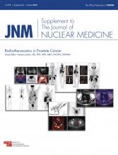
This special supplemental issue of The Journal of Nuclear Medicine focuses on the current state of affairs and outlook for the use of molecular imaging and targeted radionuclide therapy in prostate cancer. The recent strides in understanding the complex biology of prostate cancer and advances in the design and production of targeted radiotheranostics have fueled much exciting work in translational research in this important clinical setting. These efforts are anticipated to facilitate precision and personalized medical care for the individual patient with suspected or known prostate cancer.
Following this introductory overview, this supplemental issue incorporates 18 articles by leading international authorities in nuclear medicine, radiology, medical oncology, radiation oncology, and urology. Wolfgang Weber and Michael Morris from the Memorial Sloan Kettering Cancer Center in New York start off the issue by presenting a brief review of the progress that has been made over the past several years in molecular imaging and radionuclide therapy of prostate cancer (1). They point out the challenges associated with comparing the diagnostic performance of various molecular imaging agents, the effect of new imaging results on shifting the clinical state of disease and its consequence on clinical trials, and how emerging targeted radionuclide therapies could fit into the current treatment regimen armamentarium.
Next, a team of 3 authors from the University of Southern California Norris Comprehensive Cancer Center in Los Angeles representing medical oncology, radiation oncology and urology review the unmet clinical needs that can be addressed uniquely by the advances in molecular imaging and its anticipated effect on the evolution of clinical management in prostate cancer (2). The supplemental issue then continues with a review of specific imaging modalities and agents. Frank Chen and colleagues from the University of Southern California review the use of ultrasonography, including the newer techniques with contrast agents and elastography in prostate biopsy and imaging evaluation after focal treatment of localized prostate cancer (3).
Given that bone is a major site of metastatic disease from prostate cancer, the next article, by the University of Southern California and Stanford groups, focuses on bone-targeted scintigraphic imaging and radionuclide therapy for bone pain palliation and direct bone marrow tumor cell killing (4). Then I begin the discussion on PET with an article on imaging glucose metabolism with the most common PET radiotracer, 18F-FDG, and imaging cellular proliferation with PET radiotracers that track the thymidine salvage pathway of DNA synthesis (5). An article from the University of California, Los Angeles, follows with a discussion on the potential use and limitations of 11C-acetate as a PET imaging agent tracking tumor lipogenesis (6). Another PET radiotracer for imaging tumor lipogenesis is choline, which may be radiolabeled with either 11C or 18F. There is relatively robust published material on the use of radiolabeled choline in all clinical phases of prostate cancer. The next 4 articles presented are from colleagues in Europe with the most experience in the clinical use of radiolabeled choline. The article from the investigators at Rostock University in Germany focuses on diagnosis and initial staging (7). The Italian researchers from Milan and Bologna summarize the data on the most common use of radiolabeled choline in the imaging evaluation of biochemical recurrence after definitive therapy for primary prostate cancer (8). Another paper from Italy, led by Stefano Fanti, describes the potential value of choline in treatment planning for salvage radiotherapy and salvage lymph node dissection as well as response assessment of systemic therapies and outcome prediction (9). The last of the choline papers is from the St. Vincent’s Hospital in Austria and compares 18F-fluorocholine and 18F-NaF in the imaging assessment of bone metastases from prostate cancer (10).
Over the past few years, there have been major exciting developments in the synthesis and evaluation of PET radiotracers based on other biologic targets in prostate cancer. David Schuster from Emory University in Atlanta is the lead author on an article on the use of radiolabeled amino acids, in particular, anti-1-amino-3-18F-fluorocyclobutane-1-carboxylic acid (fluciclovine), which received approval from the U.S. Food and Drug administration on May 27, 2016, for PET imaging in men with biochemical recurrence after definitive treatment for primary prostate cancer (11). The 4-author team of investigators from Germany, Japan, and Stanford present the next paper, which is on the potential clinical utility of radiotracers targeted to the gastrin-releasing peptide receptors (12). Then, the group from Memorial Sloan Kettering Cancer Center, led by Steven Larson, discusses the development and evaluation of androgen receptor axis–based radiotracers in the biologic context of castration-resistant metastatic prostate cancer (13). The next 3 articles focus on the exciting results from theranostics based on prostate-specific membrane antigen (PSMA) as the biomarker. First, Ali Afshar-Oromieh and colleagues from Heidelberg, Germany, provide an overview of PSMA ligands in the imaging and targeted radionuclide therapy of prostate cancer (14). The second article in this series—from the groups of investigators at Johns Hopkins University, the University Hospital of Cologne, and Duke University—focuses on small-molecule PSMA inhibitors, in particular those radiolabeled with 18F (15). The third and final paper on PSMA theranostics, led by Richard Baum, reviews in general terms the Bad Berka group’s overall experience with over 400 administrations of PSMA-based radioligand therapy in men with castration-resistant metastatic prostate cancer (16). As the authors contend, further prospective randomized clinical trials will be needed to compare the effectiveness of PSMA-based radioligand therapy to other current and novel treatments in metastatic prostate cancer and to address such issues as treatment timing, sequence, and combination.
The supplemental issue continues with an article from colleagues at Massachusetts General Hospital on the potential role of nanoparticles in prostate cancer theranostics (17). The final article in this supplemental issue is from the investigators at the U.S. National Institutes of Health, led by Peter Choyke, on the potential utility of integrated PET/MRI systems in providing combined multiparametric MR and physiologic PET imaging data in one imaging session (18). Based on the information presented in this special supplemental issue, it is reasonable to envision that in the near future, theranostics will become commonplace in the personalized care of men with prostate cancer. However, a greater number of prospective studies will be needed, particularly in the areas of comparative effectiveness and cost effectiveness, and important issues such as availability, accessibility, and the hurdles of regulatory approval and adequate reimbursement will also need to be worked out.
In closing, I would like to thank all the authors and reviewers for sharing their expertise in the preparation of a concise yet comprehensive review of this important subject matter. I also appreciate the administrative help of the JNM editorial office staff, Susan Alexander and Josh Wachtel, and the advice of the JNM editor, Dominique Delbeke. My hope is that JNM readers will find this supplemental issue on prostate cancer helpful, interesting, and stimulating.
- © 2016 by the Society of Nuclear Medicine and Molecular Imaging, Inc.
REFERENCES
- Received for publication September 1, 2016.
- Accepted for publication September 1, 2016.


