


Article Figures & Data
Figures
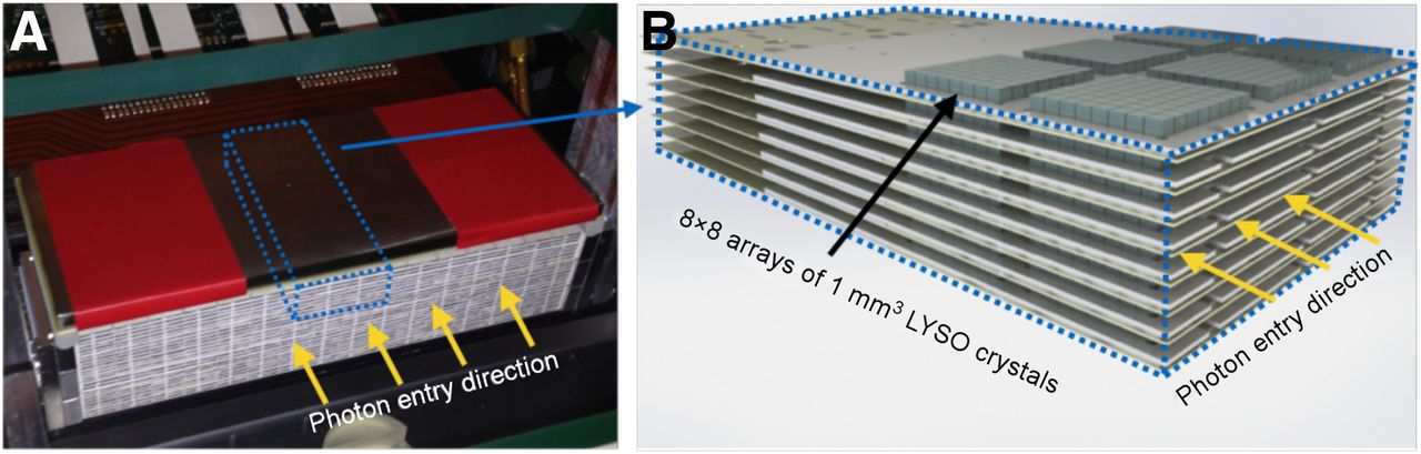
- FIGURE 1.
Stanford’s 1-mm-resolution, 3-dimensional position-sensitive PET scintillation detectors. (A) One panel from actual system, showing edge-on photon entry from imaging FOV. (B) Magnified section depicting edge-on orientation of detectors with respect to incoming photons, allowing for direct measurement of one or more photon DOI locations. LYSO = lutetium-yttrium oxyorthosilicate.
- FIGURE 2.
Correlation between crystal size and reconstructed spatial resolution along one dimension for breast-dedicated PEM/PET system designs presented in Table 1 that use discrete crystal elements.
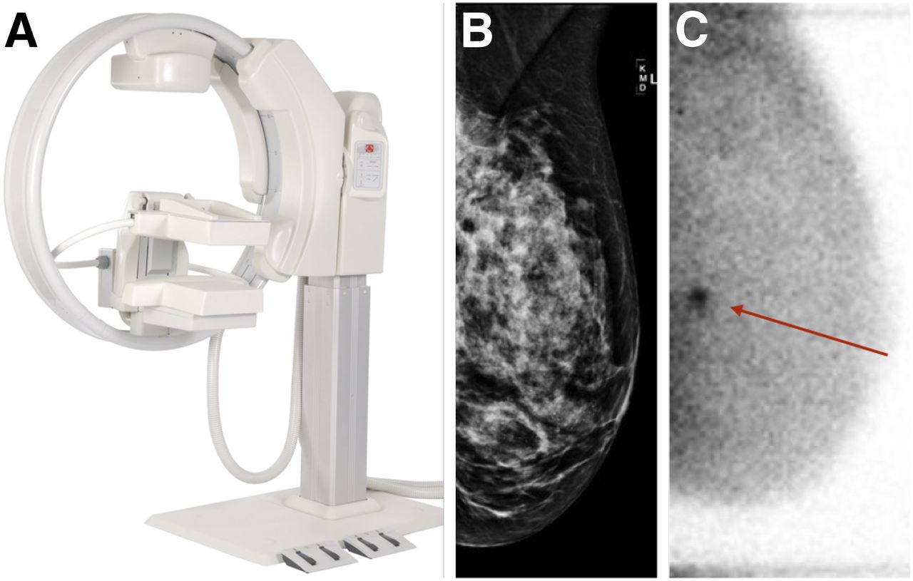
- FIGURE 3.
(A) Gamma Medica’s LumaGEM MBI system. (B) Negative routine digital mammogram interpretation for asymptomatic 55-y-old woman with heterogeneously dense breasts. (C) Referral for MBI secondary screening shows lesion with high uptake. Biopsy showed it to be invasive ductal carcinoma. (Panel A courtesy of Gamma Medica, Inc.; panels B and C courtesy of Dr. Robin Sermis, ProMedica Toledo Hospital.)
- FIGURE 4.
(A) Duke’s SPECT/CT system. (B) Coronal view (C) and volume rendering of patient scan taken with 790-MBq injection of 99mTc-sestamibi. Large arrows point to lesion surgically confirmed as ductal carcinoma in situ. Small arrow points to posteriorly located biopsy clip. Myocardial uptake inside chest wall and external scanner fiducial markers outside breast periphery are both visible in C. (Courtesy of Dr. Martin Tornai, Duke Multi-Modality Imaging Lab.)
- FIGURE 5.
(A) Naviscan’s PEM Flex Solo II system. (B) Biopsy guidance with attachments to align and guide needle. (C) PEM scan of patient with microinvasive ductal carcinoma in situ. (D) Image of alignment line source and lesion during biopsy. (E) Image of removed sample. (Courtesy of Dr. Kathy Schilling, Boca Raton Regional Hospital.)


Tables
- TABLE 1
Selected Published BD PET System Specifications and Imaging Performance Statistics
Spatial resolution at center of FOV (mm) Group Geometry FOV (mm) Crystal type Crystal size (mm) Photodetector Radial (x) Tangential (y) Axial (z) DOI resolution Energy resolution Timing resolution/window Sensitivity (%) PEMi (14) Annular 110 d, 128 a LYSO 1.9 × 1.9 × 15 PS PMT 1.58* 1.41 1.31 None NA NA/6 ns 6.88% Shimadzu (8,41) Annular (O) 180 d, 155.5 a (O) LGSO 1.44 × 1.44 × 4.5 PS PMT 1.6* 1.7 2.0 4.5 mm 16.9% (both) 1.2 ns/NA (both) 16.3% (O) C-ring (C) 179 d, 105 a (C) 6.9% (C) MAMMI (2,42) Annular, translatable 170 d, 170 a‡ LYSO Monolithic, 40 × 40 × 10 PS PMT 1.6† 1.8 1.9 4 mm 18% NA/5 ns 3.6% Texas HOTPET (43) Annular, reconfigurable 540 d, 210 a (breast); 830 d, 130 a (whole body) BGO 2.68 × 2.68 × 18 PMT 2.7 (breast), 3.3 (whole body) (averaged)* NA 2.6 (both) None NA NA/15 ns 9.2% (breast); 4.2% (whole body) Brookhaven PET/MR (10) Annular, multimodal 145.3 d, 96.46 a LYSO 2.2 × 2.2 × 15 APD 1.2† 1.1 NA None NA NA NA Lawrence Berkeley (16) Rectangular 82 x, 60 y, 50 z LSO 3 × 3 × 30 PMT and SiPD 1.9 mm† (direction unclear) NA NA 3.8 mm 24%–51% along crystal 3.4 ns/6 ns 4.94% West Virginia University PEM/PET (38) Rotating panel, rectangular 150 x, 150 y, 150 z LYSO 2 × 2 × 15 PS PMT 2.01† 2.04 1.84 None NA 3.5 ns 6.88% Clear-PEM (4,5) Rotating panel 162 x, 141 y LYSO 2 × 2 × 20 APD 1.4†§ (direction unclear) NA 1.4 2.5 mm 13% 4 ns 4.3%§ UC Davis PET/CT (6) Rotating panel, multimodal 119 x, 119 y LSO 3 × 3 × 20 PS PMT 2.70† 2.73 2.17 None 25% NA/12 ns 1.64% M.D. Anderson (13) Dual-panel 200 x, 120 y LYSO 1.54 × 1.54 × 10 PMT 1.19* 2.01 4.10 None 17% NA/7.5 ns 8.9% (avg separation) PEM I (19) Dual-panel 72 x, 72 y BGO 1.9 × 1.9 × 6.5 PS PMT 2.8* (direction unclear) NA NA 6.5 mm 53% 12 ns/NA 3% Pisa (44) Dual-panel 100 x, 100 y LYSO 1.9 × 1.9 × 16 PS PMT NA NA NA None 20% 9.1 ns/NA NA Thomas Jefferson Lab (45) Dual-panel 150 x, 200 y LGSO 3.03 × 3.03 × 10 PS PMT 4.1 mm (10° acceptance)* NA NA None NA NA 0.07% (10°) 4.7 mm (40° acceptance) NA NA 1.35% (40°) maxPET (20) Dual-panel 150 x, 150 y LSO 3 × 3 × 20 PS PMT 2.26 (intrinsic), 4 (imaging)* NA NA None 21.6% 8.1 ns/NA 0.57% Stanford (11,46) Dual-panel 160 x, 100 y LYSO 0.9 × 0.9 × 1 PS APD 0.9 mm† NA NA 1 mm 10.6% 15.7 ns/NA NA PEM Flex Solo II (47) Dual-panel, translatable 240 x, 163 y‡ LYSO 2 × 2 × 13 PS PMT 1.94† 1.59 6.45 None NA NA/12 ns 0.15% (normalized) West Virginia University PEM (7,48) Dual-panel or rotating panel 100 x, 100 y GSO 3.1 × 3.1 × 10 PS PMT 5.5† (rotating) 5.0 (rotating) NA (rotating) None 20% NA/10 ns 0.016% (3° acceptance) 3.7 (static) 3.7 (static) 8.9 (static) 0.07% (10° acceptance) University of Pennsylvania BPET (12) Dual-panel, curved 280 x, 210 y (active area) NaI (Tl) Curved plate detector, 35 × 23 cm surface area PMT 3.8† NA NA None 10% NA 0.34% (scanner incomplete) ↵* Filtered backprojection.
↵† Iterative reconstruction.
↵‡ FOV with translating detector heads.
↵§ Simulated.
d = diameter; a = axial; x/y/z = linear orthogonal axes; LYSO = lutetium-yttrium oxyorthosilicate; LGSO = lutetium gadolinium oxyorthosilicate; BGO = bismuth germanate; LSO = lutetium oxyorthosilicate; GSO = germanium oxyorthosilicate; PS = position sensitive; PMT = photomultiplier tube; APD = avalanche photodiode; SiPD = silicon photodiode.
- TABLE 2
Selected Published BD Single-Photon Radionuclide Imaging System Specifications and Imaging Performance Statistics
System Geometry FOV (mm) Patient orientation Crystal type Collimator type Collimator hole diameter/hole length/septal thickness (mm) Spatial resolution (mm) Sensitivity (cps/MBq) Energy resolution at 140 keV Dilon Diagnostics 6800 (49) Single-panel projection 200 x, 200 y Compression NaI Parallel-hole 1.22/25/0.15 4.2 (S/C, 3 cm) 31 (EW, 126–154 keV) 9.5% 1.22/36/0.375 4.0 (S/C, 3 cm) 16 (EW, 126–154 keV) Gamma Medica LumaGEM 3200S (50) Dual-panel projection 200 x, 160 y Compression CdZnTe Parallel-hole 1.22/9.4/0.15 5.6 (S/C, 3 cm) 510 (EW, 110–154 keV) 3.8% 2.5/25/0.30 4.8 (S/C, 3 cm) 176 (EW, 110–154 keV) GE Healthcare Discovery NM750b (50) Dual-panel projection 200 x, 200 y Compression CdZnTe Parallel-hole 2.1/21/0.40 4.6 (S/C, 3 cm) 318 (EW, 110–154 keV) 6.5% 2.26/34.7/0.24 4.4 (S/C, 3 cm) 149 (EW, 110–154 keV) Duke University SPECT/CT (22) Rotating 200 x, 160 y Pendant CdZnTe Parallel-hole 1.22/25.4/0.2 3.4 (S/C, 1 cm, planar); 2.7 (S/C, reconstructed at 3-mm rotation radius, sagittal and coronal) 37.9 (EW, 129–151 keV) 6.8% University of Naples SPECT/CT (21) Rotating 70 x, 70 y Pendant CdTe Pinhole 1.2-mm effective aperture diameter 5.1 (S/C, 3 cm); 7.2 (S/C, 5 cm) Not applicable Not applicable University of Virginia breast tomosynthesis (23,51) Limited rotation 203 x, 152 y Compression NaI (Tl) Parallel-hole 1.778/19.99/0.305 3.2 (intrinsic) 147 (EW, 126–154 keV) 17.5% x/y = linear orthogonal axes; EW = energy window; S/C = source-to-collimator distance.


