


Article Figures & Data
Figures
- FIGURE 1.
Visualization of VOI (red) fused on digital reference smoothed to PET resolution. AU = arbitrary units.
- FIGURE 2.
(A) MR and CT images of phantom. (B) MR-based μ-maps and digital reference. CT images show all phantom structures, whereas polymer structure is invisible on MR and is classified as air on MR-based μ-maps. UTE = ultrashort echo time.
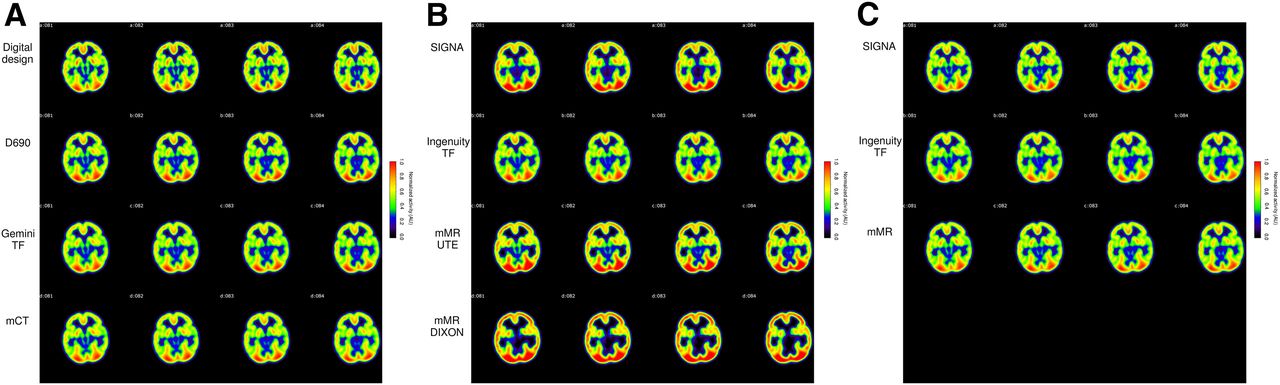
- FIGURE 3.
(A) Digital reference and PET/CT images. (B) PET/MR images with MRAC. (C) PET/MR images with CTAC. PET/CT and digital reference agree well. Effect of classifying polymer as air in MRAC can clearly be seen, whereas using CTAC for PET/MR brings PET/MR to agreement with PET/CT and digital reference. UTE = ultrashort echo time.
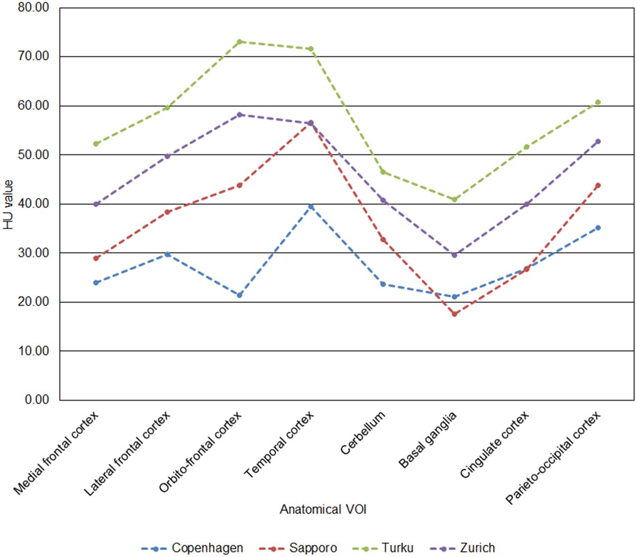
- FIGURE 4.
HUs as function of anatomic VOI at each institution. HUs show little variation, especially between Copenhagen, Turku, and Zurich, where same phantom was used.
- FIGURE 5.
Regional comparison of PET/CT and PET/MR systems (Eqs. 4 and 6). Mediofrontal cortex, lateral frontal cortex, cingulate cortex, and parietooccipital cortex agree well between systems. Ingenuity TF shows negative bias, whereas Gemini TF64 shows the largest variation between regions. MFC = medial frontal cortex; LFC = lateral frontal cortex; OFC = orbitofrontal cortex; TC = temporal cortex; Cer = cerebellum; BGa = basal ganglia; CC = cingulate cortex; POC = parietooccipital cortex.
- FIGURE 6.
Relative differences in PET/MR systems within institution by system (A) and by region (B) (Eq. 5). PET/MR systems agree well with PET/CT systems. Ingenuity TF shows the largest difference. MFC = medial frontal cortex; LFC = lateral frontal cortex; OFC = orbitofrontal cortex; TC = temporal cortex; Cer = cerebellum; BGa = basal ganglia; CC = cingulate cortex; POC = parietooccipital cortex.
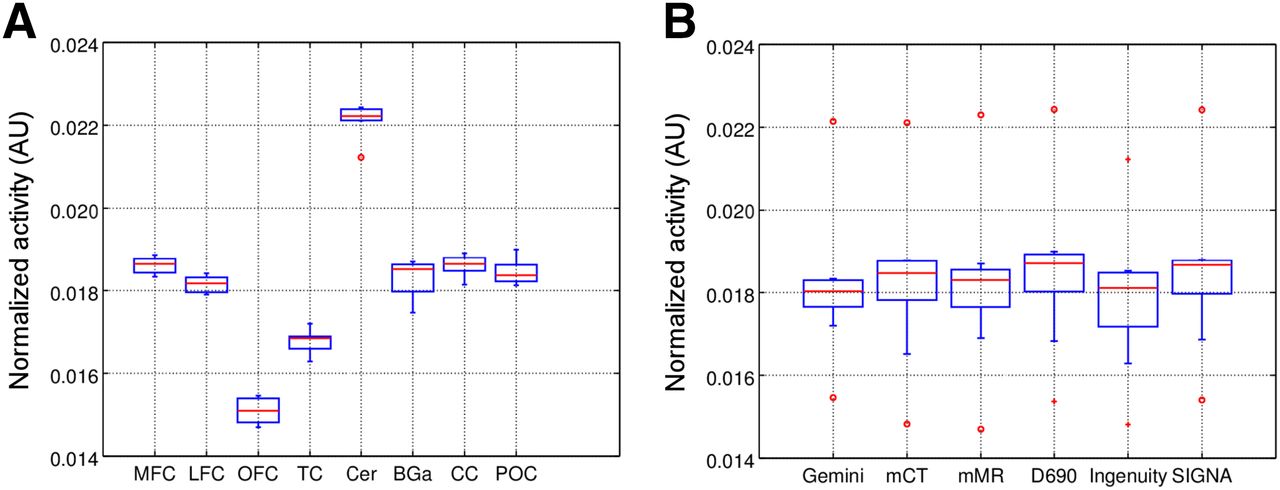
- FIGURE 7.
VOIs in box-and-whisker plot systemwise (A) and regionally (B). Excellent agreement is seen systemwise. Red bars denote median values, defining upper and lower quartiles. Whiskers indicate difference in quartiles by 1.5 times the interquartile range. Circles and crosses indicate outliers in each dataset. AU = arbitrary units; MFC = medial frontal cortex; LFC = lateral frontal cortex; OFC = orbitofrontal cortex; TC = temporal cortex; Cer = cerebellum; BGa = basal ganglia; CC = cingulate cortex; POC = parietooccipital cortex.


Tables
Institution and system Activity (MBq) Duration (min) Reconstruction algorithm (iterations/subsets) Matrix size (voxels) Voxel size (mm) Turku 97* Ingenuity TF 82 15 LOR RAMLA (10/33) 128 × 128 × 90 2 × 2 × 2 Discovery 690 48 20 3D OSEM (10/21) 256 × 256 × 47 1.38 × 1.38 × 3.27 Copenhagen 37* mMR 26 15 OP OSEM (8/21) 344 × 344 × 127 0.83 × 0.83 × 2 mCT 23 15 OP OSEM (8/24) 344 × 344 × 127 0.83 × 0.83 × 2 Zurich 78* Discovery 690 46 20 3D OSEM (3/18) 256 × 256 × 47 1.38 × 1.38 × 3.27 Signa 40 20 3D OSEM (2/28, 10/28) 256 × 256 × 89 1.17 × 1.17 × 2.8 Sapporo 50* Gemini TF64 39 15 LOR RAMLA (10/33) 128 × 128 × 90 2 × 2 × 2 ↵* Activity at injection time.
LOR = line of response; RAMLA = row action maximum likelihood; OSEM = ordered-subsets expectation maximization; OP = ordinary Poisson.
Method and institution Maximum Median Mean ± SD Automatic segmentation Turku 0.142 0.108 0.110 ± 0.012 Copenhagen 0.140 0.108 0.109 ± 0.011 Zurich 0.146 0.109 0.109 ± 0.016 Sapporo 0.162 0.108 0.112 ± 0.018 Manual VOI Turku 0.142 NA 0.0908 Copenhagen 0.140 NA 0.0912 Zurich 0.146 NA 0.0900 Sapporo 0.162 NA 0.0934 NA = not applicable.
Data are cm−1.
Supplemental Data
Files in this Data Supplement:


