


Article Figures & Data
Figures
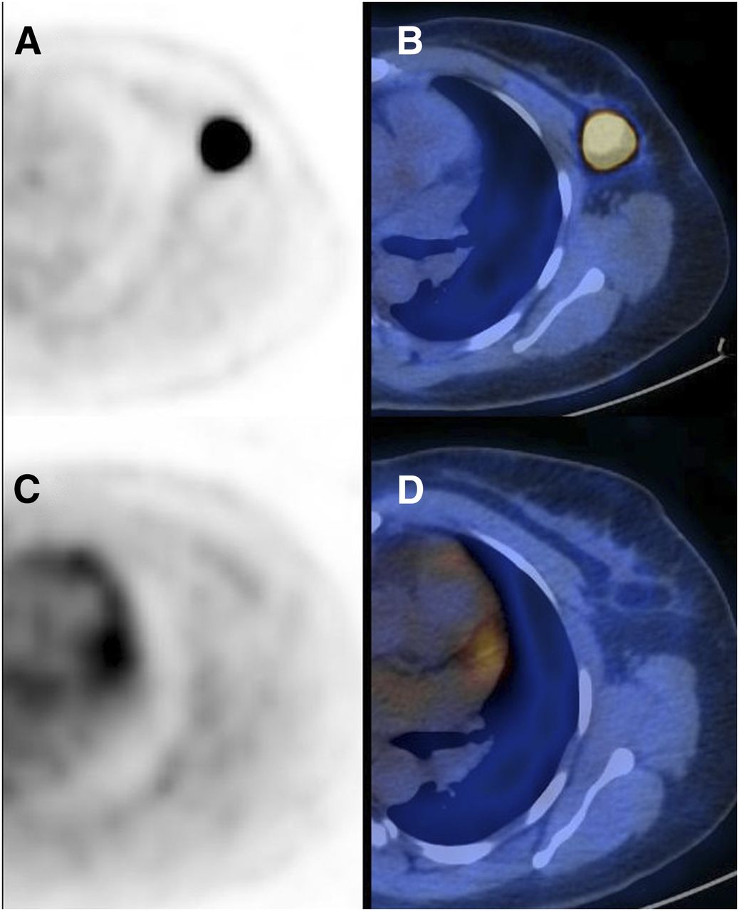
- FIGURE 1.
In 44-y-old patient with TNBC of left breast, transaxial PET (A) and PET/CT (B) images of primary tumor at baseline (SUVmax, 27.3) and after 2 cycles of SIM (C and D; SUVmax, 1.4). ΔSUVmax is −95%. No residual tumor was detected at surgery after 4 additional cycles of chemotherapy. Patient had no local or distant recurrence more than 1 y after surgery.
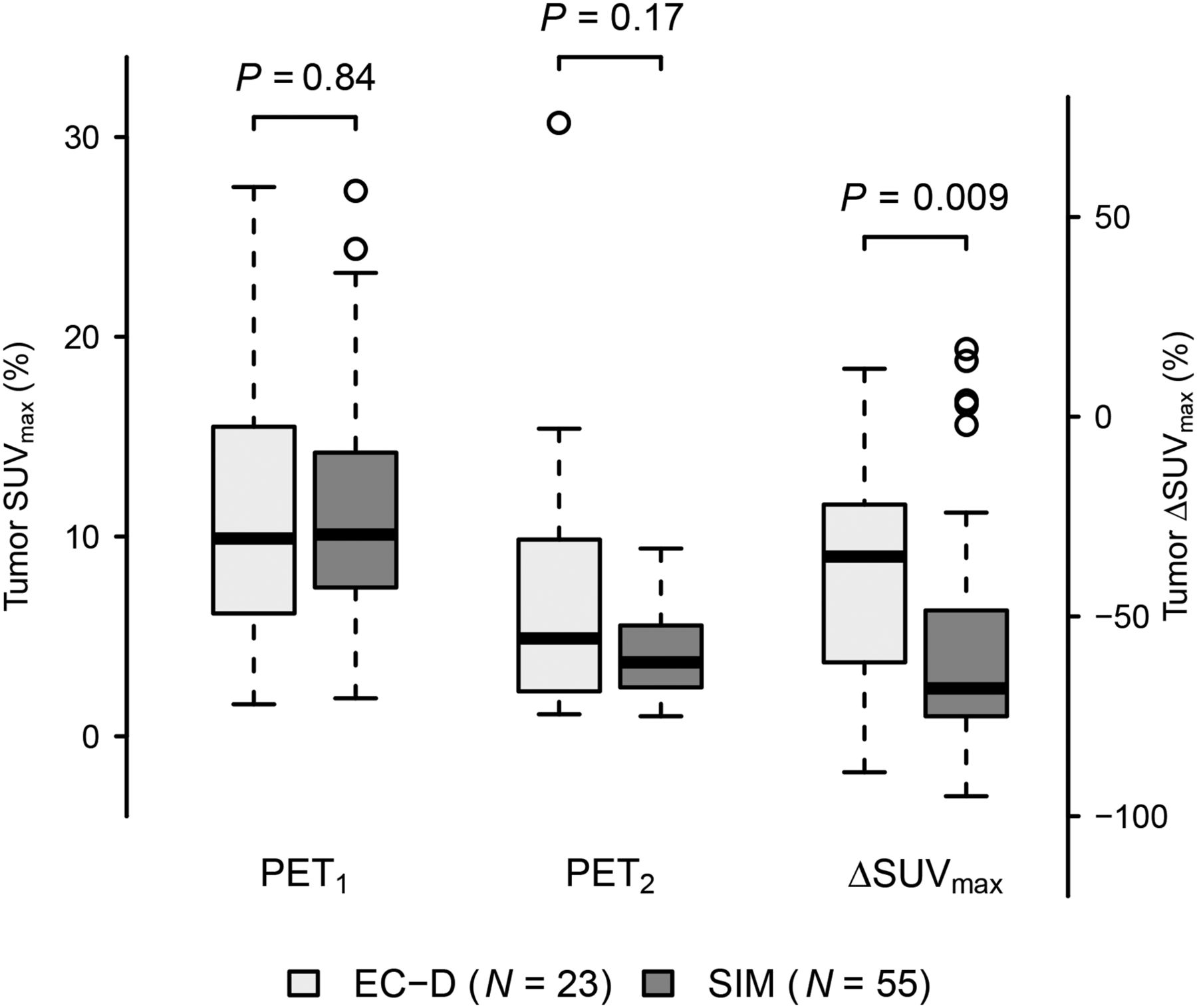
- FIGURE 2.
SUVmax of primary tumor at PET1 and PET2 and ΔSUVmax in EC-D and SIM groups.
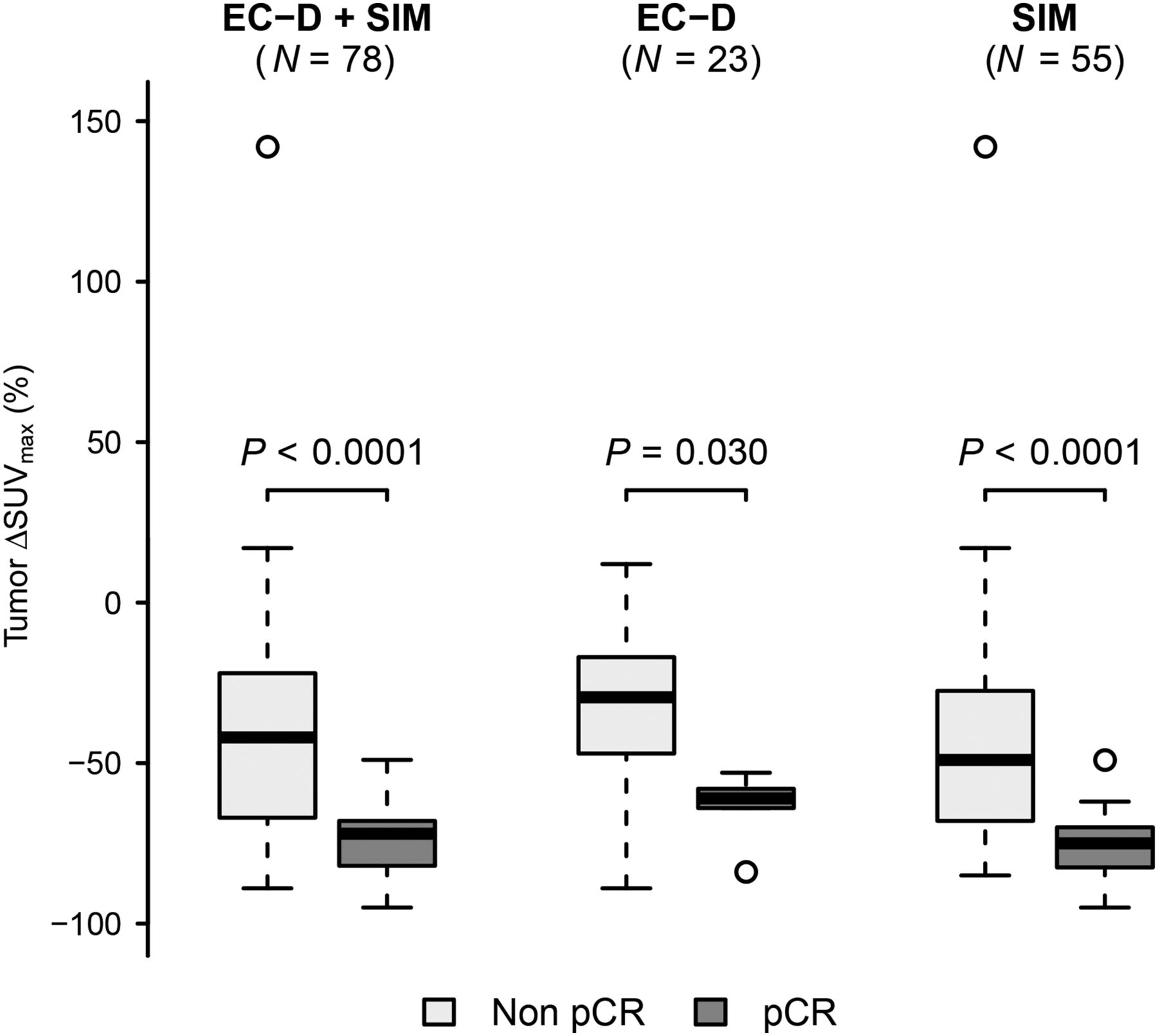
- FIGURE 3.
ΔSUVmax according to pathologic response after neoadjuvant chemotherapy (pCR vs. non-pCR) in whole population and in EC-D and SIM groups.
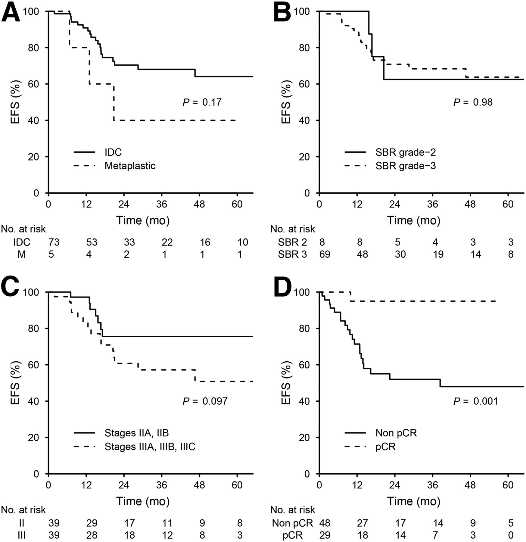
- FIGURE 4.
Kaplan–Meier curves for EFS in 78 patients according to tumor histology (A), SBR grade (B), AJCC stage (C), and pathology findings after neoadjuvant chemotherapy (D).
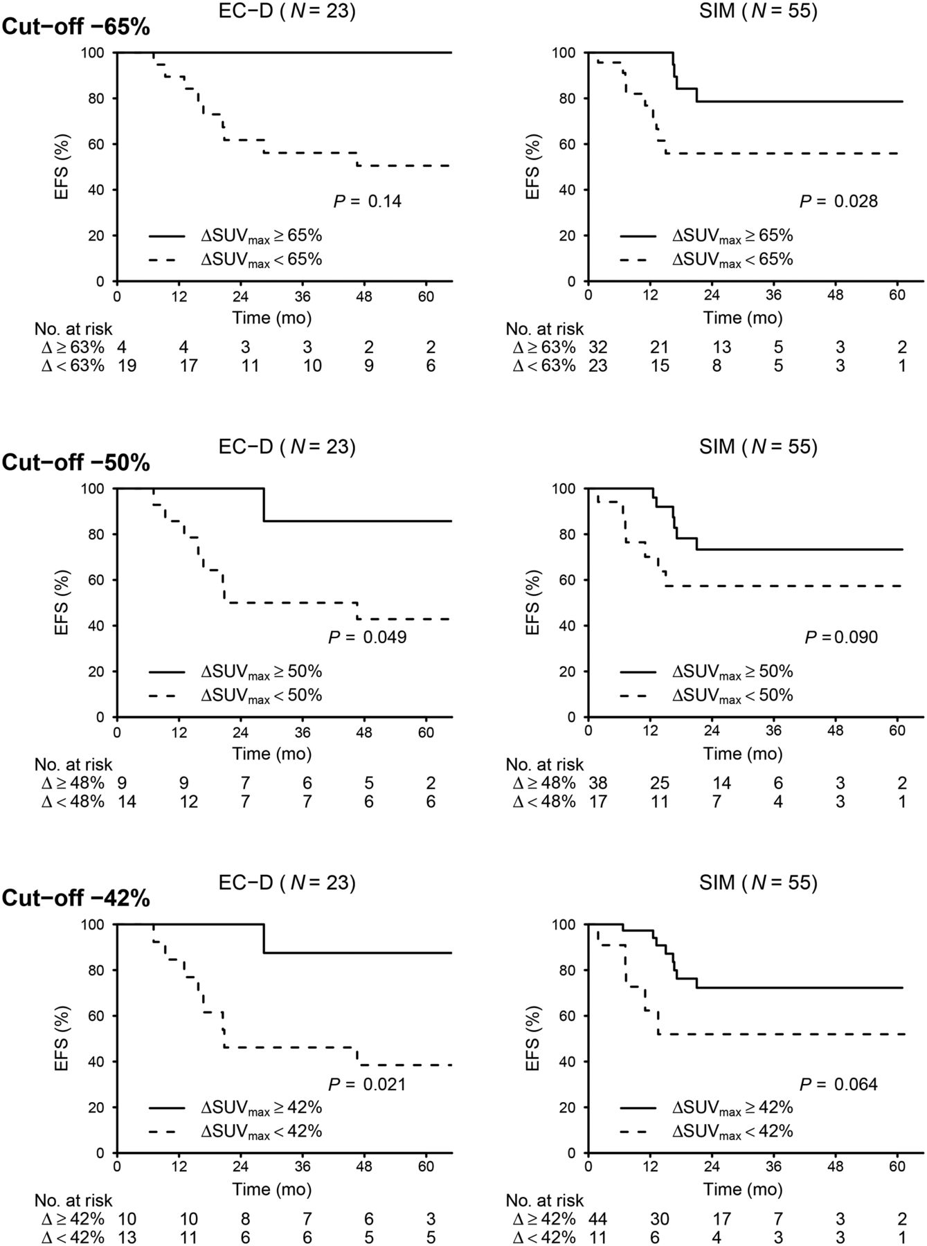
- FIGURE 5.
Kaplan–Meier curves for EFS according to metabolic response after 2 courses of neoadjuvant chemotherapy. Analysis was performed with 3 different ΔSUVmax cutoffs.


Tables
Variable Whole population EC-D group SIM group P Patients (n) 78 (100) 23 (29) 55 (71) Median age (y) 51 (27–78) 55 (38–78) 49 (27–71) 0.21 Median tumor size (mm) 50 (18–170) 50 (22–160) 45 (18–170) 0.46 Histology (n) 0.15 Invasive ductal carcinoma 73 (94) 20 (87) 53 (96) Metaplastic 5 (6) 3 (13) 2 (4) SBR grade (n) 0.039* 2 8 (10) 5 (23) 3 (5) 3 69 (90) 17 (77) 52 (95) Tumor classification† (n) 0.93 T1 1 (1) 0 (0) 1 (2) T2 36 (46) 10 (43) 26 (47) T3 22 (28) 7 (30) 15 (27) T4 19 (24) 6 (26) 13 (24) Lymph node classification† (n) 0.57 N0 32 (41) 11 (48) 21 (38) N1 27 (35) 6 (26) 21 (38) N2 15 (19) 4 (17) 11 (20) N3 4 (5) 2 (9) 2 (4) AJCC stage† (n) 0.83 IIA 21 (27) 7 (30) 14 (25) IIB 18 (23) 4 (17) 14 (25) IIIA 18 (23) 5 (22) 13 (24) IIIB 17 (22) 5 (22) 12 (22) IIIC 4 (5) 2 (9) 2 (4) Type of surgery (n) 0.62 Breast-conserving surgery 34 (44) 9 (39) 25 (46) Mastectomy 43 (56) 14 (61) 29 (54) Pathologic response (n) 0.078 Non-pCR 49 (63) 18 (78) 31 (56) pCR 29 (37) 5 (22) 24 (44) - TABLE 2
Clinical, Histologic, and Immunohistochemical Factors and PET Parameters According to Pathologic Response
Variable Non-pCR pCR P Patients (n) 49 (63) 29 (37) Median age (y) 50 (27–78) 51 (33–70) 0.78 Median tumor size (mm) 50 (18–160) 40 (21–170) 0.16 Histology (n) 0.15 Invasive ductal carcinoma 44 (90) 29 (100) Metaplastic 5 (10) 0 (0) Grade (n) 0.022* 2 8 (17) 0 (0) 3 40 (83) 29 (100) T-score (n) 0.003* T1 1 (2) 0 (0) T2 17 (35) 19 (66) T3 20 (41) 2 (7) T4 11 (22) 8 (28) N-score (n) 0.019* N0 19 (39) 13 (45) N1 13 (27) 14 (48) N2 14 (29) 1 (3) N3 3 (6) 1 (3) Stage (n) 0.031* IIA 9 (18) 12 (41) IIB 12 (24) 6 (21) IIIA 16 (33) 2 (7) IIIB 9 (18) 8 (28) IIIC 3 (6) 1 (3) SUVmax At PET1 Tumor 9 (2–28) 13 (5–27) 0.004* Axilla (n = 58) 6 (1–21) 5 (1–16) 0.22 Target 11 (2–28) 13 (5–27) 0.066* At PET2 Tumor 5 (1–31) 3 (1–10) 0.013* Axilla (n = 58) 2 (1–17) 1 (0.5–4) 0.001* Target 5 (1–31) 3 (1–10) 0.001* Difference (ΔSUVmax %) Tumor −42 (−89 to 142) −72 (−95 to −49) <0.0001* Axilla (n = 58) −53 (−90 to 0) −74 (−94 to 0) 0.21 Target −48 (−90 to 17) −74 (−95 to −49) <0.0001* ↵* Statistically significant.
Data in parentheses are percentage or range.
SUVmax AUC 95% confidence interval At PET1 Tumor 0.70 0.57–0.81 Axilla (n = 58) 0.60 0.45–0.74 Target* 0.62 0.49–0.74 Tumor + axilla† 0.71 0.59–0.82 At PET2 Tumor 0.67 0.55–0.79 Axilla (n = 58) 0.76 0.62–0.88 Target 0.72 0.6–0.83 Tumor + axilla 0.76 0.65–0.87 Difference (ΔSUVmax %) Tumor 0.86 0.77–0.93 Axilla (n = 58) 0.69 0.54–0.84 Target 0.82 0.72–0.9 Tumor + axilla 0.86 0.77–0.93 - TABLE 4
Comparison of Preparation Procedures, Some Patient Characteristics, and Instrumental Factors Between EC-D and SIM Groups
Variable EC-D group (n = 23) SIM group (n = 55) P Weeks between PET1 and PET2 8 (6–14) 6 (4–12) <0.0001 Weeks between PET1 and surgery 28 (10–37) 17 (14–37) <0.0001 Uptake time (min) PET1 69 (51–93) 70 (57–95) 0.49 PET2 64 (55–95) 67 (55–109) 0.25 Injected 18F-FDG dose (MBq) PET1 317 (249–486) 359 (248–476) 0.22 PET2 335 (272–503) 359 (210–494) 0.50 Patient weight (kg) PET1 65 (55–99) 70 (49–100) 0.22 PET2 66 (55–103) 68 (48–110) 0.68 Patient glycemia (mmol/L) PET1 5.8 (4.4–8.3) 5.3 (3.6–10.9) 0.082 PET2 5.4 (4.0–7.5) 5.5 (3.7–9.8) 0.71 Data in parentheses are range.
- TABLE 5
Performance of Various ΔSUVmax Cut-Offs in Predicting Non-pCR in EC-D and SIM Groups
EC-D SIM Cutoff R NR Acc Se Sp PPV NPV R NR Acc Se Sp PPV NPV −75 13 87 74 89 20 80 33 29 71 75 90 54 72 81 −70 17 83 70 83 20 79 25 47 53 82 81 83 86 77 −65 17 83 70 83 20 79 25 58 42 78 68 92 91 69 −60 30 70 74 78 60 88 43 62 38 78 65 96 95 68 −55 35 65 78 78 80 93 50 64 36 76 61 96 95 66 −50 39 61 83 78 100 100 56 69 31 71 52 96 94 61 −45 44 57 78 72 100 100 50 76 24 67 42 100 100 57 −40 48 52 74 67 100 100 46 82 18 62 32 100 100 53 R = metabolic responders (percentage of patients with ΔSUVmax ≥ cutoff); NR = metabolic nonresponders; Acc = accuracy; Se = sensitivity; Sp = specificity; PPV = positive predictive value; NPV = negative predictive value.
Data are percentages (n = 23 for EC-D group and 55 for SIM group).


Jump to section
Related Articles
Cited By...
- The Current and Future Roles of Precision Oncology in Advanced Breast Cancer
- 68Ga-Labeled Fibroblast Activation Protein Inhibitor PET/CT for the Early and Late Prediction of Pathologic Response to Neoadjuvant Chemotherapy in Breast Cancer Patients: A Prospective Study
- Association of Pathologic Complete Response with Long-Term Survival Outcomes in Triple-Negative Breast Cancer: A Meta-Analysis
- Now Is the Time to Use 18F-FDG PET/CT to Optimize Neoadjuvant Treatment in Triple-Negative Breast Cancer!
- Bevacizumab Induces Acute Hypoxia and Cancer Progression in Patients with Refractory Breast Cancer: Multimodal Functional Imaging and Multiplex Cytokine Analysis
- Complete Metabolic Response on Interim 18F-Fluorodeoxyglucose Positron Emission Tomography/Computed Tomography to Predict Long-Term Survival in Patients with Breast Cancer Undergoing Neoadjuvant Chemotherapy