


Article Figures & Data
Figures
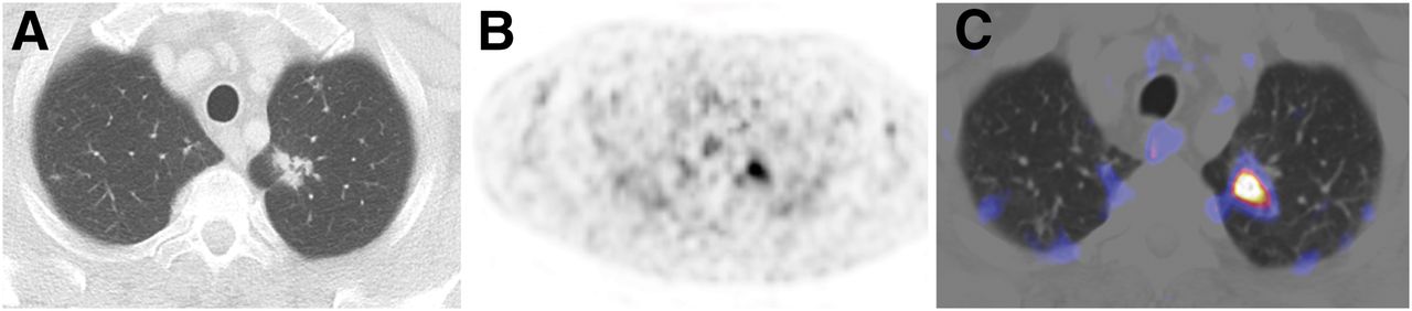
- FIGURE 1.
Example PSMA PET/CT scan of patient with pulmonary PC metastasis. (A) CT shows irregularly shaped lesion in left upper lobe in PC patient after radical prostatectomy, no known metastases, and PSA of 1.58 ng/mL. Lesion was subsequently biopsied and diagnosed as pulmonary PC metastasis. (B) PSMA PET is positive, with SUVmax of 7.1. (C) PET/CT fusion image.
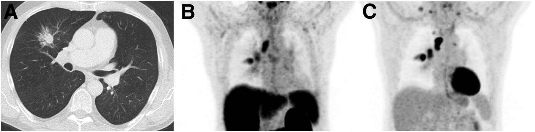
- FIGURE 2.
Example PET/CT scans of patient with primary lung cancer (adenocarcinoma). (A) CT shows spiculated lesion in right upper lobe in PC patient after prostatectomy and suspected local recurrence; PSA was 4.09 ng/mL. (B) PSMA PET exhibits focal tracer enhancement in pulmonary lesion (SUVmax, 5.3) and in 2 mediastinal lymph nodes, which were histologically confirmed as primary lung cancer and associated lymph node metastases. This was the only case for which lymph node uptake was evaluated. (C) 18F-FDG PET as established imaging method of choice for lung cancer shows similar enhancement in primary tumor and 2 mediastinal lymph node metastases.
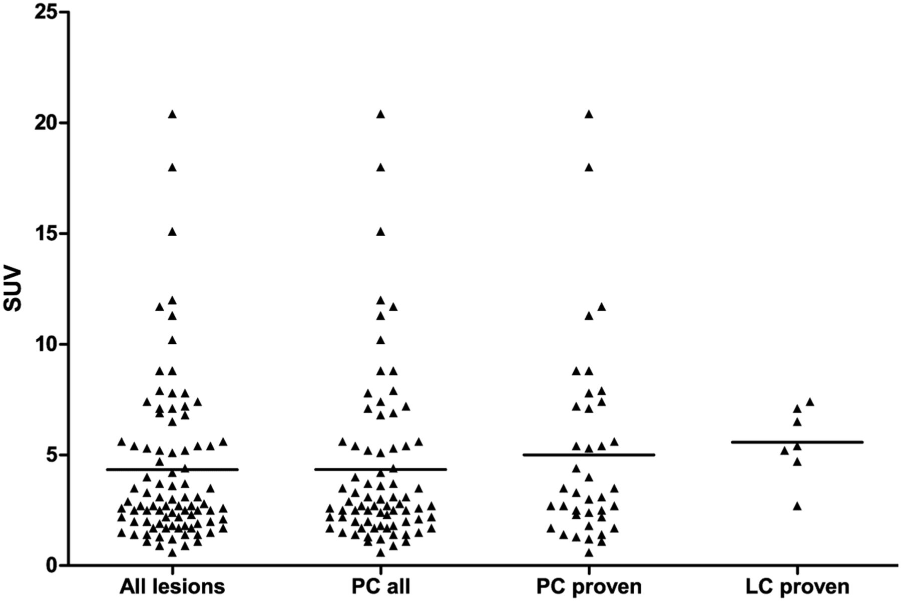
- FIGURE 3.
Distribution of SUVmax for different lesion classes. No significant difference was shown when comparing SUVmax distributions between groups: PC all (n = 76) consisting of PC-proven and PC highly probable vs. lung cancer–proven (n = 7) (P = 0.408) and PC-proven (n = 39) only vs. lung cancer–proven (P = 0.780).
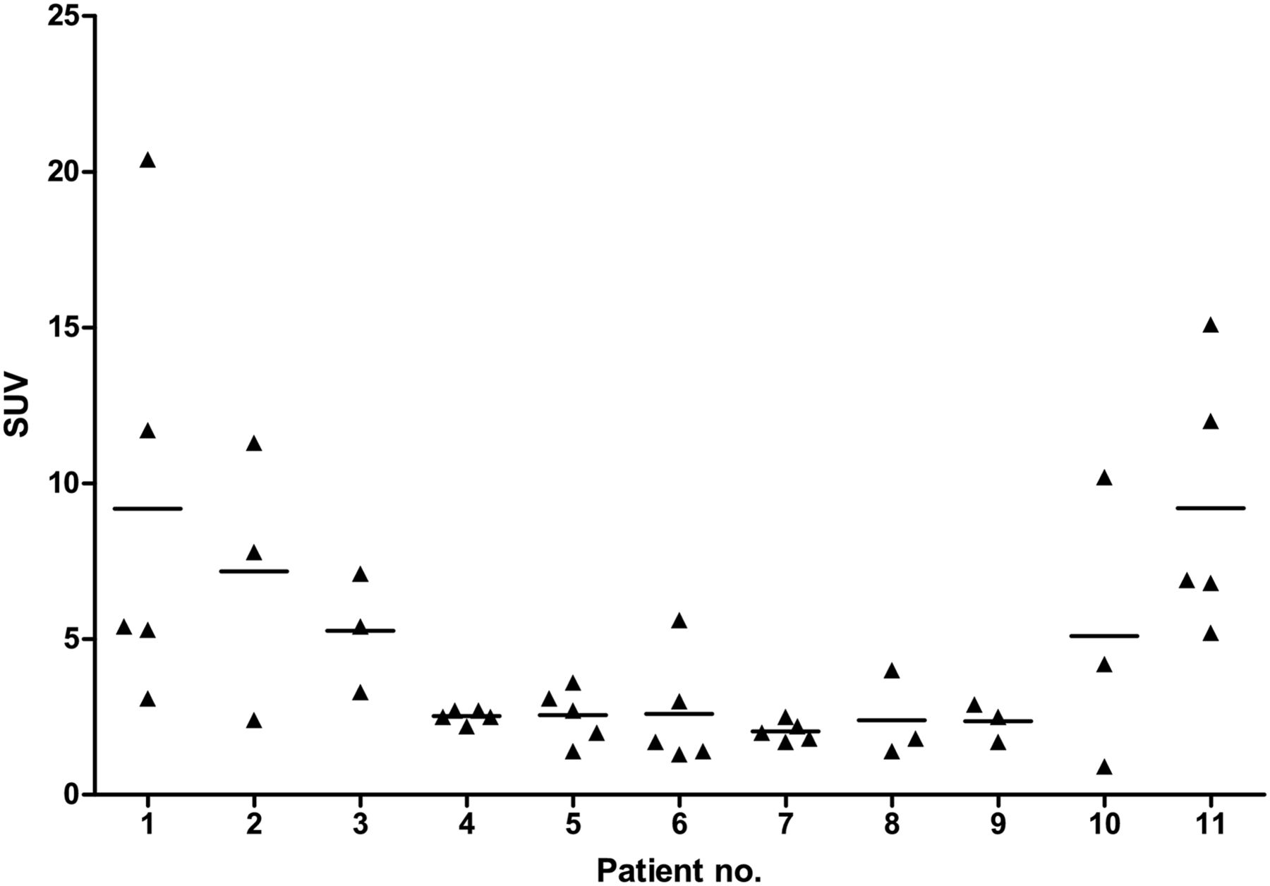
- FIGURE 4.
Intraindividual variability of PSMA uptake in pulmonary PC metastases. Shown is distribution of SUVmax for proven or probable pulmonary PC metastases in patients exhibiting 3 or more lesions.
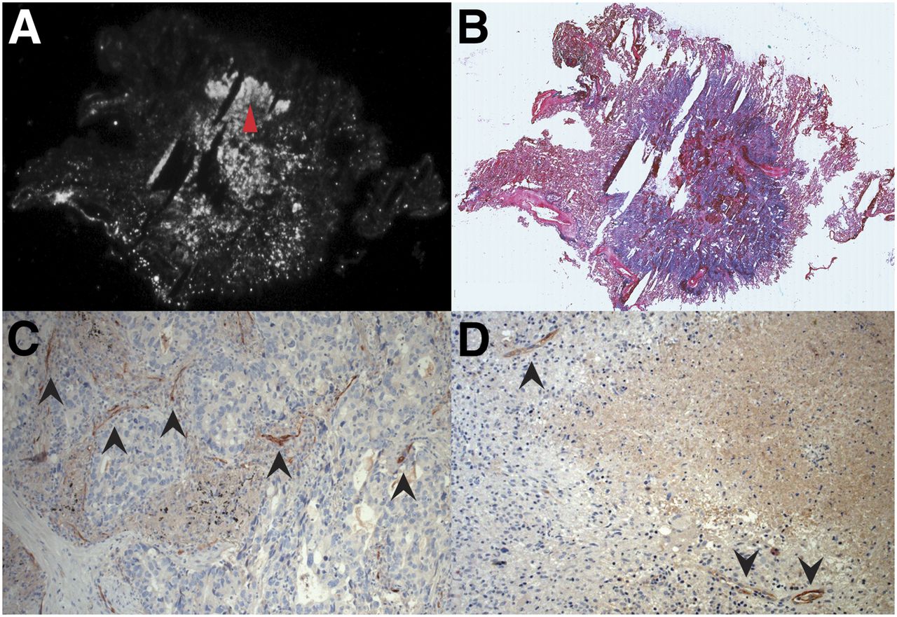
- FIGURE 5.
Autoradiography and PSMA immunohistochemistry of exemplary case of primary lung cancer and PSMA immunohistology of tuberculous lesion. (A) PSMA autoradiography of primary lung carcinoma from imaged cohort, which exhibited strong signal in PSMA PET (SUVmax, 5.2). (B) Hematoxylin and eosin stain showing tumor contours. (C) PSMA immunohistochemistry of same tumor, showing detail of tumor region with high cell density and high signal in autoradiography (red arrowhead in A), exhibiting no tumor cells with clear PSMA expression but PSMA in neovasculature (black arrowheads). (D) PSMA immunohistochemistry of case of active tuberculosis from our histologic database, showing PSMA-positive blood vessels (black arrowheads).
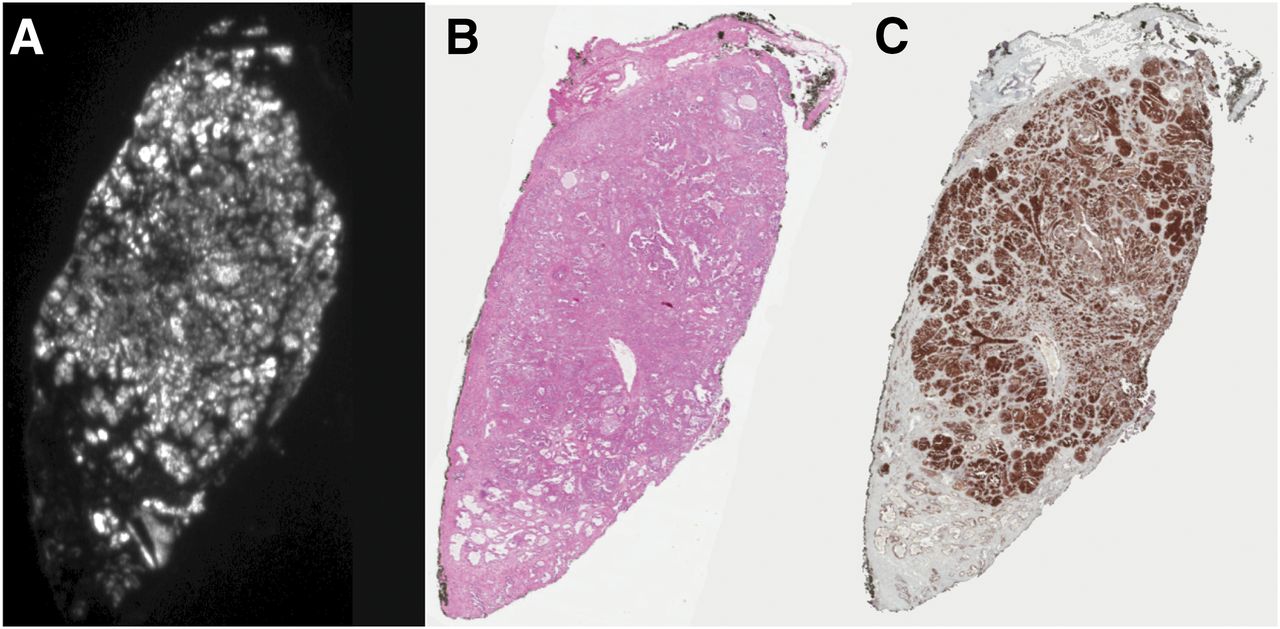
- FIGURE 6.
68Ga-PSMA autoradiography and PSMA immunohistochemistry of prostate carcinoma PC samples were used to establish autoradiography and immunohistology protocols. (A) PSMA autoradiography. (B) Hematoxylin and eosin stain. (C) Immunohistochemistry with anti-PSMA antibody 7E11 (aliquot kindly provided by Dr. Jan Grimm, Memorial Sloan Kettering Cancer Center).
Tables
Characteristic n Age (y) 68.8 (50–83) PSA (ng/mL) 5.67 (0.2–18,000) Injected dose (MBq) 151 (97–236) Classification PC-proven 18 By histology 7 By therapy response 11 Other entity–proven 8 Non–small cell lung cancer 7 Tuberculosis 1 PC highly probable 15 Unclear 4 Data in parentheses are ranges.
Characteristic All PC all PC-proven Lung cancer–proven Other/unclear No. of lesions 89 76 39 7 6 Localization Right upper lobe 22 16 10 5 1 Right lower lobe 19 17 9 1 1 Middle lobe 4 4 1 0 0 Left upper lobe 25 22 14 1 2 Left lower lobe 19 17 5 0 2 Configuration Smooth 41 39 14 0 2 Lobulated 11 10 5 0 1 Irregular 30 26 19 1 3 Speculated 7 1 1 6 0 Mean CT diameter ± SD (mm) 12.5 ± 6.3 11.8 ± 5.6 13.2 ± 6.9 21.0 ± 9.2 11.7 ± 1.8 Mean SUVmax ± SD 4.3 ± 3.7 4.4 ± 3.9 5.0 ± 4.4 5.5 ± 1.9 2.7 ± 2.5


Jump to section
Related Articles
Cited By...
- Comparison of 3 Interpretation Criteria for 68Ga-PSMA11 PET Based on Inter- and Intrareader Agreement
- 68Ga-PSMA-HBED-CC Uptake in Cervical, Celiac, and Sacral Ganglia as an Important Pitfall in Prostate Cancer PET Imaging
- Imaging of Nonprostate Cancers Using PSMA-Targeted Radiotracers: Rationale, Current State of the Field, and a Call to Arms
- Detection of Synchronous Primary Malignancies with 68Ga-Labeled Prostate-Specific Membrane Antigen PET/CT in Patients with Prostate Cancer: Frequency in 764 Patients
- PSMA Ligands for PET Imaging of Prostate Cancer
- What Medical, Urologic, and Radiation Oncologists Want from Molecular Imaging of Prostate Cancer
- Evaluation of Prostate Cancer with PET/MRI