
Main menu

  • Home
  • Content
    • Current
    • Ahead of print
    • Past Issues
    • JNM Supplement
    • SNMMI Annual Meeting Abstracts
    • Continuing Education
    • JNM Podcasts
  • Subscriptions
    • Subscribers
    • Institutional and Non-member
    • Rates
    • Journal Claims
    • Corporate & Special Sales
  • Authors
    • Submit to JNM
    • Information for Authors
    • Assignment of Copyright
    • AQARA requirements
  • Info
    • Reviewers
    • Permissions
    • Advertisers
  • About
    • About Us
    • Editorial Board
    • Contact Information
  • More
    • Alerts
    • Feedback
    • Help
    • SNMMI Journals
  • SNMMI
    • JNM
    • JNMT
    • SNMMI Journals
    • SNMMI

User menu

  • Subscribe
  • My alerts
  • Log in
  • My Cart

Search

  • Advanced search
Journal of Nuclear Medicine
  • SNMMI
    • JNM
    • JNMT
    • SNMMI Journals
    • SNMMI
  • Subscribe
  • My alerts
  • Log in
  • My Cart
Journal of Nuclear Medicine

Advanced Search

  • Home
  • Content
    • Current
    • Ahead of print
    • Past Issues
    • JNM Supplement
    • SNMMI Annual Meeting Abstracts
    • Continuing Education
    • JNM Podcasts
  • Subscriptions
    • Subscribers
    • Institutional and Non-member
    • Rates
    • Journal Claims
    • Corporate & Special Sales
  • Authors
    • Submit to JNM
    • Information for Authors
    • Assignment of Copyright
    • AQARA requirements
  • Info
    • Reviewers
    • Permissions
    • Advertisers
  • About
    • About Us
    • Editorial Board
    • Contact Information
  • More
    • Alerts
    • Feedback
    • Help
    • SNMMI Journals
  • View or Listen to JNM Podcast
  • Visit JNM on Facebook
  • Join JNM on LinkedIn
  • Follow JNM on Twitter
  • Subscribe to our RSS feeds
Review ArticleContinuing Education

Molecular Imaging of Gastroenteropancreatic Neuroendocrine Tumors: Current Status and Future Directions

Christophe M. Deroose, Elif Hindié, Electron Kebebew, Bernard Goichot, Karel Pacak, David Taïeb and Alessio Imperiale
Journal of Nuclear Medicine December 2016, 57 (12) 1949-1956; DOI: https://doi.org/10.2967/jnumed.116.179234
Christophe M. Deroose
1Nuclear Medicine, University Hospitals Leuven, Leuven, Belgium
  • Find this author on Google Scholar
  • Find this author on PubMed
  • Search for this author on this site
Elif Hindié
2Nuclear Medicine, Haut-Lévêque Hospital, University of Bordeaux, France
3LabEx TRAIL, University of Bordeaux, France
  • Find this author on Google Scholar
  • Find this author on PubMed
  • Search for this author on this site
Electron Kebebew
4Endocrine Oncology Branch, National Cancer Institute, NIH, Bethesda, Maryland
  • Find this author on Google Scholar
  • Find this author on PubMed
  • Search for this author on this site
Bernard Goichot
5Internal Medicine, Strasbourg University Hospitals, Strasbourg, France
  • Find this author on Google Scholar
  • Find this author on PubMed
  • Search for this author on this site
Karel Pacak
6Section on Medical Neuroendocrinology, Eunice Kennedy Shriver NICHD, NIH, Bethesda, Maryland
  • Find this author on Google Scholar
  • Find this author on PubMed
  • Search for this author on this site
David Taïeb
7Nuclear Medicine, La Timone University Hospital, Aix-Marseille University, Marseille, France
8European Center for Research in Medical Imaging, Marseille, France
9INSERM UMR1068, Marseille, France
  • Find this author on Google Scholar
  • Find this author on PubMed
  • Search for this author on this site
Alessio Imperiale
10Biophysics and Nuclear Medicine, Strasbourg University Hospitals, Strasbourg, France; and
11ICube, UMR 7357, University of Strasbourg/CNRS and FMTS, Faculty of Medicine, University of Strasbourg, Strasbourg, France
  • Find this author on Google Scholar
  • Find this author on PubMed
  • Search for this author on this site
  • Article
  • Figures & Data
  • Info & Metrics
  • PDF
Loading

Article Figures & Data

Figures

  • Tables
  • FIGURE 1.
    • Download figure
    • Open in new tab
    • Download powerpoint
    FIGURE 1.

    Head-to-head comparison of 111In-pentetreotide SSTR scintigraphy (A) and 68Ga-DOTATATE (B) PET/CT in patient with metastatic low-grade cecal NET evaluated before PRRT. In liver, retroperitoneal and thoracic lymph nodes, and bones, PET/CT shows multiple metastases, many of which are undetectable on SSTR scintigraphy.

  • FIGURE 2.
    • Download figure
    • Open in new tab
    • Download powerpoint
    FIGURE 2.

    68Ga-DOTATATE PET/CT results (A: anterior PET maximum-intensity projection, B: axial CT scan, C: axial PET/CT scan) in patient referred for preoperative staging of low-grade duodenal NET (white arrows) appearing as nodular thickening of lateral wall of duodenum with contrast enhancement and intense radiotracer uptake. 68Ga-DOTATATE PET/CT also shows additional pathologic focal uptake in epigastric region corresponding to synchronous duodenal G1 NET (black arrow).
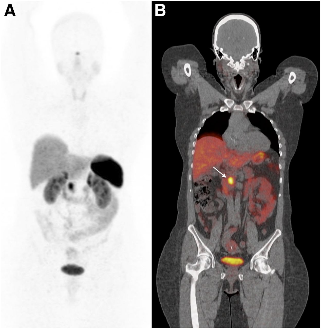
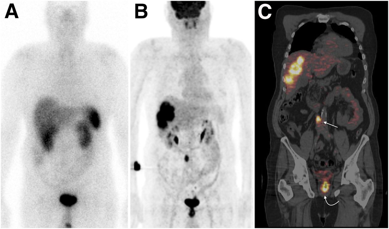
  • FIGURE 3.
    • Download figure
    • Open in new tab
    • Download powerpoint
    FIGURE 3.

    68Ga-DOTATATE PET/CT results (A: anterior PET maximum-intensity projection, B: coronal PET/CT scan) in patient with nonfunctional G1 NET of pancreatic head referred for primary staging. Tumor exhibited highly elevated uptake of 68G-DOTATATE (arrow) without locoregional or distant metastasis.
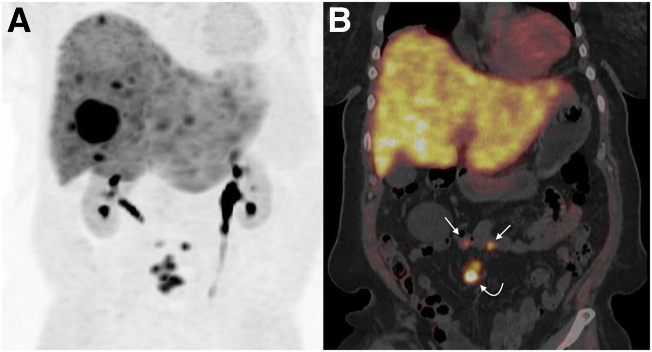
  • FIGURE 4.
    • Download figure
    • Open in new tab
    • Download powerpoint
    FIGURE 4.

    18F-FDOPA PET/CT results (A: anterior PET maximum-intensity projection, B: coronal PET/CT scan) in patient with carcinoid syndrome, retractile mesenteric lesions (curved arrow), and hepatic metastases of low-grade NET of unknown origin. Conventional imaging and 111In-pentetreotide SSTR scintigraphy failed to detect primary site. 18F-FDOPA PET/CT depicted 2 pathologic foci in ileum (straight arrows). Pathologic examination after surgery confirmed diagnosis of bifocal ileal G1 NET.
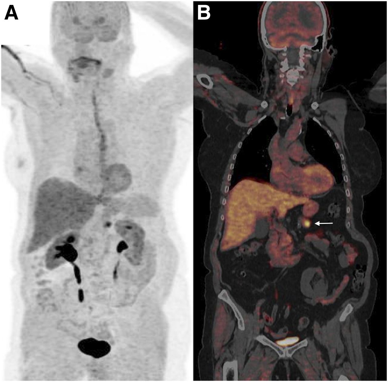
  • FIGURE 5.
    • Download figure
    • Open in new tab
    • Download powerpoint
    FIGURE 5.

    18F-FDOPA PET/CT after carbidopa premedication (A: anterior PET maximum-intensity projection, B: coronal PET/CT scan) in patient with hyperinsulinemic hypoglycemia. Insulinoma (arrow) was clearly identified by PET/CT. Normal pancreatic parenchyma has low uptake because of premedication by carbidopa.
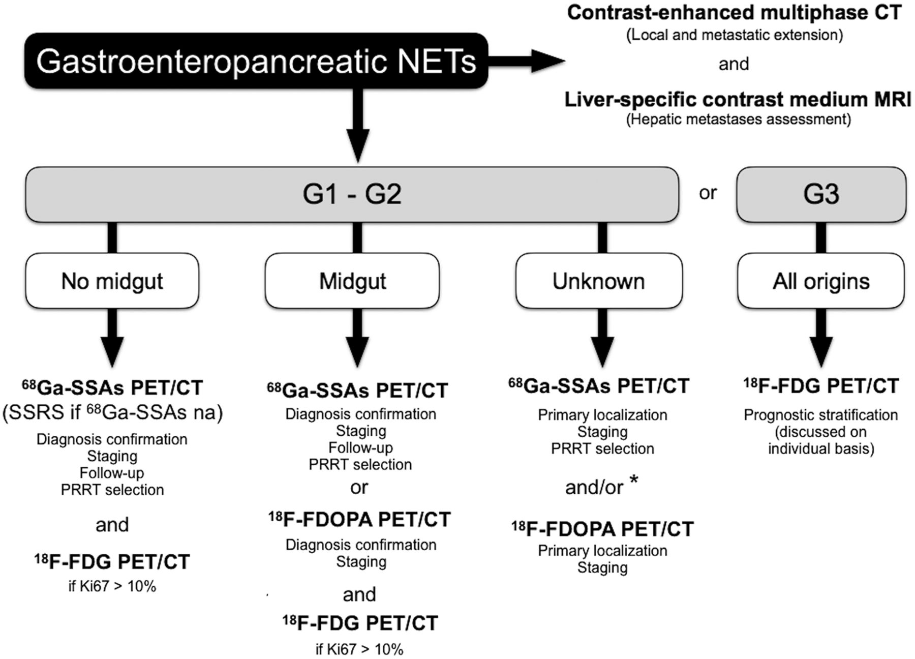
  • FIGURE 6.
    • Download figure
    • Open in new tab
    • Download powerpoint
    FIGURE 6.

    Proposed diagnostic imaging algorithm for patients with GEP NETs. na = not available; SSRS = SSTR scintigraphy. *Based on presumption of origin and hormonal secretion if present.

  • FIGURE 7.
    • Download figure
    • Open in new tab
    • Download powerpoint
    FIGURE 7.

    Typical example of flip-flop phenomenon in molecular imaging of patient with hepatic metastasis from G3 NET of unknown origin and referred before therapeutic strategy planning. Shown are 111In-pentetreotide SSTR scintigram (A), anterior 18F-FDG PET maximum-intensity projection (B), and coronal PET/CT scan (C). 18F-FDG PET/CT showed intense uptake by hepatic lesions and allowed detection of primary rectal tumor (curved arrow) and retroperitoneal lymphatic metastasis (straight arrow). These high-grade lesions showed no uptake on SSTR scintigraphy.

Tables

  • Figures
    • View popup
    TABLE 1

    Currently Available Endoscopic and Anatomic-Imaging Techniques for GEP NET Investigation

    CharacteristicTAUSEUSVideo capsuleCT*MRI*
    UseDetection of primary GI NET (solid organs only)Detection of gastric, duodenal and rectal primary NETs; diagnostic biopsyDetection of esophageal, gastric, duodenal, and small-bowel primary NETsStaging and follow-up (first-choice modality); identification of primary site; evaluation of local extent; assessment of metastasesDetection and assessment of liver metastases (first-choice modality)
    SensitivityLimited; high interoperator variabilityHighModerateCan be enhanced by enterography and enteroclysisHigh for bone marrow metastases; can be enhanced by enterography and enteroclysis
    Radiation exposureNoNoNoYesNo
    OtherIs widely availableIs invasiveCan analyze entire bowelIs widely availableUses gadolinium chelate, which is safer than CT iodine agents as regards allergic reactions and nephrotoxicity
    • ↵* Multiplanar contrast-enhanced images.

    • TAUS = transabdominal ultrasound; EUS = endoscopic ultrasound.

    • View popup
    TABLE 2

    Currently Available Functional-Imaging Techniques for GEP NET Investigation

    Characteristic111In-pentetreotide SPECT/CT123I-MIBG SPECT/CT68Ga-SSA PET/CT18F-FDOPA PET/CT18F-FDG PET/CT
    UsePrimary staging; restaging; patient selection before PRRTPatient selection before 131I-MIBG radiometabolic treatmentPrimary staging; restaging; patient selection before PRRT; imaging when primary site is unknownPrimary staging; imaging when primary site is unknown (based on presumption of ileal origin); (restaging?)Prognostic stratification; imaging of high-grade G2/G3 NETs
    Spatial resolutionLow (>10 mm)Low (>10 mm)High (5 mm)High (5 mm)High (5 mm)
    Procedure length2 d2 d1 d1 d1 d
    Radiation exposureModerateMildMildMildMild
    OtherIs approved for NET imagingHas low sensitivity for GEP NETsWill soon replace conventional SSTR scintigraphyMay be less sensitive than 68Ga-SSA PET/CT for nonileal GEP NETsIs widely available
PreviousNext
Back to top

In this issue

Journal of Nuclear Medicine: 57 (12)
Journal of Nuclear Medicine
Vol. 57, Issue 12
December 1, 2016
  • Table of Contents
  • Table of Contents (PDF)
  • About the Cover
  • Index by author
Print
Download PDF
Article Alerts
Sign In to Email Alerts with your Email Address
Email Article

Thank you for your interest in spreading the word on Journal of Nuclear Medicine.

NOTE: We only request your email address so that the person you are recommending the page to knows that you wanted them to see it, and that it is not junk mail. We do not capture any email address.

Enter multiple addresses on separate lines or separate them with commas.
Molecular Imaging of Gastroenteropancreatic Neuroendocrine Tumors: Current Status and Future Directions
(Your Name) has sent you a message from Journal of Nuclear Medicine
(Your Name) thought you would like to see the Journal of Nuclear Medicine web site.
Citation Tools
Molecular Imaging of Gastroenteropancreatic Neuroendocrine Tumors: Current Status and Future Directions
Christophe M. Deroose, Elif Hindié, Electron Kebebew, Bernard Goichot, Karel Pacak, David Taïeb, Alessio Imperiale
Journal of Nuclear Medicine Dec 2016, 57 (12) 1949-1956; DOI: 10.2967/jnumed.116.179234

Citation Manager Formats

  • BibTeX
  • Bookends
  • EasyBib
  • EndNote (tagged)
  • EndNote 8 (xml)
  • Medlars
  • Mendeley
  • Papers
  • RefWorks Tagged
  • Ref Manager
  • RIS
  • Zotero
Share
Molecular Imaging of Gastroenteropancreatic Neuroendocrine Tumors: Current Status and Future Directions
Christophe M. Deroose, Elif Hindié, Electron Kebebew, Bernard Goichot, Karel Pacak, David Taïeb, Alessio Imperiale
Journal of Nuclear Medicine Dec 2016, 57 (12) 1949-1956; DOI: 10.2967/jnumed.116.179234
Twitter logo Facebook logo LinkedIn logo Mendeley logo
  • Tweet Widget
  • Facebook Like
  • Google Plus One
Bookmark this article

Jump to section

  • Article
    • Abstract
    • OVERVIEW OF GEP NETS
    • CONVENTIONAL MORPHOLOGIC PROCEDURES
    • RADIONUCLIDE IMAGING
    • TOWARD PERSONALIZED MEDICINE
    • CONCLUSION
    • Footnotes
    • REFERENCES
  • Figures & Data
  • Info & Metrics
  • PDF

Related Articles

  • This Month in JNM
  • Erratum
  • PubMed
  • Google Scholar

Cited By...

  • 18F-Labeled Somatostatin Analogs as PET Tracers for the Somatostatin Receptor: Ready for Clinical Use
  • 18F-Labeled Somatostatin Analogs as PET Tracers for the Somatostatin Receptor: Ready for Clinical Use
  • 18F-DOPA PET/CT at the Forefront of Initial or Presurgical Evaluation of Small-Intestine Neuroendocrine Tumors
  • 64Cu-DOTATATE PET/CT for Imaging Patients with Known or Suspected Somatostatin Receptor-Positive Neuroendocrine Tumors: Results of the First U.S. Prospective, Reader-Masked Clinical Trial
  • NCCN Guidelines Insights: Neuroendocrine and Adrenal Tumors, Version 2.2018
  • New Challenges in Nuclear Endocrinology
  • Molecular Imaging of Human Embryonic Stem Cells Stably Expressing Human PET Reporter Genes After Zinc Finger Nuclease-Mediated Genome Editing
  • Expression of Gastrin-Releasing Peptide Receptor in Breast Cancer and Its Association with Pathologic, Biologic, and Clinical Parameters: A Study of 1,432 Primary Tumors
  • Association between neuroendocrine tumors biomarkers and primary tumor site and disease type based on total 68Ga-DOTATATE-Avid tumor volume measurements
  • Google Scholar

More in this TOC Section

  • Approaches to Imaging Immune Activation Using PET
  • Large Language Models and Large Multimodal Models in Medical Imaging: A Primer for Physicians
  • Precision Oncology in Melanoma: Changing Practices
Show more Continuing Education

Similar Articles

Keywords

  • gastroenteropancreatic neuroendocrine tumors (GEP NETs)
  • PET/CT
  • 68Ga-somatostatin analogues
  • 18F-FDOPA
  • 18F-FDG
SNMMI

© 2025 SNMMI

Powered by HighWire