


Abstract
Molecular imaging is indispensable for determining the fate and persistence of engrafted stem cells. Standard strategies for transgene induction involve the use of viral vectors prone to silencing and insertional mutagenesis or the use of nonhuman genes. Methods: We used zinc finger nucleases to induce stable expression of human imaging reporter genes into the safe-harbor locus adeno-associated virus integration site 1 in human embryonic stem cells. Plasmids were generated carrying reporter genes for fluorescence, bioluminescence imaging, and human PET reporter genes. Results: In vitro assays confirmed their functionality, and embryonic stem cells retained differentiation capacity. Teratoma formation assays were performed, and tumors were imaged over time with PET and bioluminescence imaging. Conclusion: This study demonstrates the application of genome editing for targeted integration of human imaging reporter genes in human embryonic stem cells for long-term molecular imaging.
In the development of stem cell–based therapies, long-term noninvasive imaging of grafted cells is indispensable for determining their fate and viability. Therefore, noninvasive imaging reporter genes are a suitable tool for long-term tracking of grafted stem cells (1).
Previous research has attempted to deliver exogenous genes into the genome of embryonic stem cells (ESCs) and induced pluripotent stem cells using various strategies (2). However, long-term follow-up of grafted cells is difficult because of signal silencing (3,4). Furthermore, random and multiple integrations in the host cell genome can perturb the biologic properties and stability of cells, resulting in a nonisogenic cell population (5).
Genome-editing approaches have recently been developed for an efficient targeting approach without random or multiple integrations within the genome of the host cells (6). One example is the use of zinc finger nucleases (ZFNs) (7–10), which contain specific recognition sites of up to 36 base pairs (bp) coupled to a FokI endonuclease domain, generating DNA double-strand breaks on dimerization. These breaks are repaired by either nonhomologous end joining or homologous recombination if a donor plasmid carrying the genes of interest is flanked by sequences homologous to the regions next to the double-strand break. The sequence of interest is then introduced into the host cell genome (5,7).
ZFNs have been designed and used to target the adeno-associated virus integration site 1 (AAVS1) locus, encoding the ubiquitously expressed protein phosphatase 1 regulatory subunit 12C (PPP1R12C) gene located on chromosome 19 (11,12). The AAVS1 locus is a safe-harbor locus, as the integration of target genes in this locus does not evoke pathologic responses (13) or perturb the proliferation, karyotype, or expression of pluripotency genes in ESCs and induced pluripotent stem cells (7,14). In addition, when ESCs are differentiated, the inserted gene is not silenced.
Here, the AAVS1 locus of ESCs was used to introduce a construct carrying reporter genes for noninvasive imaging. Enhanced green fluorescent protein (eGFP) was introduced for histologic validation. Firefly luciferase (Fluc) was included for bioluminescence imaging (BLI) (15). Finally, a radionuclide imaging reporter gene was included in the construct: either the human sodium iodide symporter (hNIS) or the human somatostatin receptor subtype 2 (hSSTr2). Because the insertion site lies downstream of exon 1 of a transcribed gene, a promoterless puromycin resistance cassette was included in the donor construct to maximize the selection efficiency of correctly targeted cells (14).
The paradigm reporter gene for PET is the herpes simplex virus type 1 thymidine kinase (HSV1-TK) (16–19). This reporter gene has allowed noninvasive documentation in animal models of in vivo gene transfer, cell monitoring, and protein–protein interactions (20) and has also been used in clinical applications (17–19). Its gene product might be immunogenic; furthermore, if treatment with antiviral drugs is necessary, the HSV1-TK–mediated conversion of the prodrug to its cytotoxic form causes unwanted cell death. Finally, substrates for HSV1-TK might require nucleoside transporters to incorporate the PET probe in the cells, and alterations in transporter status could influence the PET signal.
hNIS is commonly used for SPECT, PET, and radionuclide therapy of the thyroid gland because of its ability to transport radioactive forms of iodine and other negatively charged ions into thyroid cells (21,22). hNIS is principally present in the thyroid. Other tissues, such as the stomach mucosa, mammary glands, or salivary glands, also express hNIS at lower levels (22). This lower uptake implies a low background signal for radionuclide imaging and partly contributes to the interest in the application of hNIS as a PET reporter gene (23).
hSSTr2 has been imaged for years in the clinic for detection of neuroendocrine tumors using 111In-pentetreotide or 68Ga-labeled peptides such as 68Ga-DOTATATE (24). These molecules are commercially available or are in widespread use for neuroendocrine tumor imaging (25). This receptor is predominantly expressed in the gastrointestinal tract (including the pancreas), the spleen, and the pituitary gland (24,26).
The goal of this study was to generate a stably expressing ESC line for noninvasive, longitudinal multimodality imaging with human PET reporter genes through genome editing with a ZFN-mediated approach.
MATERIALS AND METHODS
Cell Culture
The human ESC line H9 (WA09) was purchased from WiCell Research Institute. ESCs were maintained feeder-free using mTeSR1 medium (StemCell Technologies).
Generation of Reporter Cell Lines
The pZ:puro-CAGGS-eGFP-P2A-Fluc-T2A-hNIS-plasmid and the pZ:puro-CAGGS-eGFP-P2A-Fluc-T2A-hSSTr2-plasmid were generated using the pZ-donor AAVS1 vector (CompoZr Targeted Integration Kit; Sigma Aldrich).
ESCs were resuspended in nucleofection solution 2 (Amaxa; Lonza) with 10 μg of donor plasmid and 3 μg of ZFN messenger RNA per 2 × 10⁶ cells. The cells were electroporated using program F16. After a week of puromycin selection, individual clones were expanded, resulting in 2 reporter cell lines: hNIS+ and hSSTr2+ ESCs.
Polymerase Chain Reaction (PCR) Genotyping
Genomic DNA was extracted using the QIAamp DNA mini kit (Qiagen) following the manufacturer’s protocol.
To check for reporter gene incorporation, junction assays were performed at the 5′ and 3′ end of the target locus. Random integrations were checked using primers at 5′ and 3′ sites next to the cassette. The wild-type (WT) allele was amplified with primers at the sites adjacent to the insertion site to determine whether the integration was mono- or biallelic. Primer sequences can be found in Table 1.
Primers Used for PCR Genotyping
Southern Blotting
Genomic DNA was digested with EcoRI and loaded onto a 0.7% agarose gel. Fragments were transferred to a nylon membrane (Zeta-Probe; Biorad) that was ultraviolet-crosslinked, and prehybridization was performed. Digoxigenin (DIG; Roche) labeling of the probe targeting the homology arm was done by PCR using the donor vector. Hybridization of the membrane with the probe was performed using the DIG High Prime DNA Labeling and Detection Starter Kit II (Roche) according to manufacturer’s instructions.
Hepatic Differentiation
Hepatic differentiation of ESCs was performed as previously described (27).
In Vitro Validation of Reporter Gene Expression
In vitro BLI was performed as previously described (28).
ESCs were plated, washed, and incubated with tracer solution (0.74 MBq/mL) for 1 h. 99mTcO4− and iodide (124I−) (Perkin Elmer) were used for hNIS+ ESCs, and 68Ga-DOTATATE for hSSTr2+ ESCs. The cells were washed 3 times, and tracer concentration in the cell fraction was measured using a γ-counter (Wizard; Perkin Elmer). Values were corrected for cell numbers. hNIS blocking was performed with NaClO4 (0.74 MBq/mL 99mTcO4− in 10 μM NaClO4), and hSSTr2 blocking was performed with cold octreotide (0.74 MBq/mL 68Ga-DOTATATE in 0.08, 0.25, 0.74, 2.2, 6.7, 20, and 60 nM octreotide). Efflux of 99mTcO4− was measured by incubating cells with 99mTcO4− for 1 h, followed by incubation with tracer-free medium for 5, 15, 30, 60, and 120 min. Saturation experiments were performed by incubation of hSSTr2+ ESCs with different solutions of 68Ga-DOTATATE (0.015, 0.022, 0.11, 0.37, or 2.22 MBq/mL). Each condition included a blocking with cold octreotide (20 nM). The cells were incubated for 10 min, and activity in the cells was measured after 3 washing steps. On the basis of activity inside the cell and specific activity, the amount of 68Ga-DOTATATE bound to the cells was calculated.
Real-Time Quantitative PCR
Gene expression analysis of pluripotency markers and ESC differentiation toward the hepatic lineage were performed using real-time quantitative PCR as previously described (27). Primer sequences are listed in Table 2.
Primers Used for Real-Time Quantitative PCR
Immunostaining
Staining was performed as described previously (28). Goat anti-eGFP (1:500; Abcam), goat anti-Oct4 (1:200; Abcam), rabbit anti-Nanog (1:200; Abcam), goat anti-SOX2 (1:50; Santa Cruz Biotechnology), and mouse anti-TRA1-60 (1:50; Santa Cruz) were used. Antirabbit, antigoat, and antimouse Alexa Fluor 594 (1:500 in 1% bovine serum albumin; Thermo Fisher) or antigoat Alexa Fluor 488 (1:500; Thermo Fisher) secondary antibodies were used.
Animal Preparation
All animal protocols were approved by the Ethical Committee of KU Leuven. Eight-week-old female BALB/c Rag2−/−ɣc−/− mice were anesthetized with 2% isoflurane (Isoflurane ISP; Rothacher), and 3–5 × 10⁶ ESCs were resuspended 1:1 in phosphate-buffered saline and Matrigel (BD Biosciences). hSSTr2+ ESCs were injected on the left flank, and hNIS+ ESCs contralaterally (n = 8).
BLI
BLI was performed as previously described (28).
68Ga-DOTATATE Preparation
68Ga-DOTATATE was prepared by heating 68Ga-chloride (400–800 MBq) at pH 4–4.4 with 30 μg of DOTATATE (Bachem), according to a previously published method, with adaptation (29). 68Ga-chloride was obtained by elution of a 68Ge/68Ga generator with diluted HCl solution followed by purification over a Dowex column (Sigma-Aldrich/Fluka). The reaction mixture was purified over a Sep-Pak C18 column (Waters) and formulated into a clinical-grade injectable solution.
Small-Animal PET
Imaging was performed on days 1, 22, and 63 after intravenous injection of approximately 3.7 MBq of 68Ga-DOTATATE or 124I− per mouse (n ≥ 5), with static acquisitions of 20 min using a Focus 220 microPET system (Siemens Medical Solutions USA). Time after injection was 1 h for 68Ga-DOTATATE and 2 h for 124I−.
Images were reconstructed with a maximum a posteriori algorithm and analyzed using PMOD 3.0 (PMOD Technologies). Images were converted to SUV.
A manually delineated volume of interest was positioned around the teratomas. Ratios were calculated comparing the signal to the contralateral tumor.
Histologic Processing
Masson trichrome staining was performed using standard protocols and analyzed to identify the presence of derivatives of the 3 germ layers.
Statistical Analysis
Data are presented as mean ± SEM. ANOVA was performed. P values of less than 0.05 were considered statistically significant. Data were processed using Prism, version 5.00 (GraphPad Software), for Windows (Microsoft).
RESULTS
Generation of Reporter Cell Lines
Two reporter gene constructs were generated, flanked by 800-bp stretches of homology to the ZFN target site AAVS1, downstream of exon 1 of the protein phosphatase 1 regulatory subunit 12C gene on chromosome 19 (Fig. 1A). The reporter genes were eGFP and Fluc, allowing BLI, and either hNIS or hSSTr2 for PET or SPECT.
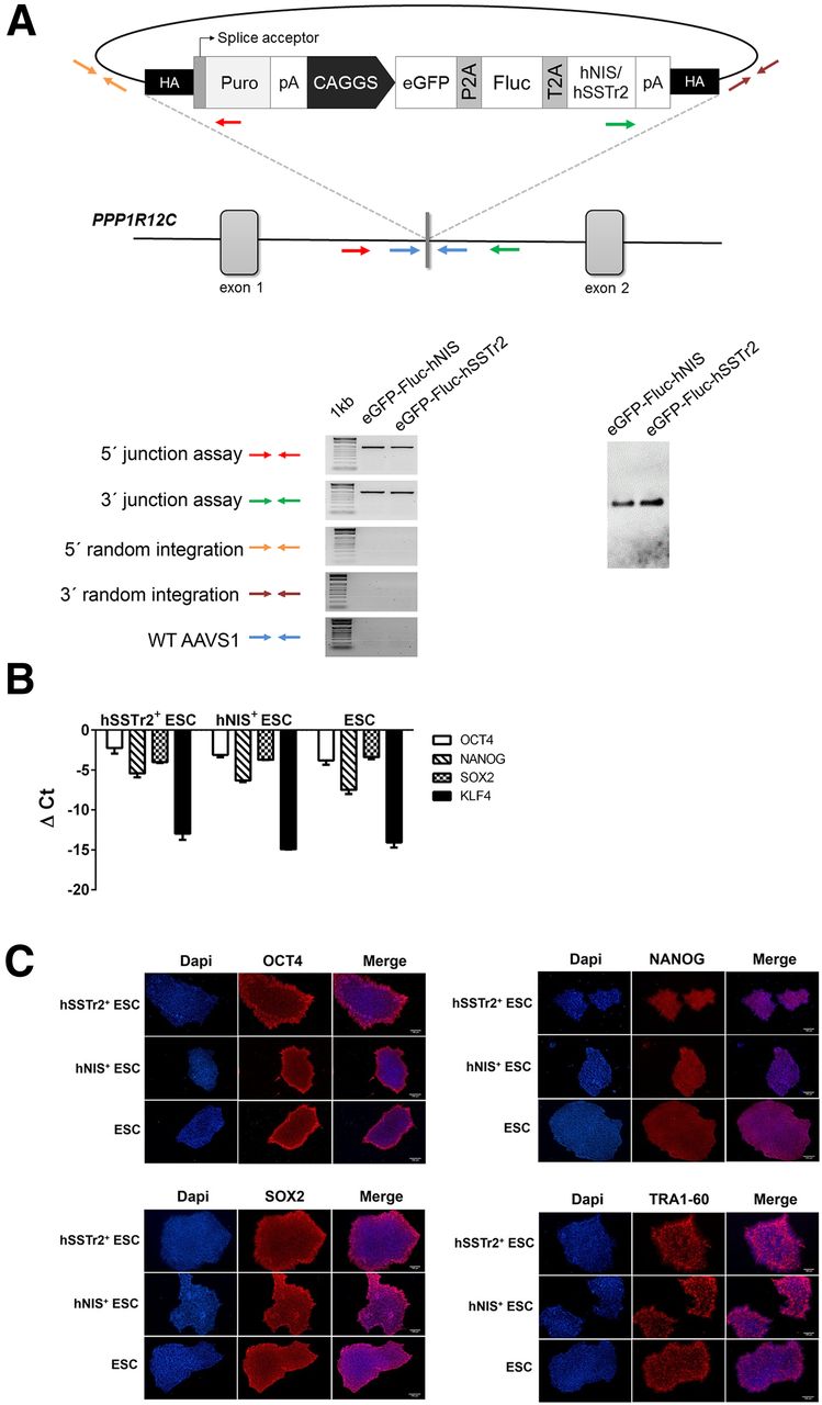
Correct integration of transgenes. (A) Recombinant lines incorporating transgenes showed correct integration, and southern blot analysis confirmed correct integration of construct. (B) Real-time quantitative PCR showed no significant difference in expression of pluripotency markers OCT4, NANOG, SOX2, and KLF4 after genetic engineering. (C) Immunocytochemistry for OCT4, NANOG, SOX2, and TRA1-60 showed similar expression in all lines. CAGGS = cytomegalovirus enhancer/chicken β-actin promoter; Ct = threshold cycle; Dapi = 4′,6-diamidino-2-phenylindole; HA = homology arms; pA = polyA tail; P2A = porcine teschovirus-1 2A; Puro = puromycin resistance gene; T2A = thoseaasigna virus 2A.
PCR Genotyping, Southern Blotting, and Expression of Pluripotency Markers
PCR genotyping was performed to assess proper integration of the construct and to exclude random integrations (Fig. 1A). Both hSSTr2+ and hNIS+ colonies carried the expression cassette as confirmed by the 5′ and 3′ junction assays. Single or biallelic integration of the cassette was assessed. In both clones, biallelic integration into the AAVS1 locus was observed. No random integrations had occurred.
Correct integration of the expression construct was confirmed with Southern blotting. A single band was present depicting biallelic integration (Fig. 1A). Expression of the pluripotency markers octamer-binding transcription factor 4 (OCT4), nanog homeobox (NANOG), sex-determining region Y-box 2 (SOX2), and Kruppel-like factor 4 (KLF4) was assessed and found to be the same as for WT ESCs (Fig. 1B). Immunocytochemistry for OCT4, NANOG, SOX2, and TRA1–60 showed a similar expression in all lines (Fig. 1C).
Hepatic Differentiation
WT, hSSTr2+, and hNIS+ ESCs showed a similar differentiation capacity toward the hepatic lineage (Fig. 2). Expression of the hepatocyte-specific genes hepatocyte nuclear factor 4α (HNF4α), α-fetoprotein (AFP), albumin (ALB), and α-1 antitrypsin (A1AT) increased in both cell lines. No significant difference was observed between the WT and the genome-edited ESCs. The definitive endoderm markers eomesdermin (EOMES), MIX1 homeobox-like protein 1 (MIXL1), CXCR4, and SOX17 were expressed similarly in all cell lines.

No significant difference in hepatic differentiation of hSSTr2+, hNIS+, and WT ESCs. Real-time quantitative PCR for hepatocyte-specific genes HNF4α, AFP, ALB, and A1AT and for definitive endoderm markers EOMES, MIXL1, CXCR4, and SOX17. Ct = threshold cycle.
In Vitro Validation of Reporter Gene Expression
Both before and after differentiation, genome-edited cells had significantly higher BLI signals than WT ESCs (Fig. 3A; P < 0.05).
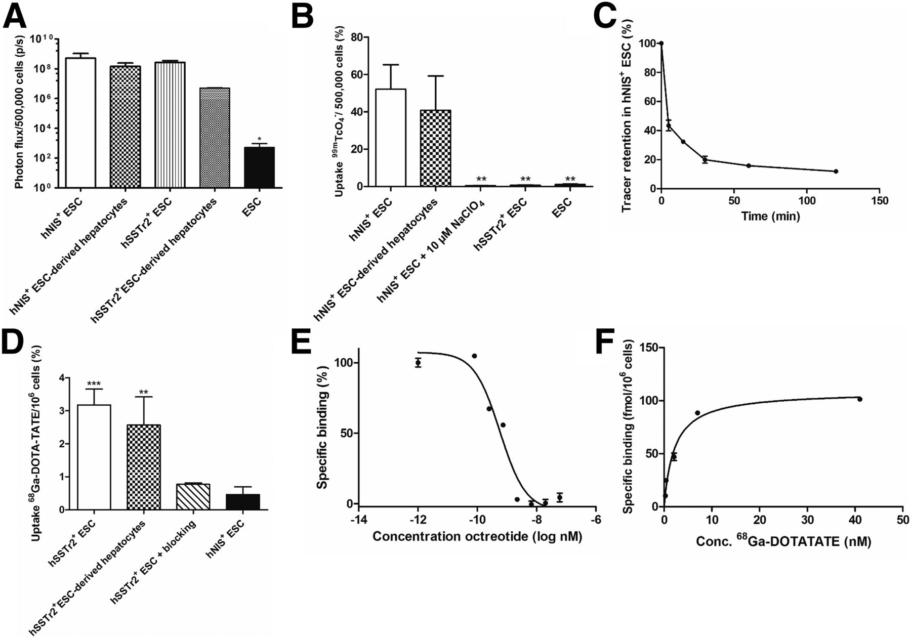
(A) In vitro BLI signals persisted after hepatic differentiation. (B) Uptake of 99mTcO4− by hNIS+ ESCs persisted after hepatic differentiation and was 50–80 times higher than in controls. NaClO4 significantly decreased tracer uptake. (C) Retention of 99mTcO4− led to significant tracer elution, with ∼20% retention. (D) Binding of 68Ga-DOTATATE in hSSTr2+ ESCs was significantly higher than in controls (hNIS+ ESCs) and persisted after hepatic differentiation. (E) Competitive inhibition using octreotide showed a half-maximal inhibitory concentration of 0.58 nM. (F) Saturation experiments revealed maximum specific binding of 111 ± 5 fmol per million cells and a dissociation constant of 3.4 ± 0.5 nM. **P < 0.01. ***P < 0.001.
The functionality of hSSTr2 and hNIS was tested with radioligand uptake experiments. After 1 h, 52.1% of 99mTcO4− was taken up per 5 × 10⁵ hNIS+ ESCs, 50–80 times more than in controls (P = 0.001). NaClO4 reduced uptake levels significantly (0.77%). hNIS+ hepatocytes maintained an uptake of 40.7%, not significantly different from undifferentiated cells (52.1%; P > 0.05) (Fig. 3B). A decreased tracer retention was observed, although after 1 h about 20% of the tracer remained intracellular (Fig. 3C).
hSSTr2+ ESCs could bind 3.18% per million cells of the 68Ga-DOTATATE, 6.9 times more than hNIS+ cells (P < 0.001). hSSTr2+ hepatocytes maintained their 68Ga-DOTATATE binding capacity. Octreotide administration caused a 4-fold reduction in 68Ga-DOTATATE uptake (0.77%; P < 0.001) (Fig. 3D). A half-maximal inhibitory concentration of 0.58 nM was demonstrated (Fig. 3E). A saturation experiment showed maximum specific binding of 111 ± 5 fmol per million cells and a dissociation constant of 3.4 ± 0.5 nM (Fig. 3F).
In Vivo Noninvasive Longitudinal Imaging
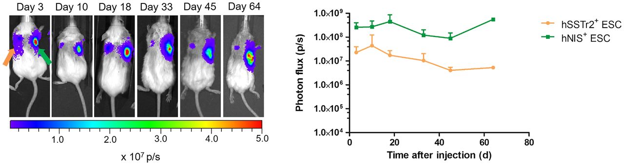
BLI of hSSTr2+ and hNIS+ teratomas showed a robust signal that increased over 70 d (Fig. 4). Small-animal PET was performed on days 1, 22, and 63 (Fig. 5). A focus of increased 68Ga-DOTATATE accumulation was observed at the hSSTr2+ teratoma. No increased 68Ga-DOTATATE uptake was seen at the site of the hNIS+ teratoma. On day 1, the hSSTr2+ ESCs showed 3.07 ± 0.84 times more signal than the hNIS+ ESCs (P < 0.01). This signal increased significantly to 5.02 ± 2.0 times greater in the hSSTr2+ ESCs on day 63 (Fig. 5A).

BLI of teratomas showed clear signal coming from both hSSTr2+ and hNIS+ ESCs and persisting over 70 d.

PET confirmed tracer uptake at correct locations. (A) 68Ga-DOTATATE accumulation observed at hSSTr2+ ESC site persisted over time (orange arrows); no signal was present at contralateral hNIS+ ESC graft (green arrows). (B) Uptake of 124I− at hNIS+ ESC site was clear and persisted over time (orange arrows); no signal was present at contralateral hSSTr2+ ESC graft (green arrows).
On the first day after engraftment of the hNIS+ ESCs, a focus of increased tracer uptake was observed, with 2.87 ± 1.0 times more accumulation of 124I− than in the hSSTr2+ teratomas (P < 0.001). On day 63 after engraftment, the signal increased significantly to 4.66 ± 0.7 times more tracer uptake in hNIS+ teratomas than in the controls (P < 0.05) (Fig. 5B).
Teratoma Assay
Histologic examination confirmed teratoma formation and thus maintenance of pluripotency after ZFN targeting. Teratomas from both lines contained cells derived from the 3 different germ layers (Supplemental Fig. 1; supplemental materials are available at http://jnm.snmjournals.org). hSSTr2+ teratomas formed neural rosettes, cartilage, and intestinal lining epithelium. hNIS+ teratomas were less dense and more fluid overall, containing neural rosettes, cartilage, and glandular tissue. Immunohistochemistry confirmed expression of hSSTr2 and hNIS in hSSTr2+ and hNIS+ teratomas, respectively.
DISCUSSION
The delivery of genetic material into cells has mainly been done through the use of viral vectors. Hence, insertional mutagenesis may occur, resulting in the disruption of the host genetic material, and there is a potential risk for oncogenesis. Furthermore, nonisogenic cell lines are generated. The primary transduced cell population is a heterogeneous mix of cells that are all genomically different from one another. Furthermore, reporter gene silencing is dependent on the integration site. These factors can result in signal loss and confound data interpretation (30,31).
In this work, we used the innovative ZFN-mediated approach for genetic engineering of ESCs. Human PET imaging reporter genes were introduced into the AAVS1 locus, which is known to be a so-called safe-harbor locus (7,13,14). AAVS1 has a continuously open chromatin structure with insulator elements that prevent epigenetic silencing and thereby allow stable transgene expression, at least when a transgene is expressed from a constitutive active promoter (7,14).
This technology is very translational, as two major limitations of stem cell imaging are tackled at once. First, ZFNs provide safe and controlled introduction of genetic material. Second, human PET reporter genes do not evoke immune responses. The tracers required for these genes are available in routine clinical use and do not require an on-site cyclotron. We used two reporter constructs for multimodal imaging, enabling the monitoring of cells after injection using BLI and PET or SPECT.
In this study, we showed the efficient introduction of the reporter cassette in both alleles of the AAVS1 locus. Very high in vitro uptake ratios were reached (∼2 orders of magnitude higher than in WT controls), and in vivo imaging was possible when the xenograft was not yet detectable by clinical examination.
hNIS is a symporter protein and thus mediates an amplification of the imaging signal (22,32). After their uptake through hNIS, no organification of tracer molecules occurs, which leads to partial leakage (28). Nevertheless, hNIS has many advantages such as its human origin, its low background expression, and the availability of several tracer molecules that are compatible. As imaging reporter proteins, receptors such as hSSTr2 bind a single ligand in a 1:1 ratio with high affinity (33). No signal amplification or tracer leakage will occur, but hSSTr2 is of human origin and is characterized by low endogenous expression outside some specific endocrine organs.
Both imaging reporter genes have their disadvantages, but their evident advantages make them very good candidates for imaging reporter genes. The choice of the reporter gene will be based mainly on the anatomic location to be imaged, depending on their endogenous expression.
BLI and small-animal PET were performed on hSSTr2+ and hNIS+ ESCs injected subcutaneously. BLI showed robust signal intensities that further increased specifically. PET signals increased significantly over time and more predominantly in hSSTr2+ ESC teratomas. hNIS+ teratomas contained more fluid-containing cysts not contributing to the signal. hSSTr2+ teratomas were denser, causing a relatively higher PET signal.
With 68Ga-DOTATATE PET, SUVmean was 0.07 ± 0.03 in hSSTr2+ and 0.03 ± 0.02 in hNIS+ teratomas (Fig. 5A). With 124I− PET, SUVmean was 0.21 ± 0.08 in hNIS+ and 0.06 ± 0.01 in SSTr2+ teratomas (Fig. 5B). Imaging of metastasis in a mouse model using melanoma cells expressing HSV1-TK imaged with 9-(4-18F-fluoro-3-hydroxymethylbutyl)guanine (18F-FHBG) resulted in a mean uptake of 3.3 percentage injected dose (%ID)/g (SUV, ∼0.9; formula from Phelps (34)). Hepatoma and rat glioma cells expressing HSV1-TK reporter led to an approximately 1.6 %ID/g and an approximately 0.7 %ID/g 1–2 wk after engraftment with 14C-labeled FIAU (2′-fluoro-2′-deoxy-β-d-arabinofuranosyl-5-iodouracil) and 18F-FHBG (35), implying approximate SUVs of 0.32 and 0.14. Prostate cancer cells expressing HSV1-TK resulted in an uptake of 0.2 %ID/g, or an SUV of 0.04 (36). Our data suggest slightly lower SUVs; however, others have used transfection or transduction to insert the reporter genes. Hence, multiple copies are inserted, leading to higher expression levels. In our work, a single copy of the reporter gene is included in the genome, and a lower expression level is inevitable. Also, in hSSTr2+ teratomas, lower SUVs were obtained for similar reasons. Furthermore, lower receptor densities were observed than in a previous report (37). To circumvent this problem, antagonists can be used, as they are able to bind more hSSTr2 sites (37,38).
Histologic examination confirmed that ZFN targeting does not alter the pluripotency of the ESCs. Furthermore, in vitro hepatic differentiation was as efficient for hSSTr2+ and hNIS+ ESCs as for WT ESCs.
Our results are in line with those of Wang et al., who introduced Fluc, monomeric red fluorescent protein, and HSV1-TK in the AAVS1 locus of ESCs and induced pluripotent stem cells (39). A high efficiency of the targeting procedure was shown, with preservation of pluripotency, differentiation capacity, and long-term gene expression. The survival of the cells was monitored with BLI but not with PET. Also, the gene product of HSV-TK is foreign to the human species, implying potential immunologic consequences and unwanted cell death.
Off-target effects have been described (40,41), but the search for novel strategies has led to the discovery of the CRISPR/Cas (clustered regulatory interspaced short palindromic repeats) system (42).
CONCLUSION
We demonstrated the introduction of human radionuclide reporter genes into the genome of human ESCs in a safe and controlled manner using the innovative ZFN-mediated strategy, thereby bypassing possible immune reactions against the imaging reporter genes. Furthermore, we performed long-term imaging of ZFN-engineered ESCs using reporter gene small-animal PET. Isogenic cell lines were generated, and these cells could be fully characterized for patient applications. It is clear that genetic engineering in the field of stem cell imaging can greatly accelerate the transition of basic research to a clinical setting, and these innovative techniques can therefore be used to explore other human PET reporter genes.
DISCLOSURE
Christophe M. Deroose and Koen Van Laere are senior clinical investigators of the Flemish Fund for Scientific Research (FWO). Cindy Casteels is a postdoctoral fellow of FWO. Bryan Holvoet and Natacha Breuls are PhD students funded by IWT. Laura Ordovas was funded by IWT (IWT/OZM/090838), IACS (BPAMER3/08/04), and the Government of Aragon (FMI048/08). Funding to Christophe M. Deroose was from FWO (G.0667.07 and G.0975.11), KU Leuven (ETH-C1900-PF and EME-C2161-GOA/11/012), IWT (IWT-HEPSTEM and IWT-HILIM-3D), BELSPO (BELSPO-IUAP-DEVREPAIR), and HeMiBio (FP7-HEALTH, project 266777). No other potential conflict of interest relevant to this article was reported.
Acknowledgments
We thank Manja Muijtjens, Pieter Berckmans, Jeanine Santermans, and Ann Van Santvoort for their help in data acquisition and processing. The radiopharmacy staff at Nuclear Medicine UZ Leuven is acknowledged for preparing the 68Ga-DOTATATE.
Footnotes
Published online Jun. 8, 2017.
- © 2017 by the Society of Nuclear Medicine and Molecular Imaging.
REFERENCES
- Received for publication January 11, 2017.
- Accepted for publication May 1, 2017.


Jump to section
Related Articles
Cited By...
- No citing articles found.