


Article Figures & Data
Figures
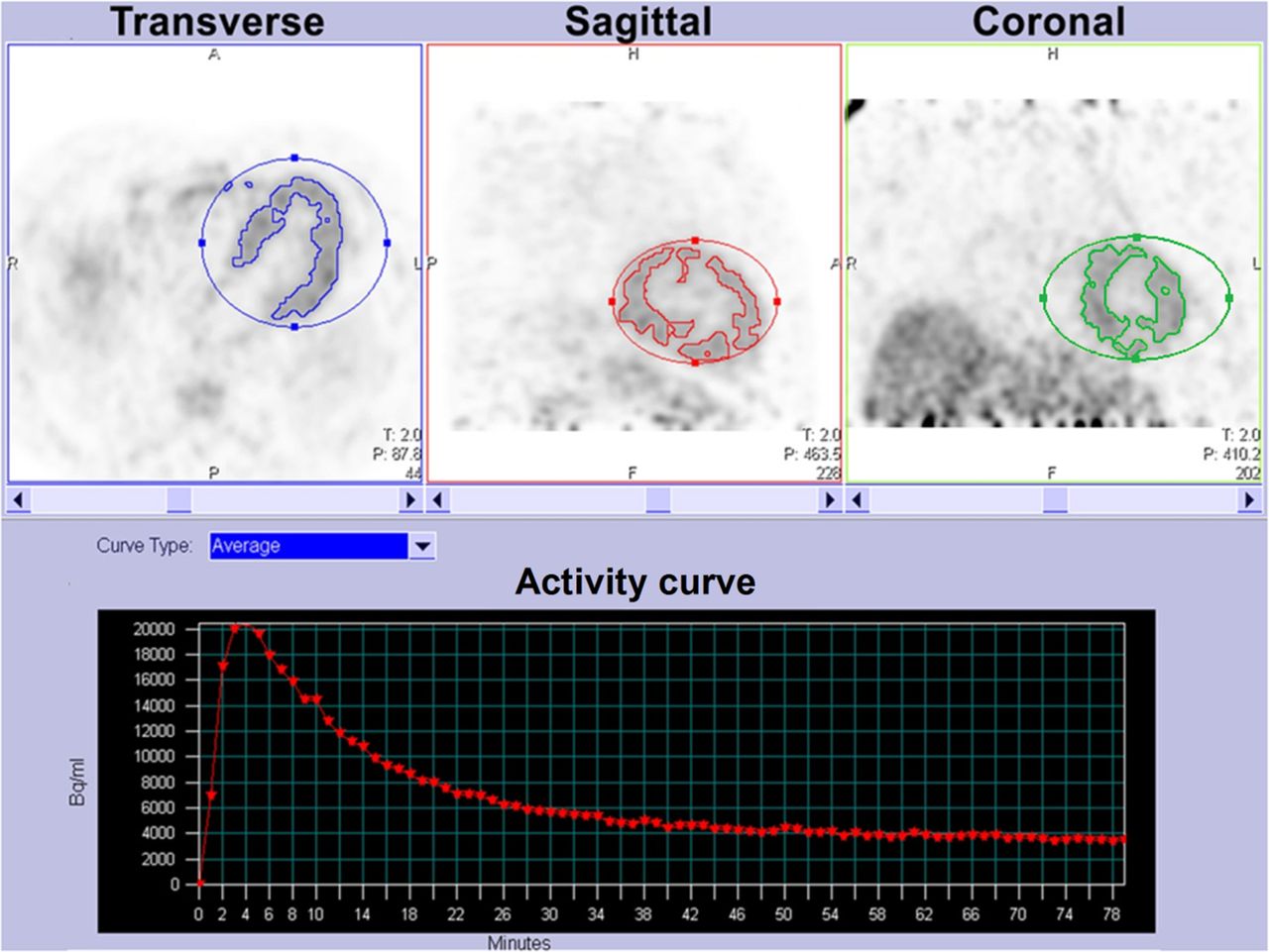
- FIGURE 1.
Example of software-automated volume-of-interest isocontouring performed on 5- to 10-min postflorbetaben images of patient with cardiac amyloidosis.
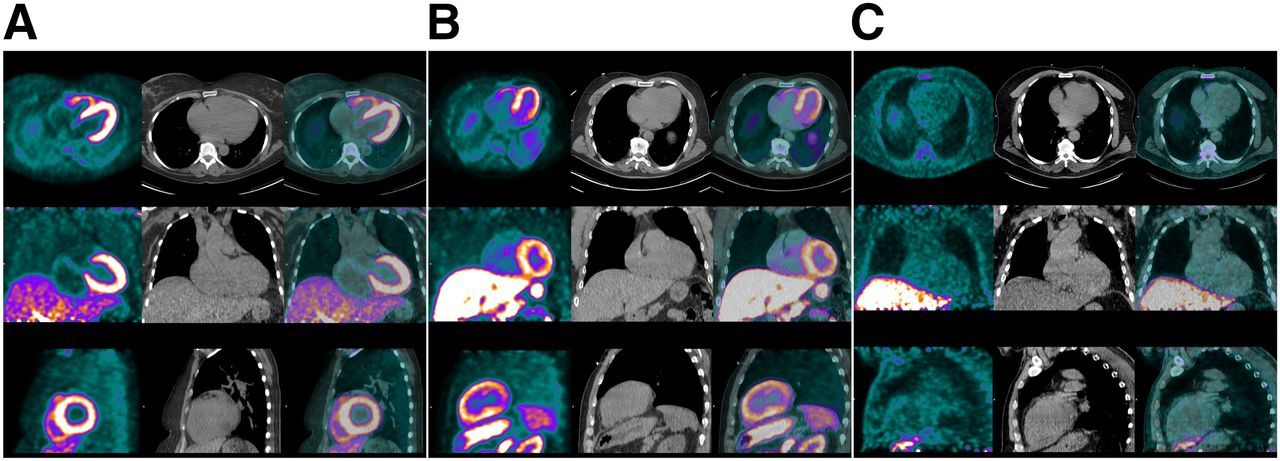
- FIGURE 2.
18F-florbetaben PET (left column in each panel), low-dose CT (middle column), and PET/CT images (right column) of representative AL patient (A), ATTR patient (B), and hypertensive control (C). There was diffuse avid 18F-florbetaben myocardial uptake in both AL and ATTR patients but little radiotracer uptake in myocardium of hypertensive control. PET images were windowed to display myocardial boundaries.
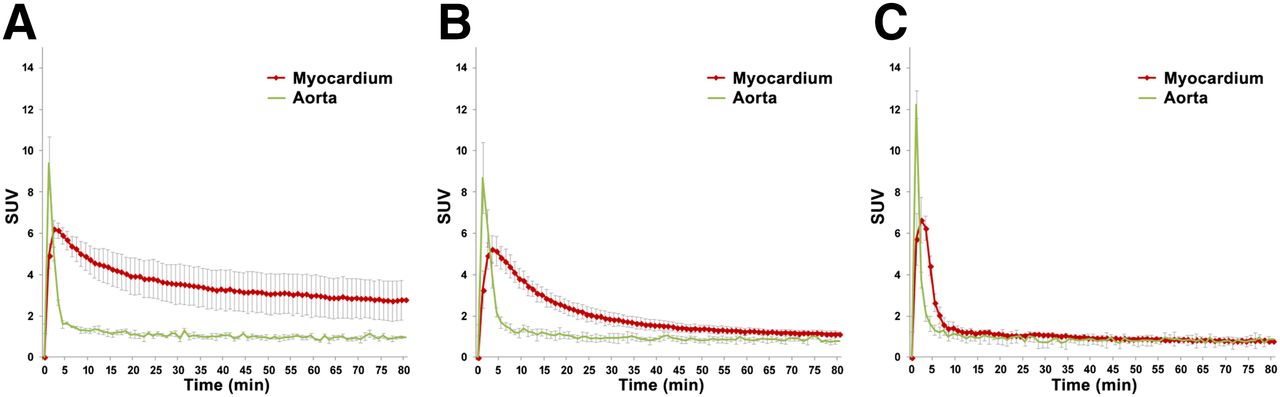
- FIGURE 3.
18F-florbetaben time–activity curves in AL patients (A), ATTR patients (B), and controls (C). Data points are mean SUVs in LV myocardium and blood-pool peak at 2−3 min after radiotracer injection; error bars represent SDs. Target-to-background ratios were significantly higher in AL and ATTR patients than in hypertensive controls after the initial 10 min and persisted until end of image acquisition. There was a trend toward higher myocardial SUV in AL than ATTR patients. There was also greater variability in myocardial SUV in AL than ATTR patients.
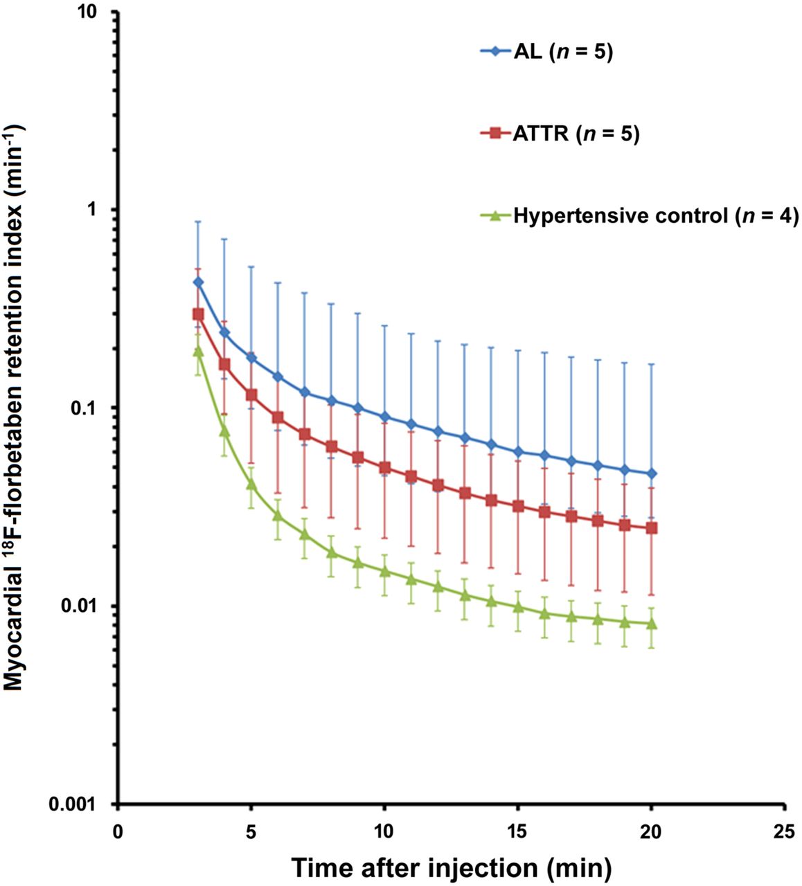
- FIGURE 4.
18F-florbetaben RI over time in AL patients, ATTR patients, and controls. Mean myocardial retention of 18F-florbetaben was higher in both AL and ATTR patients than in controls. Error bars represent ranges.
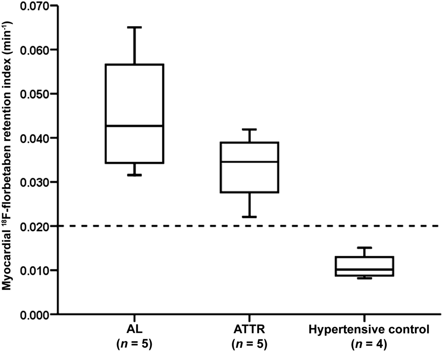
- FIGURE 5.
Box plots for myocardial 18F-florbetaben RI in AL patients, ATTR patients, and controls. Myocardial 18F-florbetaben RI was significantly higher in AL and ATTR patients. All cardiac amyloid patients and no hypertensive controls had myocardial 18F-florbetaben RI greater than 0.020 min−1.
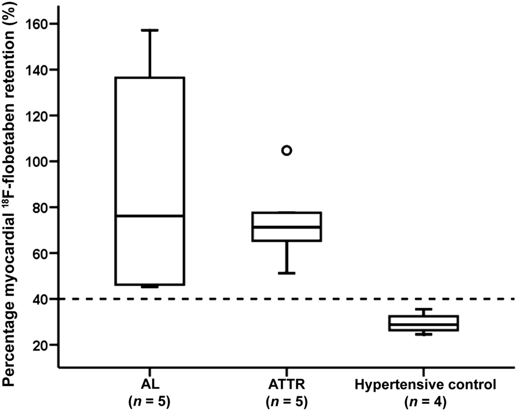
- FIGURE 6.
Box plots for percentage myocardial 18F-florbetaben retention for AL patients, ATTR patients, and controls. Percentage myocardial 18F-florbetaben retention was significantly higher in AL and ATTR patients. All cardiac amyloid patients and no hypertensive controls had myocardial retention greater than 40%.
- FIGURE 7.
Scatterplots for percentage myocardial 18F-florbetaben retention vs. LV global longitudinal strain (A) and RV free wall longitudinal strain (B), as expressed by inverse curve relationship. Scatterplots in C and D show inverse transformed percentage myocardial 18F-florbetaben retention. Triangles indicate AL patients, crosses indicate ATTR patients, and circles indicate hypertensive controls.


Additional Files
Supplemental Data
Files in this Data Supplement:


Jump to section
Related Articles
Cited By...
- Head-to-head comparison of [18F]florbetapir and [18F]FDG PET for the early detection of amyloidosis in systemic amyloidosis and plasma cell dyscrasias
- Molecular Imaging of Systemic and Cardiac Amyloidosis: Recent Advances and Focus on the Future
- Prognostic Value of Left Ventricular 18F-Florbetapir Uptake in Systemic Light-Chain Amyloidosis
- Novel Tracers for the Imaging of Cardiac Amyloidosis
- Novel Tracers for the Imaging of Cardiac Amyloidosis
- 18F-Florbetaben and PET/CT Holds Promise for the Identification and Differentiation Among Cardiac Amyloidosis Entities
- [18F]-Florbetaben PET/CT for Differential Diagnosis Among Cardiac Immunoglobulin Light Chain, Transthyretin Amyloidosis, and Mimicking Conditions
- Molecular Imaging of Cardiac Amyloidosis
- Improved Quantification of Cardiac Amyloid Burden in Systemic Light Chain Amyloidosis: Redefining Early Disease?
- Myocardial Amyloidosis: The Exemplar Interstitial Disease
- Transthyretin amyloidosis: an under-recognized neuropathy and cardiomyopathy