
Main menu

  • Home
  • Content
    • Current
    • Ahead of print
    • Past Issues
    • JNM Supplement
    • SNMMI Annual Meeting Abstracts
    • Continuing Education
    • JNM Podcasts
  • Subscriptions
    • Subscribers
    • Institutional and Non-member
    • Rates
    • Journal Claims
    • Corporate & Special Sales
  • Authors
    • Submit to JNM
    • Information for Authors
    • Assignment of Copyright
    • AQARA requirements
  • Info
    • Reviewers
    • Permissions
    • Advertisers
  • About
    • About Us
    • Editorial Board
    • Contact Information
  • More
    • Alerts
    • Feedback
    • Help
    • SNMMI Journals
  • SNMMI
    • JNM
    • JNMT
    • SNMMI Journals
    • SNMMI

User menu

  • Subscribe
  • My alerts
  • Log in
  • My Cart

Search

  • Advanced search
Journal of Nuclear Medicine
  • SNMMI
    • JNM
    • JNMT
    • SNMMI Journals
    • SNMMI
  • Subscribe
  • My alerts
  • Log in
  • My Cart
Journal of Nuclear Medicine

Advanced Search

  • Home
  • Content
    • Current
    • Ahead of print
    • Past Issues
    • JNM Supplement
    • SNMMI Annual Meeting Abstracts
    • Continuing Education
    • JNM Podcasts
  • Subscriptions
    • Subscribers
    • Institutional and Non-member
    • Rates
    • Journal Claims
    • Corporate & Special Sales
  • Authors
    • Submit to JNM
    • Information for Authors
    • Assignment of Copyright
    • AQARA requirements
  • Info
    • Reviewers
    • Permissions
    • Advertisers
  • About
    • About Us
    • Editorial Board
    • Contact Information
  • More
    • Alerts
    • Feedback
    • Help
    • SNMMI Journals
  • View or Listen to JNM Podcast
  • Visit JNM on Facebook
  • Join JNM on LinkedIn
  • Follow JNM on Twitter
  • Subscribe to our RSS feeds
Meeting ReportInstrumentation & Data Analysis

Improved PET image quantification via iterative denoising and partial volume correction

Ziyue Xu, Ulas Bagci, Mingchen Gao and Daniel Mollura
Journal of Nuclear Medicine May 2015, 56 (supplement 3) 1744;
Ziyue Xu
1Radiology and Imaging Sciences, National Institutes of Health, Bethesda, MD
  • Find this author on Google Scholar
  • Find this author on PubMed
  • Search for this author on this site
Ulas Bagci
1Radiology and Imaging Sciences, National Institutes of Health, Bethesda, MD
  • Find this author on Google Scholar
  • Find this author on PubMed
  • Search for this author on this site
Mingchen Gao
1Radiology and Imaging Sciences, National Institutes of Health, Bethesda, MD
  • Find this author on Google Scholar
  • Find this author on PubMed
  • Search for this author on this site
Daniel Mollura
1Radiology and Imaging Sciences, National Institutes of Health, Bethesda, MD
  • Find this author on Google Scholar
  • Find this author on PubMed
  • Search for this author on this site
  • Article
  • Figures & Data
  • Info & Metrics
Loading

Abstract

1744

Objectives To design an automated, accurate, robust, and efficient image enhancement algorithm for PET image to facilitate accurate diagnostic quantitative measurements of lesions

Methods We analyzed 20 PET-CT images of NEMA phantoms. An overview of our proposed framework is as follows (Fig 1A): (i) optimal segmentation based on an affinity propagation machine learning algorithm was applied for identifying the number of clusters in the PET images; (ii) segmentation/region based information was utilized to conduct edge-preserving denoising method; (iii) then, partial volume correction was performed by characterizing the geometric interaction using the uptake regions. These steps are utilized in an iterative manner until the clustering/segmentation convergences. For quantitative evaluation of image enhancement, boundaries from CT images were used as ground truths of uptake region definitions. The activity concentrations within each uptake regions were evaluated accordingly by comparing with true value from phantom parameters. Moreover, signal-to-noise ratio (SNR) was estimated from manually defined ROIs before and after enhancement.

Results For SNR of all high uptake regions before and after image preprocessing, some qualitative results were shown in Fig 1B, as shown from the three sample lines, red as before and blue as after enhancement, the system effectively promoted the image quantification accuracy for PET images. Quantitative results were given in Table 1. An average improvement of 32% was achieved. SNR was significantly promoted from 11.7 before enhancement to 360.5 after enhancement, for ROIs within the phantom body (exclude hot spheres).

Conclusions The proposed comprehensive automated framework helps to achieve better quantification of PET images in an efficient and robust manner. It provides an effective tool for PET image enhancement, and accurately delineates the uptake regions.

Figure
  • Download figure
  • Open in new tab
  • Download powerpoint

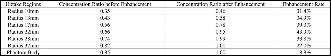
Concentration ratio (image/truth) before and after image enhancement with the enhancement rate.

Previous
Back to top

In this issue

Journal of Nuclear Medicine
Vol. 56, Issue supplement 3
May 1, 2015
  • Table of Contents
  • Index by author
Article Alerts
Sign In to Email Alerts with your Email Address
Email Article

Thank you for your interest in spreading the word on Journal of Nuclear Medicine.

NOTE: We only request your email address so that the person you are recommending the page to knows that you wanted them to see it, and that it is not junk mail. We do not capture any email address.

Enter multiple addresses on separate lines or separate them with commas.
Improved PET image quantification via iterative denoising and partial volume correction
(Your Name) has sent you a message from Journal of Nuclear Medicine
(Your Name) thought you would like to see the Journal of Nuclear Medicine web site.
Citation Tools
Improved PET image quantification via iterative denoising and partial volume correction
Ziyue Xu, Ulas Bagci, Mingchen Gao, Daniel Mollura
Journal of Nuclear Medicine May 2015, 56 (supplement 3) 1744;

Citation Manager Formats

  • BibTeX
  • Bookends
  • EasyBib
  • EndNote (tagged)
  • EndNote 8 (xml)
  • Medlars
  • Mendeley
  • Papers
  • RefWorks Tagged
  • Ref Manager
  • RIS
  • Zotero
Share
Improved PET image quantification via iterative denoising and partial volume correction
Ziyue Xu, Ulas Bagci, Mingchen Gao, Daniel Mollura
Journal of Nuclear Medicine May 2015, 56 (supplement 3) 1744;
Twitter logo Facebook logo LinkedIn logo Mendeley logo
  • Tweet Widget
  • Facebook Like
  • Google Plus One
Bookmark this article

Jump to section

  • Article
  • Figures & Data
  • Info & Metrics

Related Articles

  • No related articles found.
  • Google Scholar

Cited By...

  • No citing articles found.
  • Google Scholar

More in this TOC Section

Instrumentation & Data Analysis

  • Assessment of AI-Enhanced Quantitative Volumetric MRI with Semi-Quantitative Analysis in 18F-FDG Metabolic Imaging for Alzheimer's Diagnosis.
  • Assessment of SUV consistency in PET/CT with annulus 68Ge DQA phantom
  • Assessment of Tumor Burden in Lymphoma Patients with Deauville Score 4 Disease on Post Therapy FDG PET
Show more Instrumentation & Data Analysis

MTA II: Data Analysis & Management Posters

  • Quantitative image validation of PET-MR against stand-alone PET - A dynamic 11C-PE2I PET study.
  • Molecular Imaging And Kinetic Analysis Toolbox (MIAKAT) - A Quantitative Software Package for the Analysis of PET Neuroimaging Data
  • Validations for MR-based partial volume correction for brain PET imaging
Show more MTA II: Data Analysis & Management Posters

Similar Articles

SNMMI

© 2025 SNMMI

Powered by HighWire