


Article Figures & Data
Figures
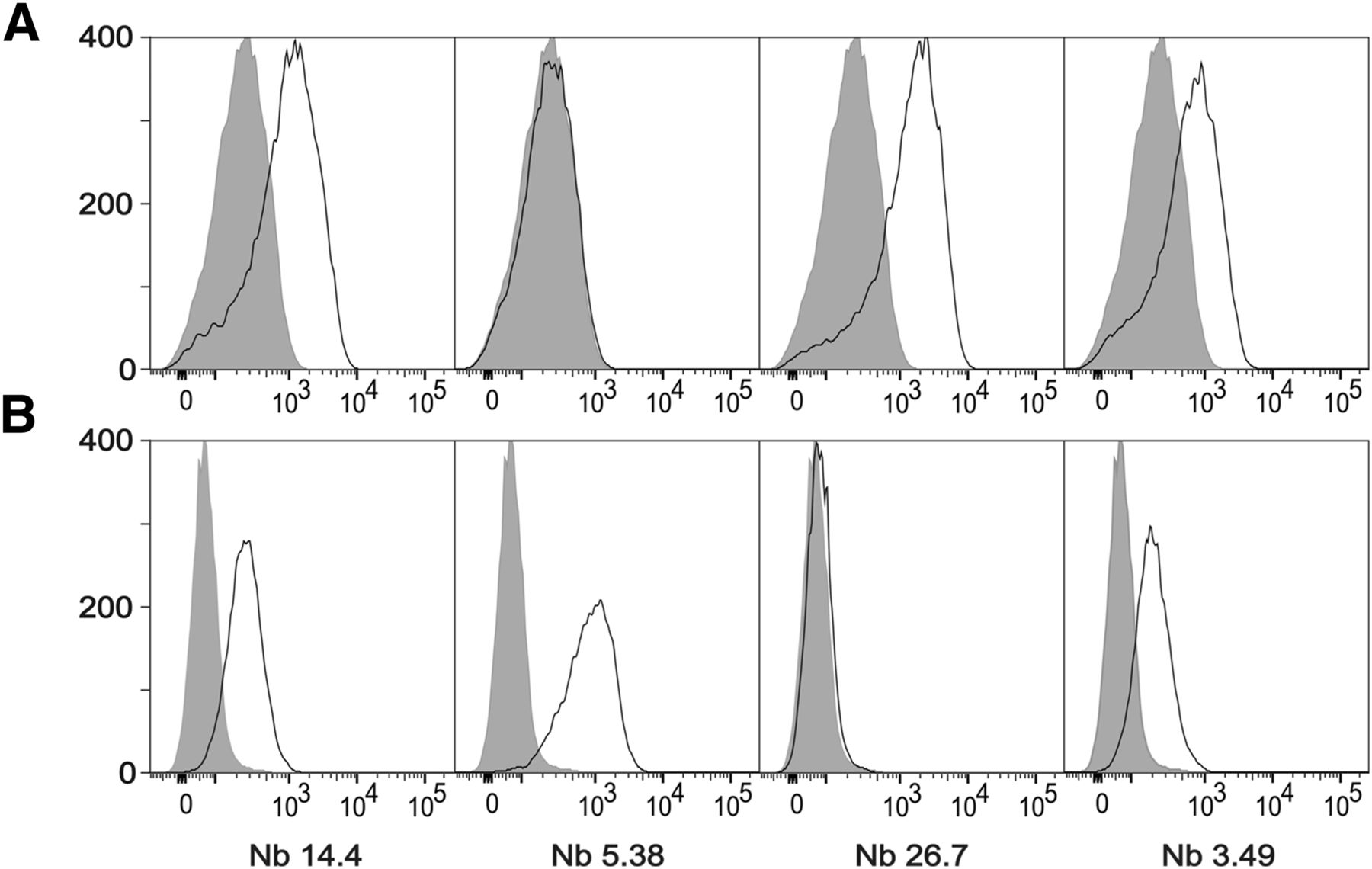
- FIGURE 1.
(A) Staining of single-cell suspensions prepared from 15-d-old 3LL-R subcutaneous tumors grown in C57BL/6 WT mice. (B) Staining on immature human dendritic cells expressing human MMR. Shaded histograms represent sdAb BCII10–negative control staining.
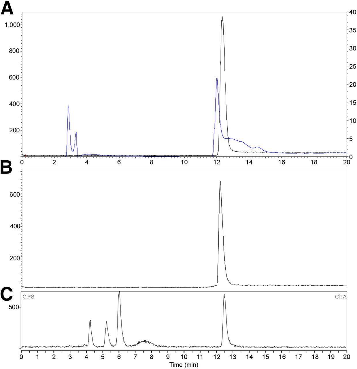
- FIGURE 2.
RP-HPLC analysis of purified 18F-FB-anti-MMR 3.49 sdAb (A; γ trace black, left axis; UV trace blue, right axis; retention time = 12.5 min) and after incubation for 3 h in phosphate-buffered saline, pH 7.4, at room temperature (B; γ trace). (C) Reversed-phase chromatogram of urine obtained 30 min after injection of 18F-FB-anti-MMR 3.49 (γ trace).
- FIGURE 3.
Comparison between uptake of 18F-FB-anti-MMR 3.49 sdAb (♦) and 99mTc-anti-MMR 3.49 sdAb (□) in spleen, liver, and tumor 3 h after injection. *Data are significantly different, P < 0.05.
- FIGURE 4.
Transverse and coronal PET/CT images of WT (left) vs. MMR-deficient (right) 3LL-R tumor–bearing mice scanned 3 h after injection of 18F-FB-anti MMR 3.49. PET signals are encoded in color scale, CT image in gray scale. Arrows point to tumor (T), kidney (K), and bladder (B). Autoradiography performed on slices from 3LL-R tumors grown in WT (left) vs. MMR-deficient (right) mice. max = maximum; min = minimum.


Tables
hMMR mMMR sdAb ka (M−1 s−1) kd (s−1) KD (nM) ka (M−1 s−1) kd (s−1) KD (nM) 14.4 1.4 × 10⁵ 1.4 × 10⁻³ 10 3.3 × 10⁴ 2.3 × 10⁻³ 68 5.38 2.0 × 10⁵ 6.6 × 10⁻⁴ 3.3 1.3 × 10⁵ 3.3 × 10⁻³ 25 26.7 5.8 × 10⁵ 7.3 × 10⁻³ 13 6.9 × 10⁵ 1.3 × 10⁻³ 1.9 3.49 4.4 × 10⁵ 8.0 × 10⁻⁴ 1.8 2.9 × 10⁵ 3.6 × 10⁻³ 12 h = human; m = mouse; ka = association rate constant; kd = dissociation rate constant; KD = equilibrium dissociation constant.
Organ/tissue 99mTc-anti-MMR 3.49 WT (n = 3) 99mTc-anti-MMR 14.4 WT (n = 3) 99mTc-anti-MMR 3.49 MMR-KO (n = 2) Heart 2.18 ± 0.15 2.77 ± 0.77 0.14 ± 0.02 Lungs 1.44 ± 0.15 0.92 ± 0.28 0.40 ± 0.08 Liver 14.79 ± 0.52 27.37 ± 3.37 0.61 ± 0.05 Spleen 4.94 ± 0.32 6.20 ± 1.69 0.22 ± 0.04 Kidney 146.61 ± 2.85 69.30 ± 11.46 234.77 ± 25.91 Muscle 0.57 ± 0.14 0.42 ± 0.11 0.06 ± 0.02 Bone 1.88 ± 0.17 1.78 ± 0.81 0.12 ± 0.01 Lymph nodes 3.04 ± 0.33 3.02 ± 0.46 0.19 ± 0.04 Blood 0.30 ± 0.03 0.13 ± 0.02 0.18 ± 0.03 Tumor 2.41 ± 0.34 1.40 ± 0.26 Not done Data were obtained at 3 h after injection and expressed as mean %IA/g ± SD.
- TABLE 3
Biodistribution of 18F-FB-Anti-MMR 3.49 sdAb in 3LL-R–Bearing WT, MMR-Deficient, and CCR-2–Deficient Mice at 3 Hours After Injection
Organ/tissue WT mice (n = 8) MMR-KO mice (n = 8) CCR2-KO mice (n = 5) Lungs 1.60 ± 0.40 0.82 ± 0.52 1.45 ± 0.14 Heart 0.81 ± 0.11 0.28 ± 0.12* 1.00 ± 0.14 Liver 2.26 ± 0.51 0.52 ± 0.30* 2.54 ± 0.31 Spleen 1.34 ± 0.31 0.38 ± 0.16* 1.71 ± 0.69 Kidney 7.98 ± 0.86 4.76 ± 2.76 7.60 ± 0.76 Muscle 0.37 ± 0.14 0.10 ± 0.07* 0.42 ± 0.27 Bone 0.67 ± 0.28 0.15 ± 0.02* 1.03 ± 0.20 Blood 1.02 ± 0.31 0.73 ± 0.31 1.16 ± 0.20 Tumor 2.40 ± 0.46 0.29 ± 0.14* 1.42 ± 0.17† ↵* Data are significantly different, P < 0.05.
Data are expressed as mean %IA/g ± SD.
Supplemental Data
Files in this Data Supplement:


Jump to section
Related Articles
Cited By...
- Phase I Study of [68Ga]Ga-Anti-CD206-sdAb for PET/CT Assessment of Protumorigenic Macrophage Presence in Solid Tumors (MMR Phase I)
- Single-Domain Antibody Theranostics on the Horizon
- Single-Domain Antibody Nuclear Imaging Allows Noninvasive Quantification of LAG-3 Expression by Tumor-Infiltrating Leukocytes and Predicts Response of Immune Checkpoint Blockade
- Single-domain antibodies for targeting, detection and in vivo imaging of human CD4+ cells
- Radionuclide Image-Guided Repair of the Heart
- Molecular Imaging of Myocardial Inflammation With Positron Emission Tomography Post-Ischemia: A Determinant of Subsequent Remodeling or Recovery
- The Immunoimaging Toolbox
- Same-Day Imaging Using Small Proteins: Clinical Experience and Translational Prospects in Oncology
- Preclinical Evaluation of 18F-Labeled Anti-HER2 Nanobody Conjugates for Imaging HER2 Receptor Expression by Immuno-PET
- An Effective Immuno-PET Imaging Method to Monitor CD8-Dependent Responses to Immunotherapy