


Article Figures & Data
Figures
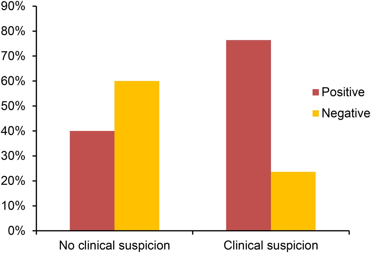
- FIGURE 1.
Added value of PET/CT in clinical assessment. PET/CT was helpful in excluding tumor in 23.6% (35/148) of the times PET/CT scan with clinical suspicion of recurrence or metastasis was obtained and identifying recurrence or metastasis in 40.0% (61/165) of times PET/CT scan with no prior clinical suspicion was obtained.
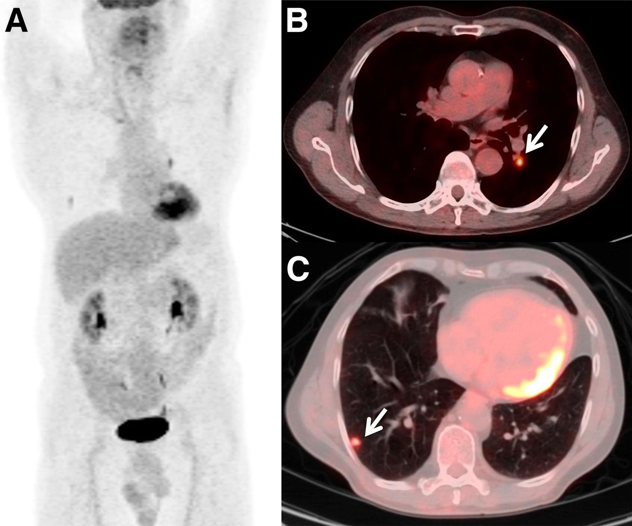
- FIGURE 2.
Positive follow-up scan obtained for routine follow-up. Anterior maximum-intensity-projection (A) and axial fused PET/CT (B and C) images of a 59-y-old man with history of rectal adenocarcinoma metastatic to liver, status after low anterior resection and adjuvant chemotherapy, who underwent a fourth follow-up PET/CT study for routine follow-up. PET/CT study demonstrated metabolically active (maximum standardized uptake value, 4.11) left hilar node (B; arrow) and hypermetabolic (maximum standardized uptake value, 2.4) right pulmonary nodule (C; arrow), consistent with metastatic disease. After study, he underwent chemotherapy with 3 cycles of 5-fluorouracil, oxaliplatin, folinic acid and bevacizumab.
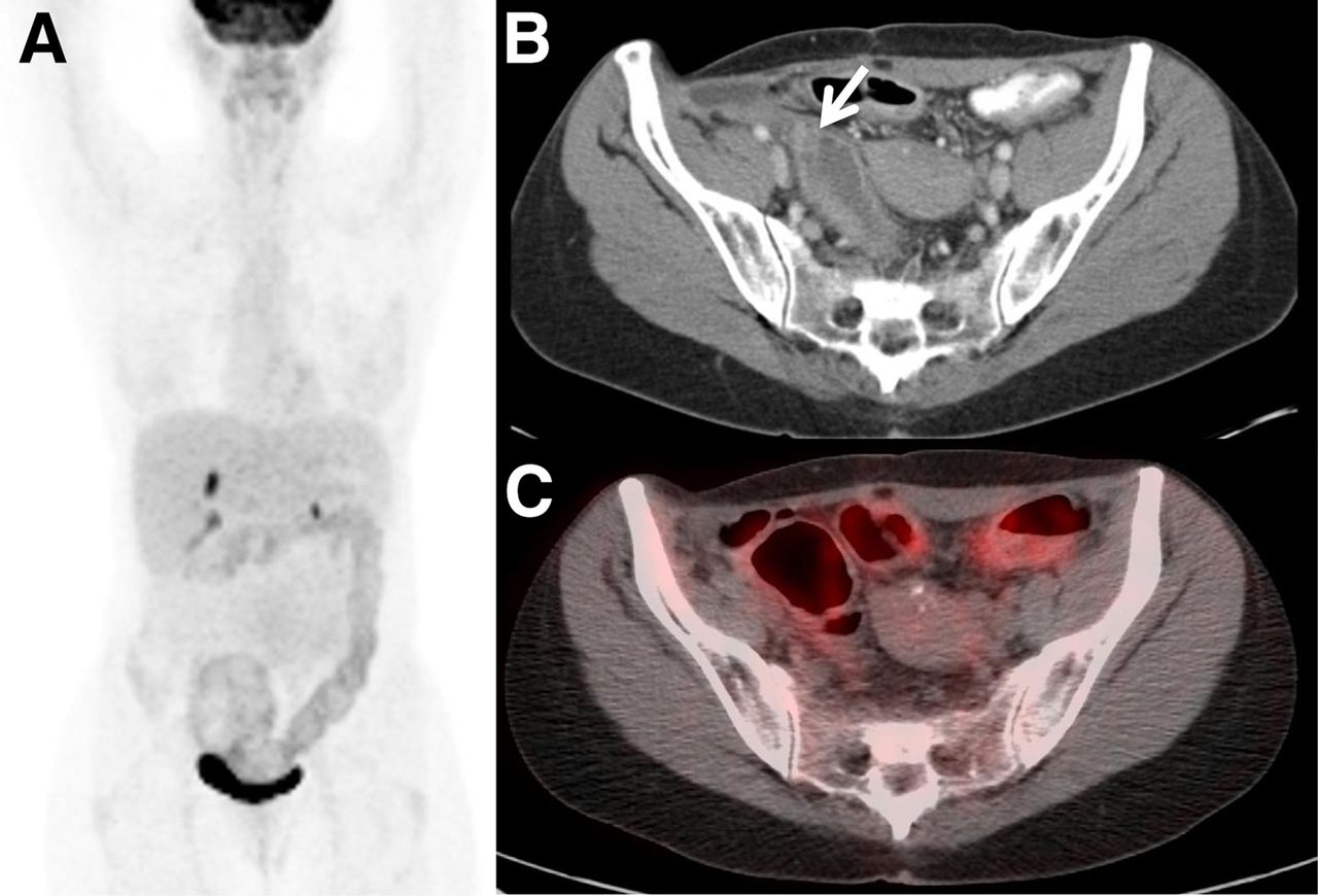
- FIGURE 3.
Negative follow-up scan obtained with clinical suspicion. Anterior maximum-intensity-projection (A), axial contrast-enhanced CT (B), and axial fused PET/CT (C) images of a 52-y-old woman with history of adenocarcinoma of colon, status after right hemicolectomy and chemotherapy, who was on follow-up. She was treated for local recurrence 2 y after diagnosis. Follow-up CT (B) performed 4 y after diagnosis revealed hypodense area in right lower quadrant, suggestive of local recurrence in rectosigmoid colon (arrow). Fourth follow-up PET/CT was ordered for further evaluation, which demonstrated no specific increase in metabolic activity to suggest disease recurrence, and patient has been on regular follow-up since.
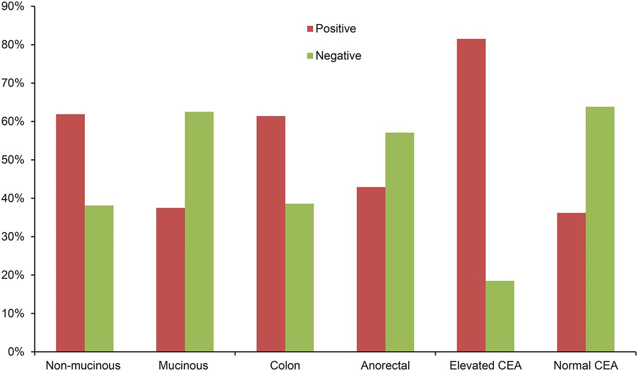
- FIGURE 4.
Tumor characteristics and PET/CT results. Among patients with nonmucinous adenocarcinoma, 61.9% PET/CT scans were positive and 38.1% scans were negative. Among patients with mucinous adenocarcinoma, 37.5% scans were positive and 62.5% scans were negative. Among patients with a colon primary, 61.4% scans were positive and 38.6% scans were negative. Among patients with an anorectal malignancy, 42.9% scans were positive and 57.1% scans were negative. Of scans with prior elevated CEA level, 81.5% scans were positive and 18.5% scans were negative. Of scans with prior normal CEA level, 36.2% scans were positive and 63.8% scans were negative.
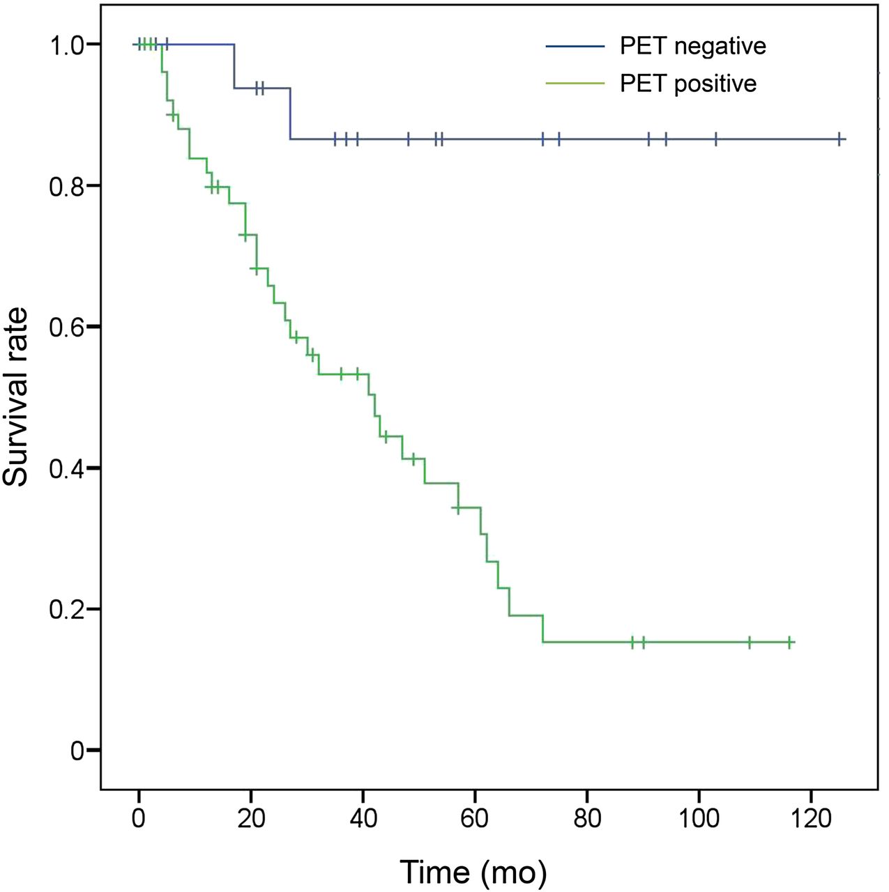
- FIGURE 5.
Kaplan–Meier survival plot by PET/CT result (patient level). OS (mo) between patients who had at least 1 positive (green line) and all negative (blue line) fourth and subsequent follow-up PET/CT scans for colorectal cancer differed significantly (log-rank, P = 0.001).

Tables
Characteristic n % Age (y)* <60 46 63 60–70 15 20.5 >70 12 16.5 Sex Female 33 45.2 Male 40 54.8 Tumor location Colon 48 65.8 Anorectal 25 34.2 Histology Adenocarcinoma 55 75.3 Squamous cell carcinoma 14 19.2 Unknown 4 5.5 Tumor differentiation Well differentiated 3 4.1 Moderately differentiated 43 58.9 Poorly differentiated 11 15.1 Unknown 16 21.9 Stage I 2 2.7 II 9 12.3 III 20 27.4 IV 35 47.9 Unknown 7 9.6 CEA level before PET/CT scan Elevated 31 42.5 Normal 10 13.7 Unknown 32 43.8 Last treatment Surgery 25 34.2 Radiation 2 2.7 Chemotherapy 37 50.7 Chemoradiation 9 12.3 PET/CT outcome Negative 20 27.4 Positive 53 72.6 Outcome Alive 39 53.4 Dead 34 46.6 *Mean ± SD, 56 ± 11 y.
PET/CT result Routine n Clinical suspicion n Total n Positive 61 (40.0) 113 (76.4) 174 (55.6) Negative 104 (60.0) 35 (23.6) 139 (44.4) Total 165 148 313 Data in parentheses are percentages.
Scan result Scan impact on treatment Positive Negative Total % No treatment to no treatment 20 (14.5) 118 (85.5) 138 44.1 Treatment to continued treatment 44 (86.3) 7 (13.7) 51 16.3 No treatment to new treatment 71 (94.7) 4 (5.3) 75 24.0 Treatment to change in treatment 22 (88.0) 3 (12.0) 25 8.0 Treatment to treatment stopped 5 (71.4) 2 (28.6) 7 2.2 Unknown 12 (70.6) 5 (29.4) 17 5.4 Data in parentheses are percentages.


