


Article Figures & Data
Figures
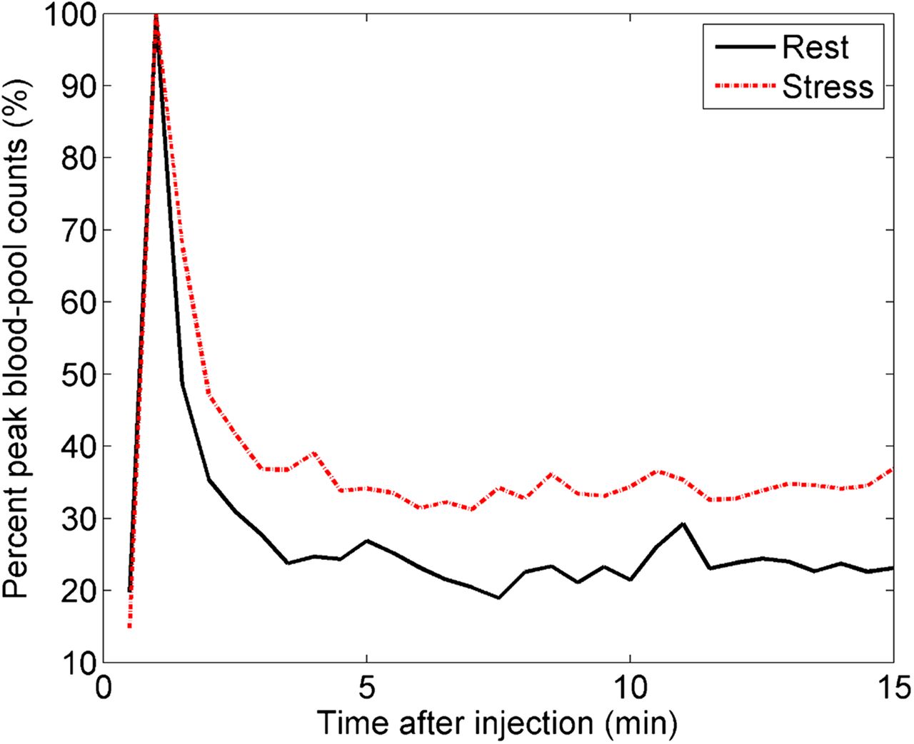
- FIGURE 1.
Blood time–activity curve for CMICE-013 based on counts from volume of interest in left ventricle. Time–activity curves do not go to zero at late times because of limited spatial resolution of SPECT and consequent spill-in from tracer in myocardial wall.
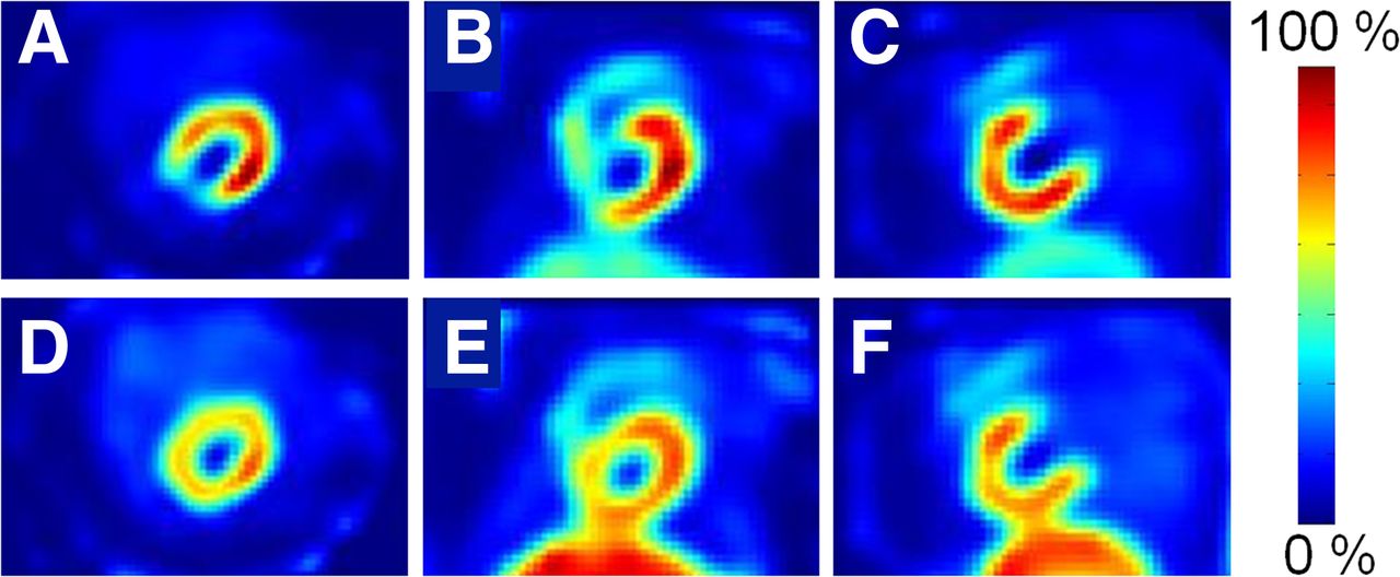
- FIGURE 2.
Representative CMICE-013 images of porcine model of stress-induced myocardial ischemia showing stress (top) and rest (bottom) from 15–30 min after injection. Images are central slices in transverse (A and D), coronal (B and E), and sagittal (C and F) views. Images show good contrast between heart and surrounding tissues, except for liver for which contrast is similar to that of standard SPECT tracers.
- FIGURE 3.
Ratios of maximum uptake in heart, compared with liver and lung at rest (A) and stress (B) for each of the tracers studied.
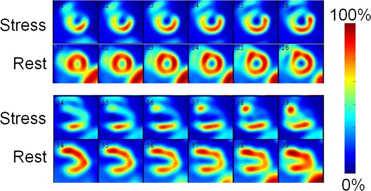
- FIGURE 4.
Representative CMICE-013 images of heart. Images are shown at rest and stress (both 15 min after injection) for short axis (top) and vertical long axis (bottom). Images show good uniformity at rest and clear definition of the occluded region during stress.
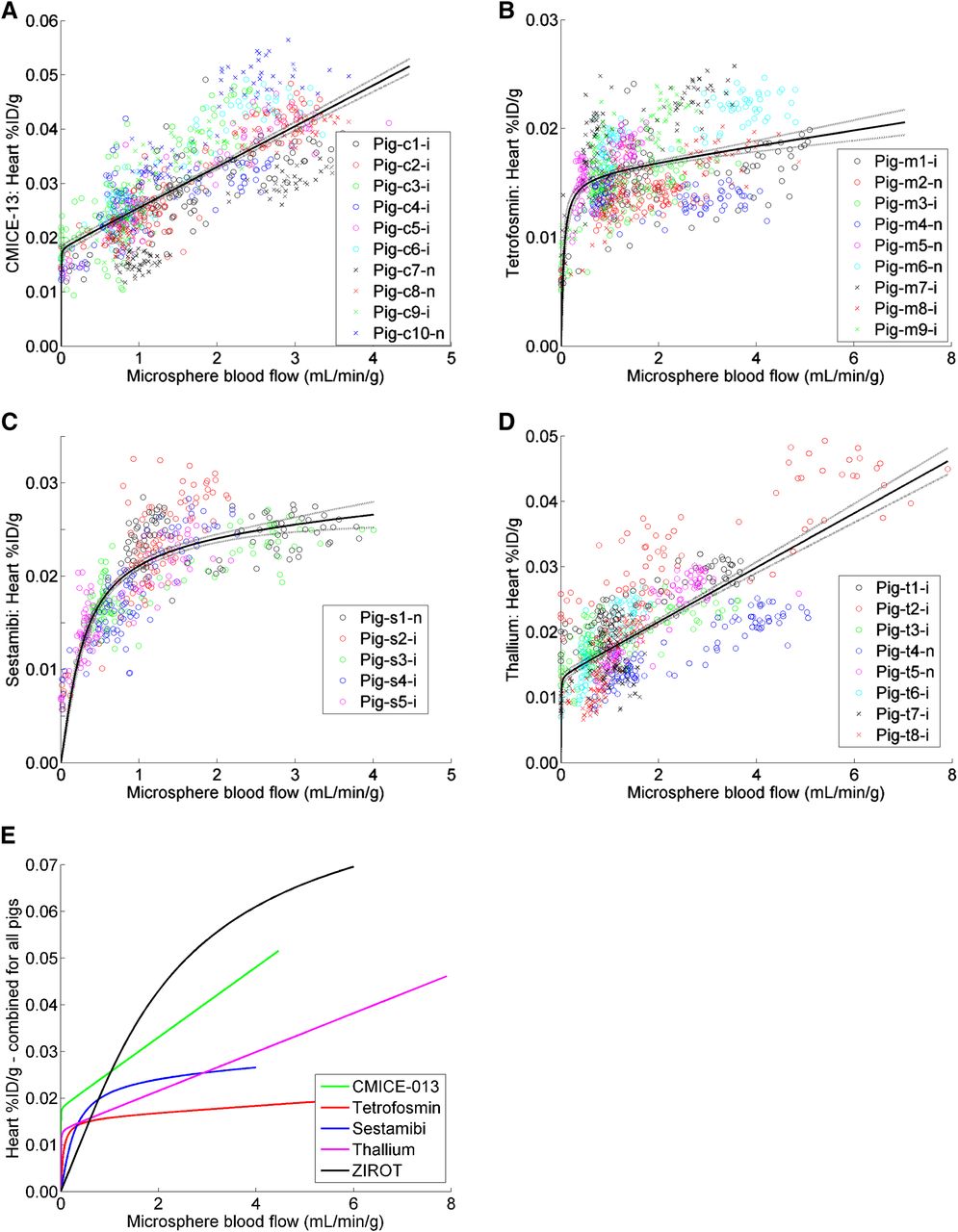
- FIGURE 5.
Tracer uptake versus microsphere flow values for each tissue sample from individual animals with CMICE-013 (A), 99mTc-tetrofosmin (B), 99mTc-sestamibi (C), and 201Tl (D). Fit to Equation 2 is shown as solid line for each dataset, with 95% confidence interval on fit represented by dotted lines. Fitted curves to combined data are replotted separately for clarity (E) along with curve derived from previously reported data for 123I-iodorotenone (ZIROT) (3) as described in supplemental materials. n = normal pigs; i = ischemic pigs.


Tables
Rest Stress Tracer Heart rate (bpm) Systolic blood pressure (mm Hg) Heart rate (bpm) Systolic blood pressure (mm Hg) CMICE-013 (n = 10) 82 ± 12 66 ± 10 82 ± 10 59 ± 4 Tetrofosmin (n = 9) 84 ± 8 71 ± 8 86 ± 5 63 ± 9 Sestamibi (n = 5) 83 ± 11 66 ± 18 83 ± 14 60 ± 16 201Tl (n = 8) 88 ± 11 69 ± 11 92 ± 8 60 ± 8 bpm = beats per minute.
Supplemental Data
Files in this Data Supplement:


Jump to section
Related Articles
Cited By...
- Radiotracers to Address Unmet Clinical Needs in Cardiovascular Imaging, Part 1: Technical Considerations and Perfusion and Neuronal Imaging
- Quantitative Assessment of Coronary Microvascular Function: Dynamic Single-Photon Emission Computed Tomography, Positron Emission Tomography, Ultrasound, Computed Tomography, and Magnetic Resonance Imaging
- State of the art in nuclear cardiology