


Article Figures & Data
Figures
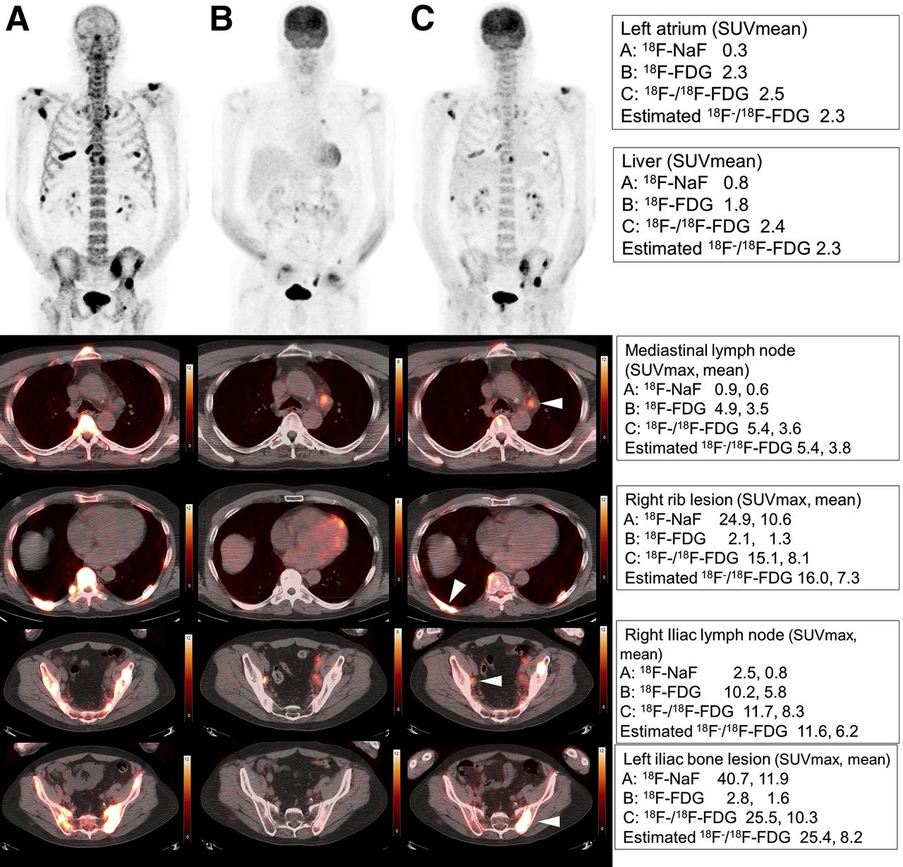
- FIGURE 1.
A 65-y-old man with metastatic prostate cancer. (A) 18F-NaF. (B) 18F-FDG. (C) 18F−/18F-FDG. SUVmax or SUVmean of left atrium, liver, mediastinal lymph node, right rib lesion, left iliac lymph node, and left iliac bone lesion were shown for 18F-NaF, 18F-FDG, and estimated 18F−/18F-FDG, respectively.
Tables
Parameter 18F−/18F-FDG 18F-FDG 18F-NaF Injection dose (MBq) 18F−/18F-FDG: 625.1 ± 85.8 478.2 ± 85.0 248.7 ± 84.0 18F-FDG: 426.9 ± 82.7 18F-NaF: 199.7 ± 35.5 Time from injection to imaging (min) 76.6 ± 13.0 75.5 ± 13.8 75.2 ± 17.7 The difference in time of starting PET/CT with 18F−/18F-FDG PET/CT (min) — 1.1 ± 13.0 1.3 ± 15.0 Time interval of PET/CT with 18F−/18F-FDG PET/CT (d) — 4.1 ± 4.7 4.1 ± 4.4 - TABLE 2
Radiopharmaceutical Uptake (SUVmean) for Studied Organs, Based on Time from Injection to Imaging
Time from injection to imaging (min) 18F−/18F-FDG 18F-FDG 18F-NaF Organ 52–60 (n = 6) 60–90 (n = 32) 90–104 (n = 11) 43–60 (n = 8) 60–90 (n = 35) 90–111 (n = 6) 39–60 (n = 10) 60–90 (n = 29) 90–117 (n = 10) Brain cortex (frontal lobe) 10.4 ± 2.4 9.7 ± 2.4 10.4 ± 2.5 10.0 ± 2.0 10.2 ± 2.5 11.2 ± 2.5 0.2 ± 0.1 0.3 ± 0.1 0.3 ± 0.1 Cerebellum 8.5 ± 1.8 8.2 ± 1.9 8.6 ± 2.0 8.2 ± 1.9 8.7 ± 1.9 9.2 ± 1.2 0.2 ± 0.1 0.2 ± 0.1 0.2 ± 0.1 Parotid grand 2.1 ± 0.9 1.9 ± 0.7 1.5 ± 0.4 1.6 ± 0.9 2.1 ± 1.1 1.4 ± 0.6 0.6 ± 0.2 0.5 ± 0.1 0.4 ± 0.1 Lung 0.7 ± 0.2 0.6 ± 0.2 0.7 ± 0.2 0.4 ± 0.2 0.5 ± 0.1 0.6 ± 0.1 0.3 ± 0.1 0.4 ± 0.2 0.4 ± 0.1 Myocardium 4.3 ± 2.4 4.8 ± 4.1 4.0 ± 4.0 3.7 ± 2.6 4.3 ± 3.9 7.7 ± 7.1 0.6 ± 0.3 0.7 ± 0.2 0.6 ± 0.2 Left atrium (background) 2.7 ± 0.7 2.3 ± 0.6 2.2 ± 0.6 1.8 ± 0.5 1.8 ± 0.4 1.9 ± 0.3 1.1 ± 0.4 1.2 ± 0.4 0.9 ± 0.3 Liver 2.6 ± 0.6 2.3 ± 0.5 2.5 ± 0.6 2.0 ± 0.5 2.2 ± 0.4 2.2 ± 0.3 0.4 ± 0.1 0.5 ± 0.2 0.4 ± 0.1 Spleen 2.2 ± 0.4 1.8 ± 0.6 2.3 ± 1.1 1.8 ± 0.6 1.7 ± 0.5 1.9 ± 0.3 0.6 ± 0.2 0.7 ± 0.2 0.5 ± 0.2 Pancreas 1.9 ± 0.3 1.7 ± 0.5 1.5 ± 0.6 1.4 ± 0.3 1.5 ± 0.4 1.2 ± 0.4 0.6 ± 0.2 0.6 ± 0.2 0.6 ± 0.2 Kidney 3.1 ± 0.8 2.7 ± 0.8 2.9 ± 1.1 2.4 ± 0.6 2.4 ± 0.6 2.0 ± 0.5 1.2 ± 0.5 1.7 ± 0.8 1.3 ± 0.5 Cecum 1.6 ± 0.4 1.7 ± 1.4 1.8 ± 1.8 0.9 ± 0.3 1.8 ± 2.0 1.0 ± 0.5 0.6 ± 0.2 0.7 ± 0.4 0.5 ± 0.1 Trapezius muscle 1.0 ± 0.3 1.0 ± 0.3 1.0 ± 0.3 0.6 ± 0.1 0.6 ± 0.1 0.7 ± 0.1 0.7 ± 0.2 0.8 ± 0.2 0.8 ± 0.2 Gluteus maximus muscle 1.0 ± 0.4 0.9 ± 0.3 1.1 ± 0.4 0.6 ± 0.2 0.7 ± 0.2 0.6 ± 0.1 0.7 ± 0.3 0.8 ± 0.2 0.7 ± 0.2 Fat tissue 0.3 ± 0.1 0.4 ± 0.2 0.4 ± 0.2 0.3 ± 0.1 0.3 ± 0.2 0.2 ± 0.1 0.2 ± 0.1 0.3 ± 0.1 0.2 ± 0.1 Cervical vertebrae 4.9 ± 2.6 4.5 ± 1.5 5.0 ± 2.7 1.3 ± 0.6 1.3 ± 0.3 1.7 ± 0.5 7.2 ± 3.1 6.9 ± 2.2 7.3 ± 2.5 Thoracic vertebrae 6.0 ± 2.9 5.0 ± 1.6 5.3 ± 2.5 1.8 ± 0.7 1.7 ± 0.6 1.6 ± 0.5 7.0 ± 2.5 7.6 ± 2.1 7.2 ± 2.0 Lumbar vertebrae 5.6 ± 2.4 4.8 ± 1.9 4.9 ± 3.0 1.5 ± 0.4 1.7 ± 0.6 1.5 ± 0.7 6.7 ± 1.6 7.1 ± 2.4 6.3 ± 2.4 Sacrum 4.9 ± 2.5 3.8 ± 1.5 4.2 ± 3.0 1.1 ± 0.3 1.2 ± 0.5 1.6 ± 0.1 5.8 ± 2.2 5.7 ± 2.3 5.3 ± 2.8 SUVmean Comparison of 18F−/18F-FDG and 18F-FDG Organ 18F−/18F-FDG 18F-FDG 18F-NaF P Correlation (r) Brain cortex (frontal lobe) 10.0 ± 2.4 10.3 ± 2.4 0.2 ± 0.1 0.32 0.62 Cerebellum 8.4 ± 1.9 8.7 ± 1.8 0.2 ± 0.1 0.51 0.49 Parotid grand 1.8 ± 0.7 1.9 ± 1.0 0.5 ± 0.2 0.77 0.75 Lung 0.6 ± 0.2 0.5 ± 0.1 0.4 ± 0.1 <0.001 0.76 Myocardium 4.5 ± 3.8 4.6 ± 4.3 0.7 ± 0.2 0.94 0.69 Left atrium (background) 2.3 ± 0.6 1.8 ± 0.4 1.1 ± 0.4 <0.001 0.71 Liver 2.4 ± 0.5 2.2 ± 0.4 0.5 ± 0.2 <0.007 0.60 Spleen 2.0 ± 0.7 1.7 ± 0.5 0.6 ± 0.2 <0.001 0.60 Pancreas 1.7 ± 0.5 1.5 ± 0.4 0.6 ± 0.2 0.001 0.46 Kidney 2.8 ± 0.8 2.4 ± 0.6 1.5 ± 0.7 0.003 0.28 Cecum 1.7 ± 1.4 1.6 ± 1.7 0.7 ± 0.3 0.30 0.75 Trapezius muscle 1.0 ± 0.3 0.6 ± 0.1 0.8 ± 0.2 <0.001 0.50 Gluteus maximus muscle 1.0 ± 0.3 0.7 ± 0.2 0.7 ± 0.2 <0.001 0.68 Fat tissue 0.4 ± 0.2 0.3 ± 0.2 0.2 ± 0.1 <0.001 0.85 Cervical vertebrae 4.7 ± 1.9 1.4 ± 0.4 7.0 ± 2.4 <0.001 0.51 Thoracic vertebrae 5.2 ± 2.0 1.7 ± 0.6 7.4 ± 2.1 <0.001 0.65 Lumbar vertebrae 4.9 ± 2.2 1.6 ± 0.6 6.8 ± 2.2 <0.001 0.71 Sacrum 4.0 ± 2.0 1.3 ± 0.5 5.7 ± 2.3 <0.001 0.66 - TABLE 4
SUVmean of 18F−/18F-FDG in Studied Organ and Differences from Estimated 18F−/18F-FDG Values
Comparison of 18F−/18F-FDG and estimated 18F−/18F-FDG Organ 18F−/18F-FDG Estimated 18F−/18F-FDG P Correlation (r) Brain cortex (frontal lobe) 10.0 ± 2.4 10.4 ± 2.4 0.17 0.62 Cerebellum 8.4 ± 1.9 8.8 ± 1.8 0.26 0.49 Parotid grand 1.8 ± 0.7 2.2 ± 1.1 <0.003 0.77 Lung 0.6 ± 0.2 0.7 ± 0.2 0.24 0.85 Myocardium 4.5 ± 3.8 5.0 ± 4.3 0.28 0.69 Left atrium (background) 2.3 ± 0.6 2.4 ± 0.6 0.16 0.80 Liver 2.4 ± 0.5 2.4 ± 0.5 0.24 0.64 Spleen 2.0 ± 0.7 2.0 ± 0.5 0.58 0.61 Pancreas 1.7 ± 0.5 1.8 ± 0.5 0.26 0.54 Kidney 2.8 ± 0.8 3.1 ± 0.8 <0.02 0.44 Cecum 1.7 ± 1.4 1.9 ± 1.9 0.58 0.76 Trapezius muscle 1.0 ± 0.3 1.0 ± 0.2 0.39 0.73 Gluteus maximus muscle 1.0 ± 0.3 1.0 ± 0.3 0.52 0.75 Fat tissue 0.4 ± 0.2 0.4 ± 0.2 0.66 0.87 Cervical vertebrae 4.7 ± 1.9 4.8 ± 1.7 0.19 0.77 Thoracic vertebrae 5.2 ± 2.0 5.3 ± 1.8 0.31 0.85 Lumbar vertebrae 4.9 ± 2.2 5.0 ± 1.9 0.47 0.85 Sacrum 4.0 ± 2.0 3.9 ± 1.6 0.94 0.80 - TABLE 5
SUVmean and SUVmax Measured from PET Scans and Estimated 18F−/18F-FDG Uptake in Lesions
P Correlation (r) Value Lesion 18F−/18F-FDG 18F-FDG 18F-NaF Estimated 18F−/18F-FDG 18F−/18F-FDG and 18F-FDG 18F−/18F-FDG and estimated 18F−/18F-FDG 18F−/18F-FDG and 18F-FDG 18F−/18F-FDG and 18F-NaF 18F−/18F-FDG and estimated 18F−/18F-FDG SUVmean All lesions* 7.3 ± 4.6 2.2 ± 1.7 10.0 ± 10.0 6.8 ± 4.2 <0.001 <0.007 0.10 0.84 0.88 Extraskeletal lesions 4.3 ± 2.4 3.7 ± 2.1 0.9 ± 0.4 4.1 ± 2.1 0.30 0.25 0.90 0.35 0.88 Skeletal lesions 8.3 ± 4.7 1.7 ± 1.0 12.6 ± 9.9 7.4 ± 4.3 <0.001 <0.001 0.50 0.86 0.92 Skeletal lesions, PC patient 7.3 ± 4.0 1.5 ± 0.8 10.2 ± 6.7 6.5 ± 3.5 <0.001 <0.003 0.23 0.83 0.85 Skeletal lesions, non–PC patient 10.5 ± 5.5 2.3 ± 1.2 18.7 ± 13.4 9.6 ± 5.2 <0.001 <0.001 0.69 0.88 0.98 Osteolytic lesions 10.9 ± 5.5 3.0 ± 1.0 14.5 ± 8.6 9.8 ± 5.1 <0.001 <0.02 0.51 0.88 0.96 Osteoblastic lesions 7.8 ± 4.4 1.5 ± 0.8 12.3 ± 10.0 7.0 ± 4.0 <0.001 <0.001 0.45 0.86 0.90 Background† 2.3 ± 0.6 1.8 ± 0.4 1.1 ± 0.4 2.4 ± 0.6 <0.001 0.48 0.70 0.72 0.81 SUVmax All lesions* 15.4 ± 11.4 4.7 ± 4.7 27.4 ± 28.8 16.9 ± 12.4 <0.001 <0.001 0.29 0.82 0.91 Extraskeletal lesions 9.5 ± 9.4 8.4 ± 6.3 2.2 ± 1.6 9.4 ± 6.7 0.61 0.87 0.94 0.91 0.94 Skeletal lesions 17.3 ± 11.3 3.4 ± 2.8 34.5 ± 28.9 19.1 ± 12.8 <0.001 <0.002 0.51 0.89 0.91 Skeletal lesions, PC patient 15.0 ± 9.6 2.7 ± 2.4 29.2 ± 23.9 17.3 ± 12.4 <0.001 <0.001 0.27 0.85 0.88 Skeletal lesions, non–PC patient 23.0 ± 13.5 5.2 ± 3.0 48.0 ± 35.8 23.8 ± 12.7 <0.001 0.19 0.69 0.91 0.98 Osteolytic lesions 21.6 ± 9.9 6.8 ± 2.3 36.8 ± 17.7 23.4 ± 8.5 <0.001 0.06 0.34 0.82 0.96 Osteoblastic lesions 16.6 ± 11.5 2.9 ± 2.5 34.2 ± 30.3 18.4 ± 13.2 <0.001 <0.001 0.52 0.90 0.90 Background† 3.3 ± 0.8 2.7 ± 0.7 1.8 ± 0.5 3.8 ± 0.9 <0.002 <0.02 0.52 0.52 0.67 Value Lesion 18F−/18F-FDG 18F-FDG 18F-NaF Estimated 18F−/18F-FDG P T/B ratio by SUVmean All lesions* 3.3 ± 2.2 1.2 ± 1.1 9.3 ± 9.7 2.8 ± 1.7 <0.001 Extraskeletal lesions 2.1 ± 1.8 2.1 ± 1.8 0.9 ± 0.4 1.9 ± 1.2 0.21 Skeletal lesions 3.6 ± 2.2 1.0 ± 0.8 11.7 ± 9.7 3.0 ± 1.8 0.006 Skeletal lesions, prostatecancer patient 3.3 ± 2.0 0.9 ± 0.9 9.4 ± 6.7 2.7 ± 1.5 <0.001 Skeletal lesions, non–prostate cancer patient 4.4 ± 2.3 1.2 ± 0.6 17.6 ± 13.2 4.0 ± 2.2 <0.005 Osteolytic lesions 4.6 ± 1.7 1.7 ± 0.6 13.9 ± 6.6 4.3 ± 2.0 0.28 Osteoblastic lesions 3.4 ± 2.2 0.9 ± 0.8 11.4 ± 10.1 2.8 ± 1.7 <0.001 T/B ratio by SUVmax All lesions* 4.8 ± 3.6 1.7 ± 1.7 14.6 ± 14.0 4.3 ± 3.0 <0.007 Extraskeletal lesions 3.3 ± 4.2 3.1 ± 2.4 1.4 ± 1.1 2.5 ± 1.9 0.09 Skeletal lesions 5.2 ± 3.3 1.2 ± 1.2 18.3 ± 13.7 4.8 ± 3.1 <0.03 Skeletal lesions, prostate cancer patient 4.7 ± 3.1 1.1 ± 1.2 16.0 ± 12.2 4.6 ± 3.1 0.61 Skeletal lesions, non–prostate cancer patient 6.2 ± 3.6 1.6 ± 1.1 24.2 ± 15.7 5.1 ± 3.1 <0.003 Osteolytic lesions 6.5 ± 3.0 2.4 ± 1.2 22.9 ± 9.8 6.4 ± 3.3 0.62 Osteoblastic lesions 5.0 ± 3.3 1.1 ± 1.1 17.6 ± 14.2 4.5 ± 3.0 0.011 ↵* All lesions: both soft-tissue lesions and bone lesions.
Supplemental Data
Files in this Data Supplement:


Jump to section
Related Articles
Cited By...
- Bone-Targeted Imaging and Radionuclide Therapy in Prostate Cancer
- Imaging Bone Metastases in Breast Cancer: Staging and Response Assessment
- Prospective Comparison of 99mTc-MDP Scintigraphy, Combined 18F-NaF and 18F-FDG PET/CT, and Whole-Body MRI in Patients with Breast and Prostate Cancer
- 18F-Fluoride PET in the Assessment of Malignant Bone Disease