
Main menu

  • Home
  • Content
    • Current
    • Ahead of print
    • Past Issues
    • JNM Supplement
    • SNMMI Annual Meeting Abstracts
    • Continuing Education
    • JNM Podcasts
  • Subscriptions
    • Subscribers
    • Institutional and Non-member
    • Rates
    • Journal Claims
    • Corporate & Special Sales
  • Authors
    • Submit to JNM
    • Information for Authors
    • Assignment of Copyright
    • AQARA requirements
  • Info
    • Reviewers
    • Permissions
    • Advertisers
  • About
    • About Us
    • Editorial Board
    • Contact Information
  • More
    • Alerts
    • Feedback
    • Help
    • SNMMI Journals
  • SNMMI
    • JNM
    • JNMT
    • SNMMI Journals
    • SNMMI

User menu

  • Subscribe
  • My alerts
  • Log in
  • My Cart

Search

  • Advanced search
Journal of Nuclear Medicine
  • SNMMI
    • JNM
    • JNMT
    • SNMMI Journals
    • SNMMI
  • Subscribe
  • My alerts
  • Log in
  • My Cart
Journal of Nuclear Medicine

Advanced Search

  • Home
  • Content
    • Current
    • Ahead of print
    • Past Issues
    • JNM Supplement
    • SNMMI Annual Meeting Abstracts
    • Continuing Education
    • JNM Podcasts
  • Subscriptions
    • Subscribers
    • Institutional and Non-member
    • Rates
    • Journal Claims
    • Corporate & Special Sales
  • Authors
    • Submit to JNM
    • Information for Authors
    • Assignment of Copyright
    • AQARA requirements
  • Info
    • Reviewers
    • Permissions
    • Advertisers
  • About
    • About Us
    • Editorial Board
    • Contact Information
  • More
    • Alerts
    • Feedback
    • Help
    • SNMMI Journals
  • View or Listen to JNM Podcast
  • Visit JNM on Facebook
  • Join JNM on LinkedIn
  • Follow JNM on Twitter
  • Subscribe to our RSS feeds
Review ArticleContinuing Education

Approaches to Reducing Radiation Dose from Radionuclide Myocardial Perfusion Imaging

Sharmila Dorbala, Ron Blankstein, Hicham Skali, Mi-Ae Park, Jolene Fantony, Charles Mauceri, James Semer, Stephen C. Moore and Marcelo F. Di Carli
Journal of Nuclear Medicine April 2015, 56 (4) 592-599; DOI: https://doi.org/10.2967/jnumed.112.115097
Sharmila Dorbala
Division of Nuclear Medicine and Molecular Imaging, Department of Radiology, the Noninvasive Cardiovascular Imaging Program, Departments of Medicine (Cardiology) and Radiology, Brigham and Women’s Hospital, Boston, Massachusetts
  • Find this author on Google Scholar
  • Find this author on PubMed
  • Search for this author on this site
Ron Blankstein
Division of Nuclear Medicine and Molecular Imaging, Department of Radiology, the Noninvasive Cardiovascular Imaging Program, Departments of Medicine (Cardiology) and Radiology, Brigham and Women’s Hospital, Boston, Massachusetts
  • Find this author on Google Scholar
  • Find this author on PubMed
  • Search for this author on this site
Hicham Skali
Division of Nuclear Medicine and Molecular Imaging, Department of Radiology, the Noninvasive Cardiovascular Imaging Program, Departments of Medicine (Cardiology) and Radiology, Brigham and Women’s Hospital, Boston, Massachusetts
  • Find this author on Google Scholar
  • Find this author on PubMed
  • Search for this author on this site
Mi-Ae Park
Division of Nuclear Medicine and Molecular Imaging, Department of Radiology, the Noninvasive Cardiovascular Imaging Program, Departments of Medicine (Cardiology) and Radiology, Brigham and Women’s Hospital, Boston, Massachusetts
  • Find this author on Google Scholar
  • Find this author on PubMed
  • Search for this author on this site
Jolene Fantony
Division of Nuclear Medicine and Molecular Imaging, Department of Radiology, the Noninvasive Cardiovascular Imaging Program, Departments of Medicine (Cardiology) and Radiology, Brigham and Women’s Hospital, Boston, Massachusetts
  • Find this author on Google Scholar
  • Find this author on PubMed
  • Search for this author on this site
Charles Mauceri
Division of Nuclear Medicine and Molecular Imaging, Department of Radiology, the Noninvasive Cardiovascular Imaging Program, Departments of Medicine (Cardiology) and Radiology, Brigham and Women’s Hospital, Boston, Massachusetts
  • Find this author on Google Scholar
  • Find this author on PubMed
  • Search for this author on this site
James Semer
Division of Nuclear Medicine and Molecular Imaging, Department of Radiology, the Noninvasive Cardiovascular Imaging Program, Departments of Medicine (Cardiology) and Radiology, Brigham and Women’s Hospital, Boston, Massachusetts
  • Find this author on Google Scholar
  • Find this author on PubMed
  • Search for this author on this site
Stephen C. Moore
Division of Nuclear Medicine and Molecular Imaging, Department of Radiology, the Noninvasive Cardiovascular Imaging Program, Departments of Medicine (Cardiology) and Radiology, Brigham and Women’s Hospital, Boston, Massachusetts
  • Find this author on Google Scholar
  • Find this author on PubMed
  • Search for this author on this site
Marcelo F. Di Carli
Division of Nuclear Medicine and Molecular Imaging, Department of Radiology, the Noninvasive Cardiovascular Imaging Program, Departments of Medicine (Cardiology) and Radiology, Brigham and Women’s Hospital, Boston, Massachusetts
  • Find this author on Google Scholar
  • Find this author on PubMed
  • Search for this author on this site
  • Article
  • Figures & Data
  • Supplemental
  • Info & Metrics
  • PDF
Loading

Article Figures & Data

Figures

  • Tables
  • Additional Files
  • FIGURE 1.
    • Download figure
    • Open in new tab
    • Download powerpoint
    FIGURE 1.

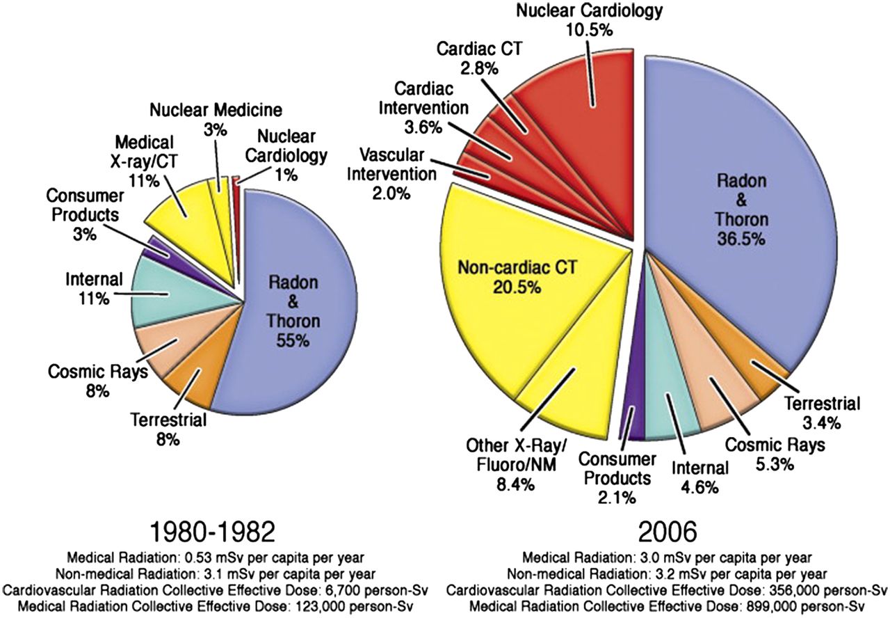
    Increasing radiation burden in United States and contributions from medical imaging. Collective dose from medical imaging increased 6-fold in 2006 compared with early 1980s. (Reprinted with permission of (2).)

  • FIGURE 2.
    • Download figure
    • Open in new tab
    • Download powerpoint
    FIGURE 2.

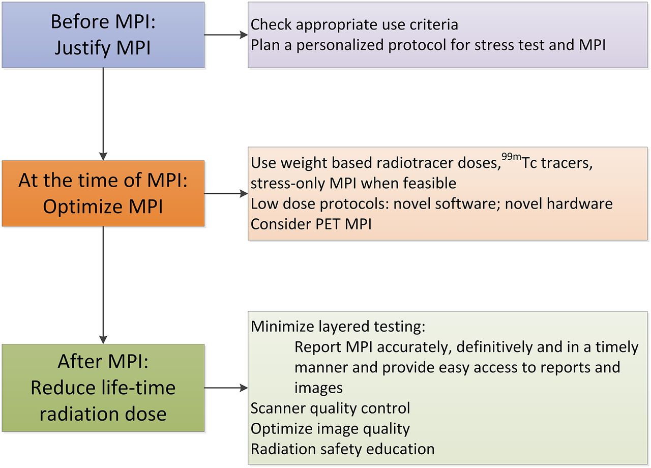
    Practical ways to implement reduced-radiation-dose MPI program.
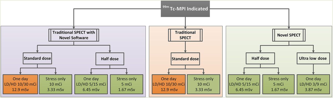
  • FIGURE 3.
    • Download figure
    • Open in new tab
    • Download powerpoint
    FIGURE 3.

    Patient-centered protocols for low-radiation-dose MPI: traditional SPECT (orange), traditional SPECT with novel software (purple), and novel SPECT scanners (green). Most MPI procedures that use novel protocols or novel technologies provide <9-mSv radiation dose from rest–stress 99mTc protocols. To achieve 50% of laboratory volume with <9-mSv dose, practices can implement several of the above options into their practice. LD = low dose; HD = high dose; 1 mCi = 37 MBq.

Tables

  • Figures
  • Additional Files
    • View popup
    TABLE 1

    Five Things Physicians and Patients Need to Know (15,16)

    SourcePoint
    SNMMIDo not perform routine annual stress testing after coronary artery revascularization.
    ASNCDo not perform stress cardiac imaging or coronary angiography in patients without cardiac symptoms unless high-risk markers are present.
    Do not perform cardiac imaging in patients who are at low risk.
    Do not perform radionuclide imaging as part of routine follow-up in asymptomatic patients.
    Do not perform cardiac imaging as preoperative assessment in patients scheduled to undergo low- or intermediate-risk noncardiac surgery.
    Use methods to reduce radiation exposure in cardiac imaging whenever possible, including not performing such tests when the benefits will likely be limited.
    • View popup
    TABLE 2

    Steps to Create Protocol Plan for MPI

    StepDescription
    1Review electronic medical records to define clinical question.
    2Check for recently performed cardiac evaluations to avoid duplicate testing and layered testing for similar clinical symptoms.
    3If clinical question is not clear or if test ordered is not the most appropriate test, discuss with referring physician for clarification.
    4Plan appropriate stress technique.
    5Plan appropriate imaging technique.
    • View popup
    TABLE 3

    Estimation of Effective Radiation Dose from Various Myocardial Perfusion Radiotracers (13)

    Administered activity
    MBqmCiEstimated dose (mSv)
    RadiopharmaceuticalEffective dose (mSv/MBq)Full doseHalf doseFull doseHalf doseFull-dose studyHalf-dose study
    82Rb rest or stress0.00171,48074040202.521.26
    13N-ammonia rest or stress0.002774037020102.01.0
    99mTc-sestamibi rest0.0079296148842.341.17
    99mTc-sestamibi stress0.00988844424128.04.0
    99mTc-tetrofosmin rest0.0069296148842.01.0
    99mTc-tetrofosmin stress0.006988844424126.133.1
    201Tl0.14148744220.7210.36
    • Recommended MPI radiotracer doses for conventional scanners are 8–12 mCi of 99mTc-sestamibi for rest imaging and 24–36 mCi for stress imaging, 40–60 mCi of 82Rb for 2D imaging and 20 mCi for 3D imaging (58), 20 mCi of 13N-ammonia for 2D imaging and 10 mCi for 3D imaging (58), and 2.5–4 mCi for 201Tl imaging (59) (1 mCi = 37 MBq). New estimates of 82Rb dose are significantly lower (0.00126 mSv/MBq) (60). Full-dose PET radiotracer is used for 2D imaging and half-dose for 3D imaging; typically, equal dose of radiotracer is administered for rest and for stress PET MPI. Average activities are listed. Estimated dose is effective dose multiplied by administered activity. Dose is calculated for rest and stress scans separately, and if attenuation correction is used, 0.3–0.7 mSv is added for CT and 0.3 mSv for radionuclide transmission scanning (13).

    • View popup
    TABLE 4

    Radiation Dose from Low-Dose Protocols for Novel SPECT Scanners

    StudyRest dose*Stress dose*No. of patientsPatient sizeRadiotracerProtocol (1 d)Study radiation dose (mSv)
    24296–481 (8–13)462.5; 925–1,332 (12.5; 25–36)717<91 kg (200 lb)99mTc-sestamibiLD stress only4.2
    HD stress only8.0
    Stress–rest11.8
    61185 (5)555 (15)131BMI, <3599mTc-sestamibiRest–stress5.8
    62640 (17.29)320 (8.65)50BMI, 19–3299mTc-tetrofosminStress–restStress only, 2.21; stress + rest, 6.62
    52185–222 (5–6)370–444 (10–12)137BMI, 39 ± 799mTc-tetrofosminStress–rest5.10–6.12
    51222 (6)740 (20)285BMI, 29 ± 599mTc-tetrofosminRest–stressRest, 1.4; stress, 4.6
    48129.5 (3.5)NA101BMI, 17.1–30.999mTc-sestamibiRest only1.2
    • ↵* Data are megabecquerels followed by millicuries in parentheses (range or mean).

    • LD = low dose; HD = high dose; BMI = body mass index (kg/m2; range, mean ± SD, or upper limit); NA = not applicable.

    • Variable scan times were used for count-based acquisition. The scanner was a Discovery NM/CT 570c (GE Healthcare) (62), D-SPECT (Spectrum Dynamics) (48), or Discovery NM530c (GE Healthcare) (24,51,52,61).

Additional Files

  • Figures
  • Tables
  • Supplemental Data

    Files in this Data Supplement:

    • Supplemental Data
PreviousNext
Back to top

In this issue

Journal of Nuclear Medicine: 56 (4)
Journal of Nuclear Medicine
Vol. 56, Issue 4
April 1, 2015
  • Table of Contents
  • Table of Contents (PDF)
  • About the Cover
  • Index by author
Print
Download PDF
Article Alerts
Sign In to Email Alerts with your Email Address
Email Article

Thank you for your interest in spreading the word on Journal of Nuclear Medicine.

NOTE: We only request your email address so that the person you are recommending the page to knows that you wanted them to see it, and that it is not junk mail. We do not capture any email address.

Enter multiple addresses on separate lines or separate them with commas.
Approaches to Reducing Radiation Dose from Radionuclide Myocardial Perfusion Imaging
(Your Name) has sent you a message from Journal of Nuclear Medicine
(Your Name) thought you would like to see the Journal of Nuclear Medicine web site.
Citation Tools
Approaches to Reducing Radiation Dose from Radionuclide Myocardial Perfusion Imaging
Sharmila Dorbala, Ron Blankstein, Hicham Skali, Mi-Ae Park, Jolene Fantony, Charles Mauceri, James Semer, Stephen C. Moore, Marcelo F. Di Carli
Journal of Nuclear Medicine Apr 2015, 56 (4) 592-599; DOI: 10.2967/jnumed.112.115097

Citation Manager Formats

  • BibTeX
  • Bookends
  • EasyBib
  • EndNote (tagged)
  • EndNote 8 (xml)
  • Medlars
  • Mendeley
  • Papers
  • RefWorks Tagged
  • Ref Manager
  • RIS
  • Zotero
Share
Approaches to Reducing Radiation Dose from Radionuclide Myocardial Perfusion Imaging
Sharmila Dorbala, Ron Blankstein, Hicham Skali, Mi-Ae Park, Jolene Fantony, Charles Mauceri, James Semer, Stephen C. Moore, Marcelo F. Di Carli
Journal of Nuclear Medicine Apr 2015, 56 (4) 592-599; DOI: 10.2967/jnumed.112.115097
Twitter logo Facebook logo LinkedIn logo Mendeley logo
  • Tweet Widget
  • Facebook Like
  • Google Plus One
Bookmark this article

Jump to section

  • Article
    • Abstract
    • THE VITAL IMPORTANCE OF RADIONUCLIDE MPI TO MANAGE CAD
    • THE NEED TO REDUCE RADIATION DOSE FROM MPI
    • RADIATION DOSE REDUCTION BEFORE MPI: APPROPRIATE USE
    • RADIATION DOSE REDUCTION DURING THE TEST: OPTIMIZE MPI
    • RADIATION DOSE REDUCTION AFTER THE TEST
    • CONCLUSION
    • Footnotes
    • REFERENCES
  • Figures & Data
  • Supplemental
  • Info & Metrics
  • PDF

Related Articles

  • This Month in JNM
  • PubMed
  • Google Scholar

Cited By...

  • Worldwide Diagnostic Reference Levels for Single-Photon Emission Computed Tomography Myocardial Perfusion Imaging: Findings From INCAPS
  • Using radiation safely in cardiology: what imagers need to know
  • Simplified approach to stress-first nuclear myocardial perfusion imaging: implementation of Choosing Wisely recommendations
  • Effective Dose in Nuclear Medicine Studies and SPECT/CT: Dosimetry Survey Across Quebec Province
  • The impact of IUGR on pancreatic islet development and {beta}-cell function
  • Trials of Quality Improvement in Imaging
  • Google Scholar

More in this TOC Section

  • Treatment Response Evaluation in Prostate Cancer Using PSMA PET/CT
  • Approaches to Imaging Immune Activation Using PET
  • Large Language Models and Large Multimodal Models in Medical Imaging: A Primer for Physicians
Show more Continuing Education

Similar Articles

Keywords

  • SPECT
  • low dose
  • PET
  • radiation dose
  • myocardial perfusion imaging
  • Quality Assurance
SNMMI

© 2025 SNMMI

Powered by HighWire