


Article Figures & Data
Figures
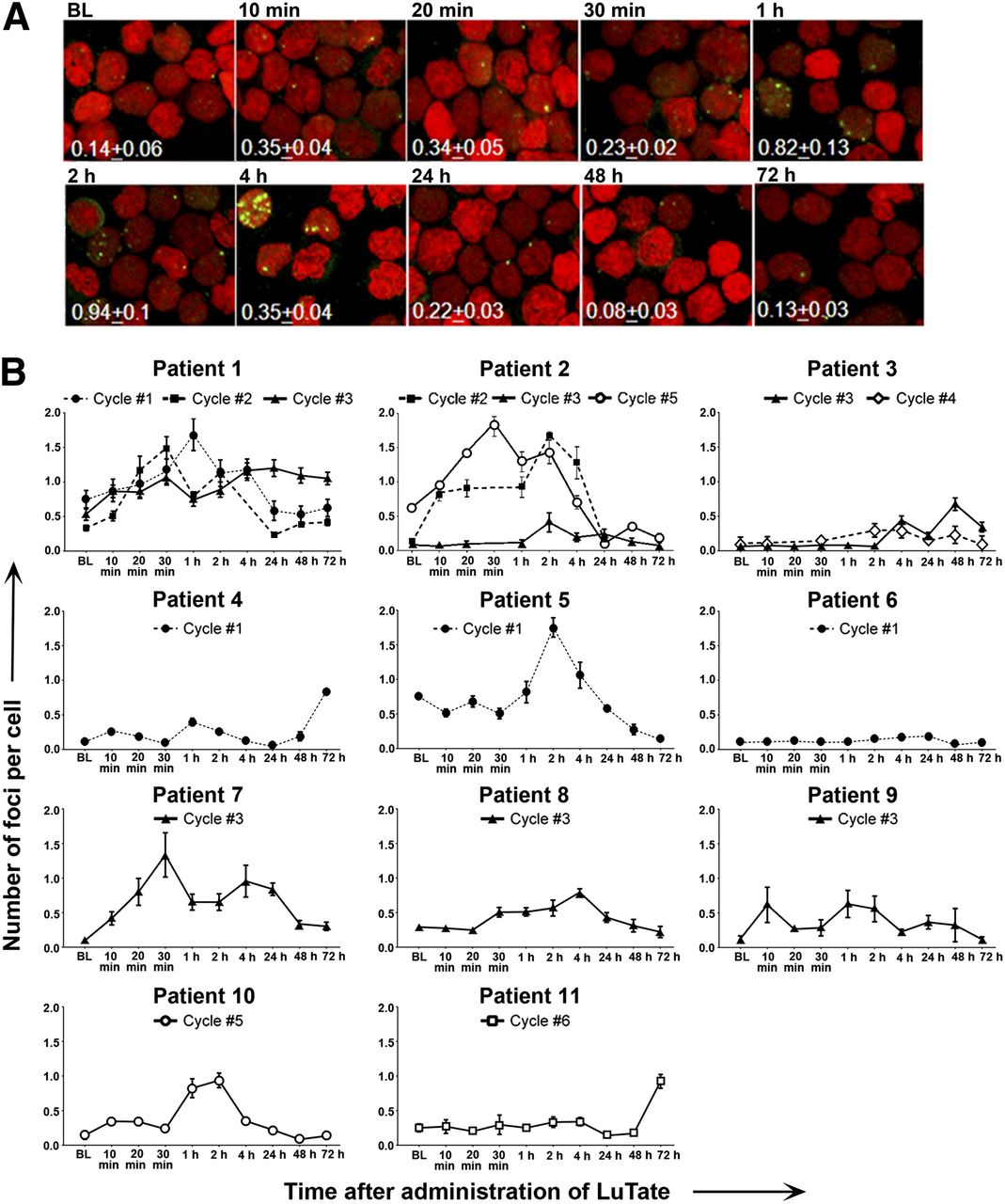
- FIGURE 1.
LuTate time–activity curve in blood up to 72 h after treatment. Each point represents single measurement of radioactivity in the blood of 11 patients at indicated times. Solid line is biexponential fit of LuTate disappearance from blood.
- FIGURE 2.
Kinetics of γ-H2AX foci formation in PBLs. (A) Representative confocal images of γ-H2AX foci (green) and nuclei (red, DNA) of patient P10.5 after administration of 8,362 MBq of LuTate. Values correspond to average number of foci per cell ± SEM. (B) Number of foci per cell in individual patients measured at baseline (BL) before therapy and 10, 20, and 30 min and at 1, 2, 4, 24, 48, and 72 h after LuTate administration in one or more cycles of therapy.
- FIGURE 3.
Analysis of γ-H2AX foci formation in PBLs. (A) Number of foci per cell vs. time after PRRT for individual patient. Black horizontal bar and error bars represent mean ± SEM of 14–16 samples. (B) Fraction of cells with no foci (blue bars), ≤3 foci (red bars), and ≥4 foci (green bars) (mean ± SEM, n = 14–16). (C) Simulation of number of foci per cell as function of time after PRRT. Symbols represent average experimental values across all patients ± SD. Simulation was implemented for radiation dose of 42 mGy delivered as pulse (blue line) or accumulated (red line) within 80-h interval. Accumulated dose was calculated according to LuTate kinetics in blood (black line). *P < 0.05, Mann–Whitney test, for baseline vs. other time points. **P < 0.01, Mann–Whitney test, for baseline vs. other time points.
- FIGURE 4.
Correlation studies. (A) Peak number of excess foci per cell in 10-min to 4-h interval vs. dose to blood at 2 h. (B) Peak number of excess foci per cell in 24- to 72-h interval vs. tumor dose. (C) Peak number of foci per cell in 10-min to 4-h interval vs. dose to bone marrow. (D) Peak number of excess foci per cell in 10-min to 4-h interval vs. lymphocyte reduction 2 wk after treatment. Data for different treatment cycles of same patient are shown as blue (P1), green (P2), and black (P3) symbols; other data points are shown as red symbols. Red lines represent linear regression for all points.


Tables
Patient blood identifier Sex Age at treatment (y) Body weight (kg) Diagnosis Primary site Treatments before PRRT 5-FU in combination with PRRT P1.1 M 46 98 Neuroendocrine Pancreas Chemotherapy + P1.2 M 47 96 Neuroendocrine Pancreas Chemotherapy + P1.3 M 47 100.4 Neuroendocrine Pancreas Chemotherapy + P2.2 M 62 82.3 Carcinoid Small bowel Nil − P2.3 M 62 82 Carcinoid Small bowel Nil + P2.5 M 62 80.1 Carcinoid Small bowel Nil + P3.3 F 46 133 Neuroendocrine Small bowel Nil + P3.4 F 46 130 Neuroendocrine Small bowel Nil + P4.1 M 55 77 Carcinoid Bowel Surgery − P5.1 F 52 79 Neuroendocrine Small bowel Chemotherapy/Sandostatin + P6.1 M 57 81 Neuroendocrine Pancreas Sandostatin − P7.3 F 49 58.6 Islet cell Pancreas Surgery + P8.3 M 32 85 Neuroendocrine Pancreas Surgery + P9.3 F 46 77 Glucagonoma Pancreas Chemotherapy/Sandostatin/surgery + P10.5 F 65 83 Gastrinoma Duodenum Chemotherapy + P11.6 M 60 114 Carcinoid Unknown Chemotherapy/Sandostatin − Patients are numbered from P1 to P11 followed by treatment cycle number. Sandostatin (Novartis AG Corp.) is a long-acting octreotide.
Patient blood identifier Octreotate-avid tumor volume at treatment (cm3) Administered activity (MBq) Cumulative absorbed dose to blood at 72 h (mGy) Cumulative absorbed dose to blood at 2 h (mGy) Absorbed dose to spleen (mGy) Absorbed dose to bone marrow (mGy) Absorbed dose to tumor (mGy)* P1.1 640 9,000 40 10 2,800 210 37,000 P1.2 700 7,000 22 7 3,600 210 31,000 P1.3 720 8,000 33 12 2,000 200 23,000 P2.2 3,100 10,000 57 16 3,200 370 30,000 P2.3 3,300 7,900 52 14 3,400 410 24,000 P2.5 3,000 7,800 37 8 ND ND ND P3.3 420 9,300 55 22 3,300 270 14,000 P3.4 240 6,500 24 11 1,700 190 13,000 P4.1 94 7,100 ND ND ND ND ND P5.1 130 6,900 ND ND 1,700 250 12,000 P6.1 1,200 7,700 45 11 10,000 190 46,000 P7.3 ND ND ND 17 ND ND ND P8.3 29 8,600 ND ND 6,100 200 6,000 P9.3 30 6,800 ND ND 3,400 280 7,000 P10.5 110 8,400 52 21 5,100 280 20,000 P11.6 900 6,700 ND ND 1,900 170 18,000 ↵* For multiple lesions, lesion with highest value was used.
ND = not determined.
Patients are numbered from P1 to P11 followed by treatment cycle number.
Patient blood identifier Baseline PBL count before first treatment (cells × 103/mm3) PBL counts before indicated treatment* (cells × 103/mm3) PBL count 2 wk after indicated treatment* (cells × 103/mm3) PBL reduction (%) before indicated treatment to 2 wk after indicated treatment PBL reduction (%) between baseline and 2 wk after indicated treatment P1.1 1.6 1.6 0.6 63 63 P1.2 1.6 0.8 0.5 38 69 P1.3 1.6 1.0 0.5 50 69 P2.2 1.4 1.0 0.5 50 64 P2.3 1.4 0.5 0.4 20 71 P2.5 1.4 0.5 0.3 40 79 P3.3 2.9 1.8 0.9 50 69 P3.4 2.9 1.1 0.9 18 69 P4.1 1.2 1.2 0.8 33 33 P5.1 1.5 1.5 0.8 47 47 P6.1 2.3 2.3 0.9 61 61 P7.3 2.5 0.5 0.3 40 88 P8.3 1.7 0.9 0.6 33 65 P9.3 1.3 0.9 0.4 56 70 P10.5 1.9 1.2 0.6 50 68 P11.6 2.3 1.7 1.5 12 34 ↵* “Indicated treatment” refers to treatment cycle number denoted by last digit in patient blood identifier.
Patients are numbered from P1 to P11 followed by treatment cycle number.
Supplemental Data
Files in this Data Supplement:


Jump to section
Related Articles
Cited By...
- Dosimetric Evaluation of the Effect of Receptor Heterogeneity on the Therapeutic Efficacy of Peptide Receptor Radionuclide Therapy: Correlation with DNA Damage Induction and In Vivo Survival
- Dissimilar DNA Damage to Blood Lymphocytes After 177Lu-Labeled DOTATOC or Prostate-Specific Membrane Antigen Therapy
- Peptide Receptor Radionuclide Therapy During the COVID-19 Pandemic: Are There Any Concerns?
- Imaging DNA Damage Repair In Vivo After 177Lu-DOTATATE Therapy
- Identification of brain metastasis genes and therapeutic evaluation of histone deacetylase inhibitors in a clinically relevant model of breast cancer brain metastasis
- The Relevance of Dosimetry in Precision Medicine
- Identification of brain metastasis genes and therapeutic evaluation of histone deacetylase inhibitors in a clinically relevant model of breast cancer brain metastasis
- Localized Synchrotron Irradiation of Mouse Skin Induces Persistent Systemic Genotoxic and Immune Responses
- Radiotherapy for Non-Small Cell Lung Cancer Induces DNA Damage Response in Both Irradiated and Out-of-field Normal Tissues
- Myeloid neoplasms after chemotherapy and PRRT: myth and reality
- DNA Damage in Peripheral Blood Lymphocytes of Thyroid Cancer Patients After Radioiodine Therapy
- Radiopeptides for Imaging and Therapy: A Radiant Future
- {gamma}-H2AX Foci in Peripheral Blood Lymphocytes to Quantify Radiation-Induced DNA Damage After 177Lu-DOTA-Octreotate Peptide Receptor Radionuclide Therapy