


Article Figures & Data
Figures
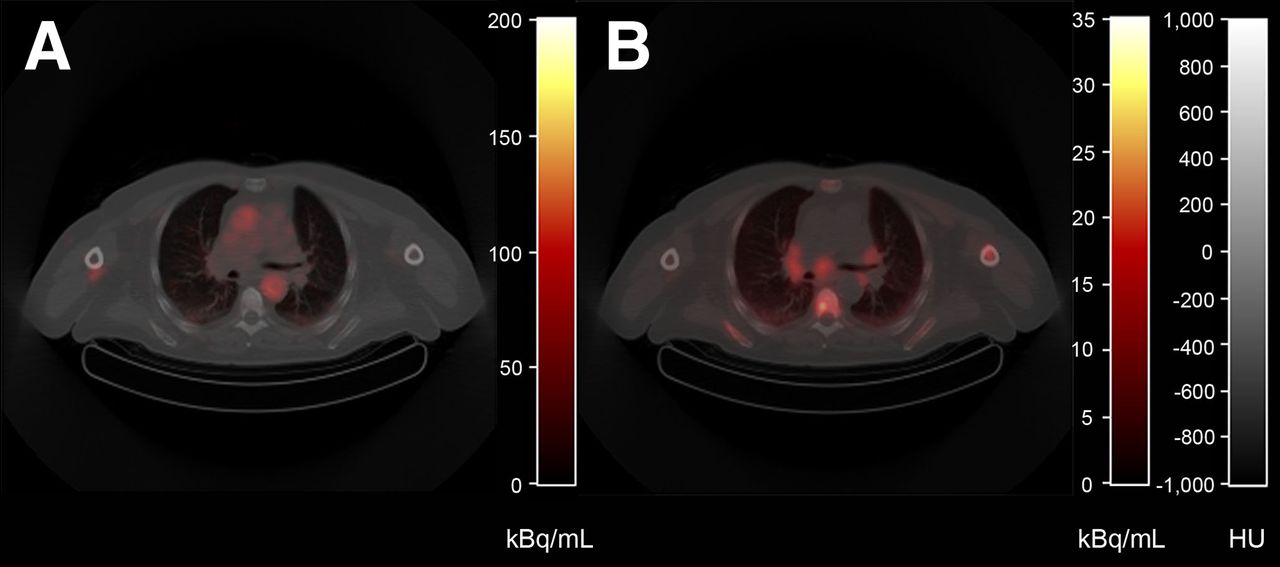
- FIGURE 1.
Typical example of PET/CT images acquired from patient diagnosed with PCa. Shown are low-dose CT (gray scale) fused with early PET image acquired from 35 to 40 s after injection displaying blood pool (A) and averaged image over 25–40 min after 18F-fluoromethylcholine injection (B) (color).
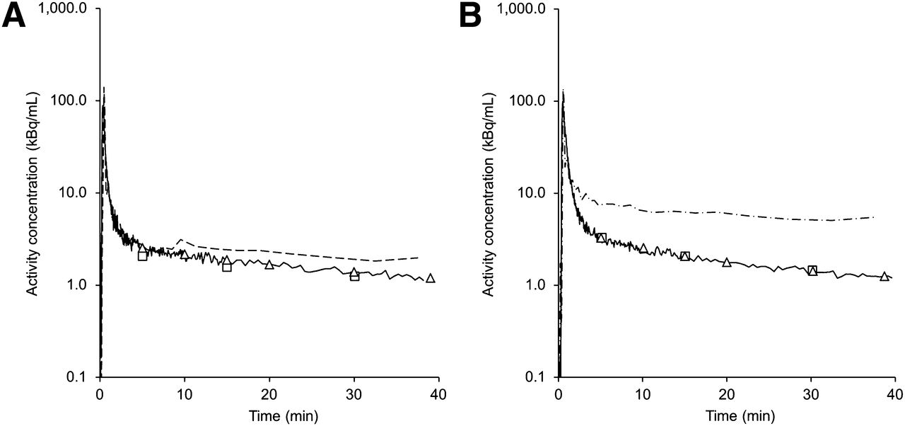
- FIGURE 2.
Two typical examples of measured whole-blood time–activity curves for patient imaged over thorax region (A) and patient imaged over abdominal region (B). Lines = calibrated blood sampler data; dashed lines (image-derived) = aortic arch; dash-dot lines (image-derived) = abdominal descending aorta; triangles = manual arterial blood samples; squares = manual venous blood samples.
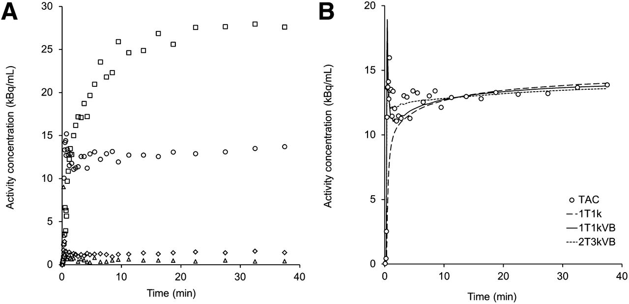
- FIGURE 3.
(A) Typical measured time–activity curves for patient shown in Figure 1. ◱ = liver; ○ = lesion; ♢ = muscle; △ = fat tissue. (B) Nonlinear regression fits to lesion time–activity curve displayed in A, using various compartment models.
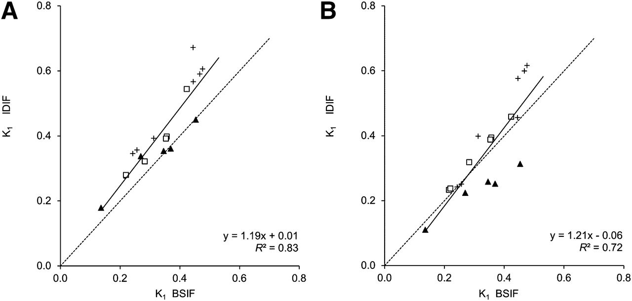
- FIGURE 4.
K1 obtained using 1T1k+VB with calibrated (A) and noncalibrated (B) IDIF-derived, compared with BSIF method. Symbols indicate IDIF origin: ▲ = aortic arch; ◱ = descending aorta; + = femoral artery.
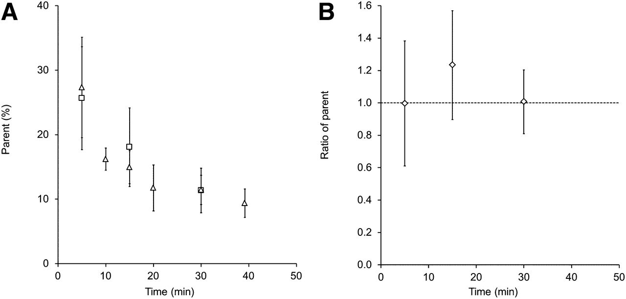
- FIGURE 5.
Manual blood sample data as function of time: mean parent fractions (△ = arterial, ◱ = venous) (A) and ratio of venous to arterial parent fractions (B). Error bars represent ± SD.
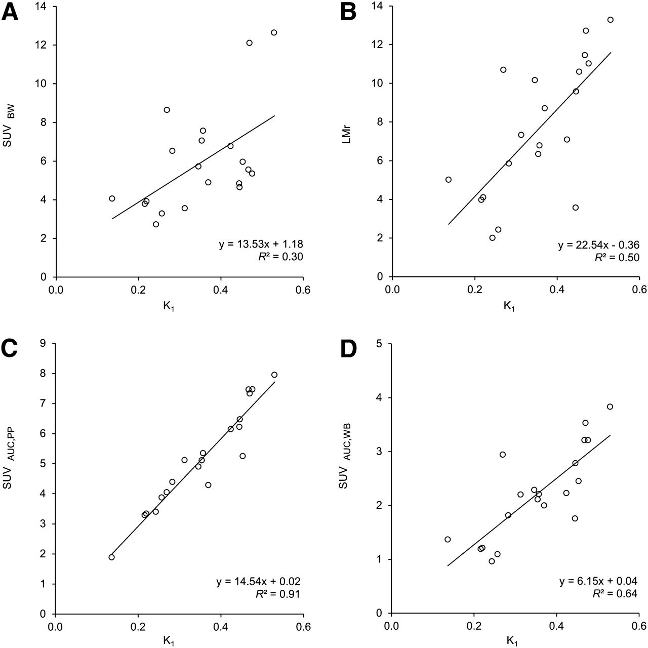
- FIGURE 6.
Correlation between simplified uptake measures and K1 (1T1k+VB). (A) SUV normalized to body weight (SUVBW), (B) LMr and lesion activity concentration in kBq at 30–40 min after injection divided by AUC of parent plasma (C), and whole-blood activity concentration (D) in MBq over 0–30 min (SUVAUC,PP and SUVAUC,WB, respectively).


Tables
Linear regression analysis* Parameter (model) Scan duration (min) Robustness (%) Slope Intercept R2 ICC* K1 (1T1k+VB) 5 100 0.98 0.05 0.82 0.84 10 100 0.98 0.03 0.94 0.95 15 100 0.99 0.02 0.97 0.98 20 100 1.01 0.01 0.99 0.99 30 100 1.00 0.00 1.00 1.00 Ki (2T3k+VB) 5 60 0.82 0.09 0.59 0.75 10 65 0.93 0.03 0.97 0.98 15 75 1.07 −0.02 0.94 0.97 20 75 1.05 −0.01 0.90 0.95 30 80 1.03 −0.01 1.00 1.00 ↵* Results for comparison of quantification parameters from short scan durations to those derived from full 40-min dynamic scan.
- TABLE 3
Comparison of Simplified Parameters to K1 Resulting from Full Kinetic Modeling with 1T1k+VB
Linear regression analysis Simplified parameter Slope Intercept R2 SUVBW 13.53 1.18 0.30 SUVBSA 315.39 27.53 0.33 SUVLBM 9.02 0.86 0.32 SUVBMI 4.65 0.20 0.34 SUVIBW 10.96 1.28 0.31 LMr 22.54 −0.36 0.50 LBr 30.38 0.32 0.44 SUVAUC,WB 5.88 −0.02 0.65 SUVAUC,PP 14.73 −0.20 0.92 SUVBW = SUV normalized to body weight; SUVBSA = SUV normalized to body surface area; SUVLBM = SUV normalized to lean body mass; SUVBMI = SUV normalized to normalized body mass index; SUVIBW = SUV normalized to ideal body weight.
Supplemental Data
Files in this Data Supplement:


Jump to section
Related Articles
Cited By...
- Simplified Methods for Quantification of 18F-DCFPyL Uptake in Patients with Prostate Cancer
- [18F]Fluorocholine and [18F]Fluoroacetate PET as Imaging Biomarkers to Assess Phosphatidylcholine and Mitochondrial Metabolism in Preclinical Models of TSC and LAM
- 11C-Choline Pharmacokinetics in Recurrent Prostate Cancer
- Repeatability of Quantitative 18F-Fluoromethylcholine PET/CT Studies in Prostate Cancer
- Simplified Methods for Quantification of 18F-Fluoromethylcholine Uptake: Is SUVAUC,PP Actually an SUV?
- Reply: Simplified Methods for Quantification of 18F-Fluoromethylcholine Uptake: Is SUVAUC,PP Actually an SUV?